0569
Towards whole-cortex enhancement with a uHDC Helmet at 3T1Radiology, Pennsylvania State College of Medicine, Hershey, PA, United States, 2HyQ Research Solutions, LLC, State College, PA, United States, 3Engineering Science and Mechanics, Pennsylvania State University, University Park, PA, United States
Synopsis
We performed a human brain imaging study using a prototype conformal helmet constructed with ultrahigh dielectric constant (uHDC) materials inserted into a standard 20-Ch head coil at 3T. We characterized the transmit and receive performance of the helmet by comparing to results using the 20-Ch and 64-Ch head coil (n=5 subjects) without the uHDC helmet. The SNR and its spatial distribution within the cerebrum using the 20-Ch Coil with uHDC Helmet is comparable to that of the 64-Ch Coil. Further improvement of the uHDC helmet insert is expected to significantly improve the performance of standard receive arrays for brain imaging.
Purpose
Prior work at 3T has focused on the development of flexible pads with permittivities up to 5501, or monolithic ceramics with permitivies in the range of 800 – 47002. Each design offers a trade-off: pads are more conformal at the expense of lower permittivity and reduced enhancement, while monolithic ceramics offer greater enhancement though it is more difficult to shape the material conformal to human body. To date, most designs have only offered regional enhancement of the B fields, whereas a more global enhancement throughout the brain would be desirable for many neuroimaging applications. Here we present a brain imaging study using a conformal helmet with high permittivity (er ~ 1000) that can be inserted into a commercial receive coil.Methods
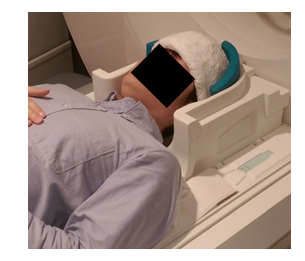
Figure 1 displays the uHDC helmet that conforms to the upper human head to the level above eye and ear and can be fitted into the standard Siemens 20-Channel head array. Acquisition: All data was acquired on a Siemens 3T PrismaFit (Siemens Healthineers, Erlangen, Germany) with a 2-Ch pTX transmit coil. B1+ maps were acquired with a Bloch-Siegert3 based sequence, with 100 mm of longitudinal coverage from the apex to the cerebellum. A small-tip angle GRE acquistion with parameters matched to the B1+ scan was acquired for calculation of B1+-normalized SNR maps4. Each subject (n = 5) was scanned under 3 reception configurations: 20-Ch Coil, 20-Ch Coil with uHDC Helmet (V1), 64-Ch Coil. One subject was scanned with the uHDC helmet in a higher permittivity configuration (V2) (er ~ 1200). Additionally, on a single subject, absolute B1+ magnitude and relative transmit phase maps were acquired on both channels of the transmit coil with the aforementioned protocols. Analysis: A distance map was calculated over the brain, the distance metric defined as the shortest Euclidean distance between a voxel and the edge of the brain mask. This distance map was utilized in a histogram analysis to quantify SNR of each receive configuration. A RF pulse optimization5 that utilized the B1+ magnitude and transmit phase maps was applied to calculate a blipped-spokes pulse off-line. This RF pulse was designed with a target flip angle of 70 degrees, 5 spokes and a duration of 7.38 ms.Results & Discussion
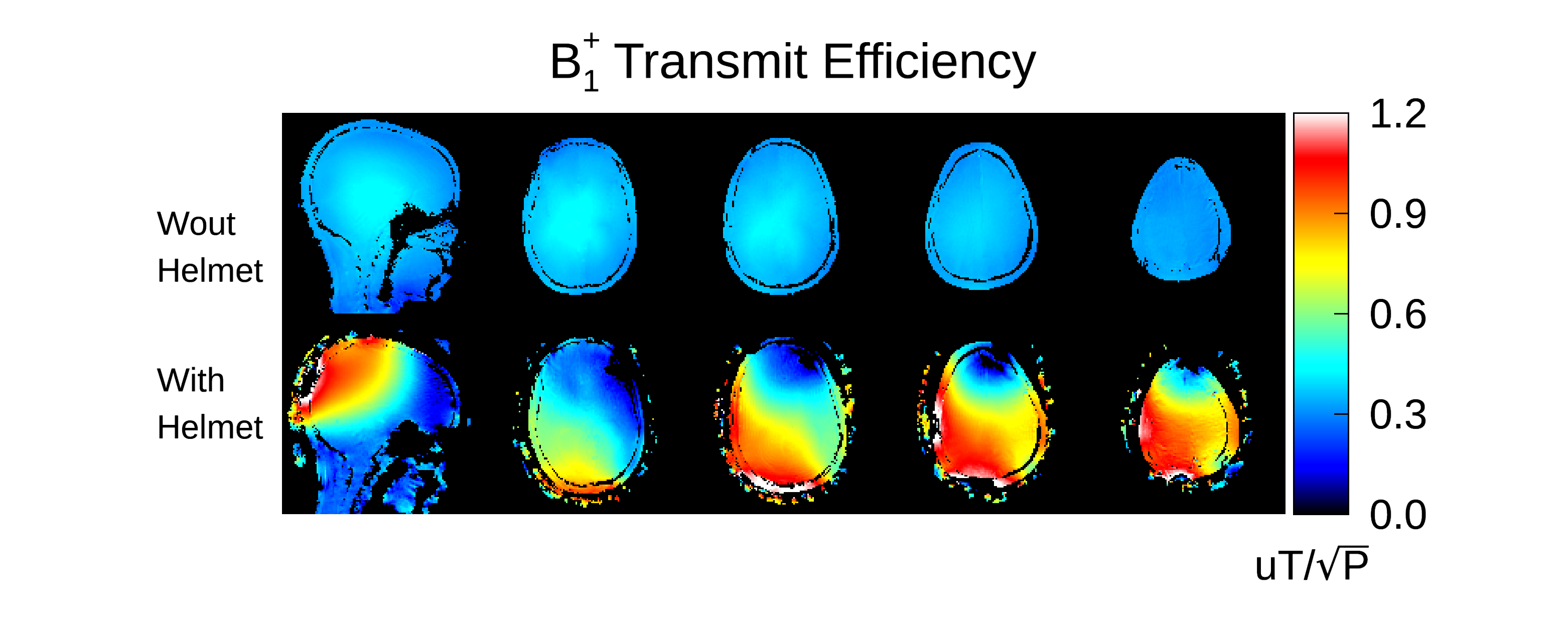
Figure 2 displays representative transmit efficiency maps from a single
subject. The helmet increased efficiency by up to a factor of 2 to 2.5 within
the brain, though the overall homogeneity of the field pattern suffered. In
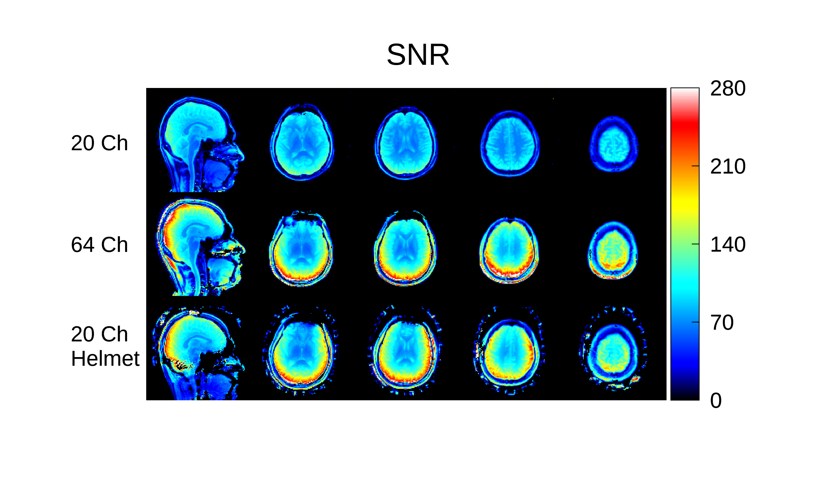
Fig. 3, SNR maps from a single subject and 3 of the receive configurations are
displayed. The figure displays a subset of the acquired slices. The SNR of the
20-Ch Coil + uHDC helmet (V1) was
enhanced strongly above the baseline configuration, almost reaching the
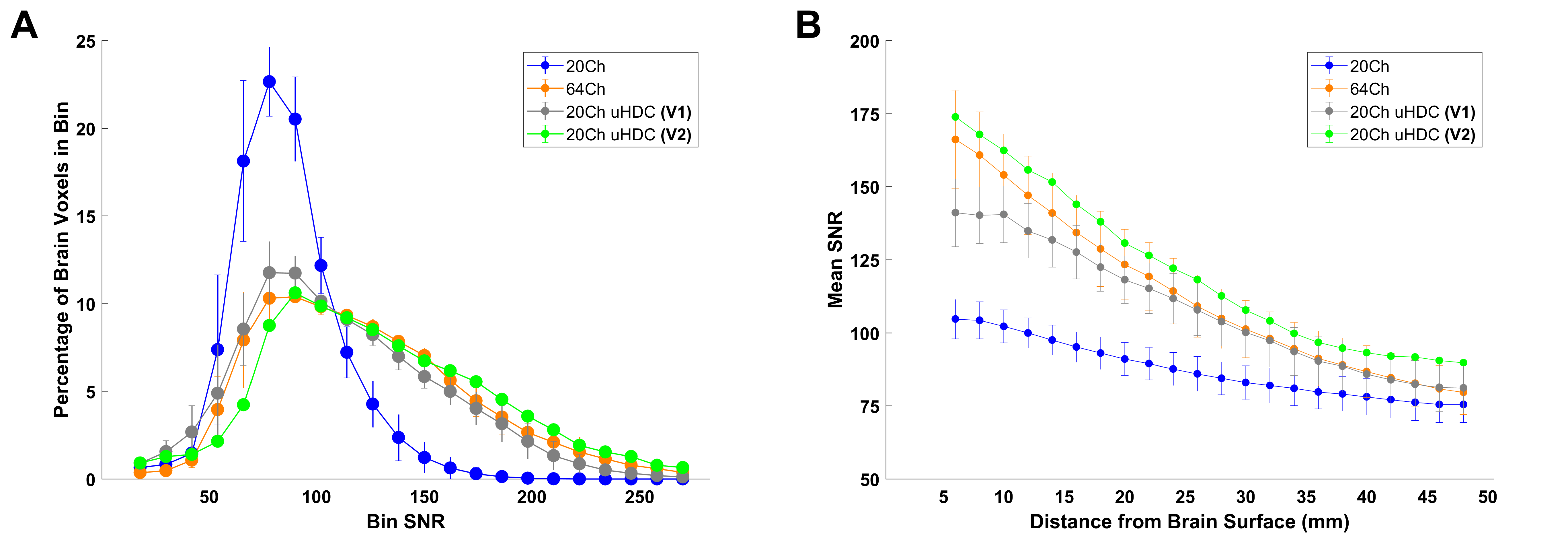
performance of the 64-Ch Coil. Figure 4 displays the results of the histogram
analysis. Fig 4a demonstrates that most of the brain voxels of the 20-Ch coil fell within the
SNR range 0–138 (95.3%), while only 4.7% of the brain voxels occurred in the 138–282 range. In order, the
20-Ch Coil + uHDC helmet (V1 & V2) and 64-Ch Coil’s percentage values were 27.13%,
34.15%, and 31.14% in the range of 138–210, and 3.34%, 8.97%, and 6.54% in the
range of 210–282 respectively. Figure 4b displays the mean SNR at a given depth
from the brain surface. Over the entire range, the 20-Ch Coil + uHDC helmet (V2) offered the best performance. From
10 mm onward, the 20-Ch Coil + uHDC helmet (V1) closely matched that of the 64-Ch Coil. Values in Fig 4.
represent mean and standard deviation among the 5 subjects, except for V2 which was acquired on a single
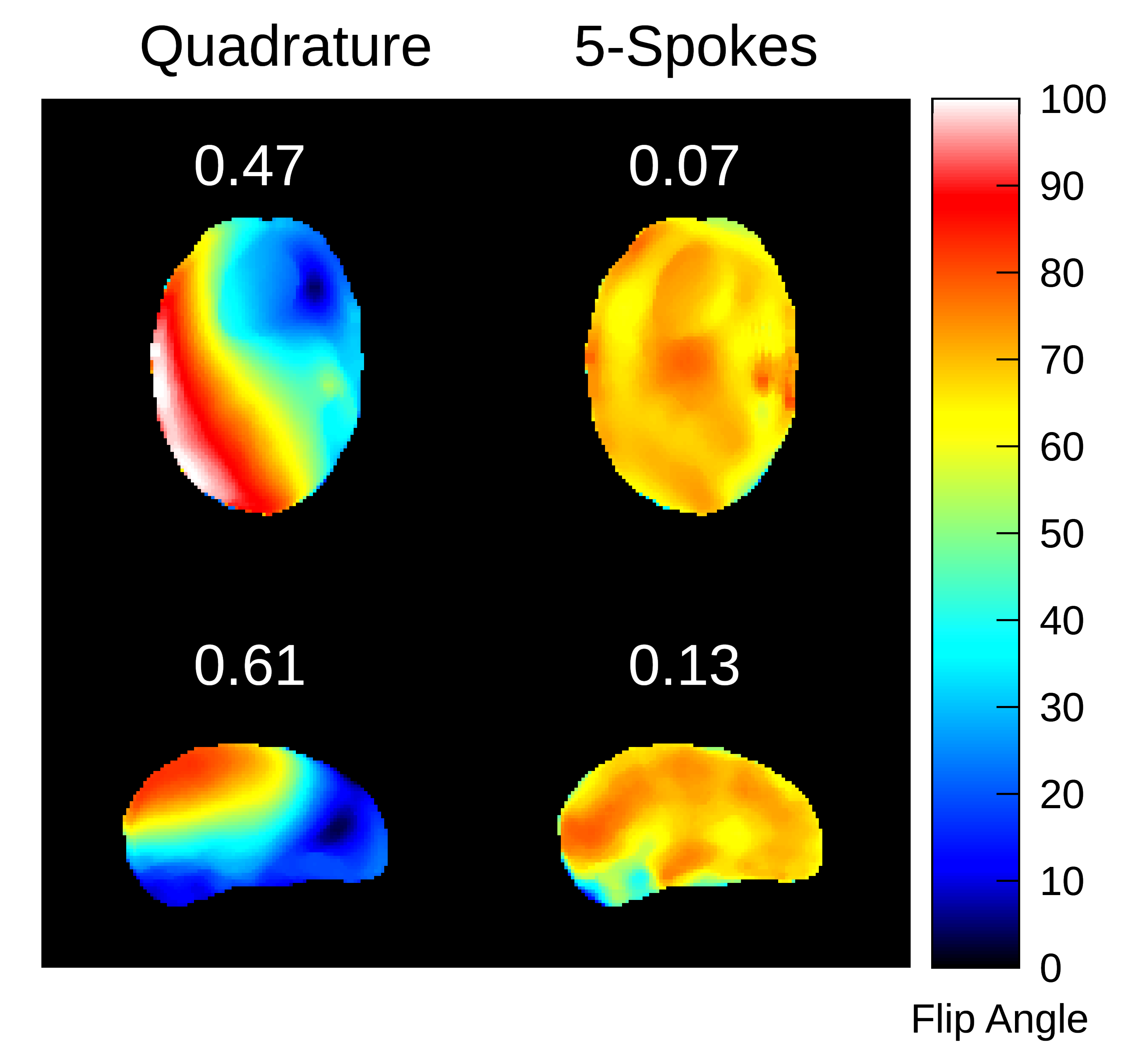
subject. In Fig. 5, results of the blipped-spokes calculation are shown, with the
coefficient of variation displayed in white above each map. The use of this RF
pulse greatly mitigated the transmit inhomogeneity induced by the uHDC helmet.Conclusion
We have presented a study of a conformal helmet constructed with uHDC materials that enhances the SNR of a 20-Ch Coil to a comparable level with the 64-Ch Coil. This helmet design represents a proof-of-concept for a uHDC helmet, and with further optimization could potentially achieve further increases in SNR of standard receive coils. The transmit field was greatly increased but become extremely inhomogeneous due to the uHDC helmet. However, this issue was resolvable with the use of a patient-specific pTX blipped-spokes pulse.Acknowledgements
This work was supported by grants from the NIH and Penn State Hershey Neuroscience Institute.References
[1] Luo et al, “Permittivity and performance of dielectric pads with sintered ceramic beads in MRI: early experiments and simulations at 3T”, MRM 2013; 70(1): 269-275
[2] Rupprecht et al, “Improvements of transmit efficiency and receive sensitivity with ultrahigh dielectric constant (uHDC) ceramics at 1.5T and 3T”, MRM 2018; 79(5): 2842-51
[3] Sacolick et al, “B1 mapping by Bloch-Siegert shift”, MRM 2010; 63(5): 1315-22
[4] Kellman et al, “Image reconstruction in SNR units: a general method for SNR measurement”, MRM 2005; 54(6): 1439-47
[5] Cao et al, “Joint Design of Large-Tip-Angle Parallel RF Pulses and Blipped Gradient Trajectories”, MRM 2016; 75: 1198-1208
Figures