0566
An 8Tx dipoles/32Rx loop coil array – Evaluation of fMRI performances in auditory cortices at 7T1LIFMET, Ecole Polytechnique Fédérale de Lausanne, Lausanne, Switzerland, 2CIBM-AIT, Ecole Polytechnique Fédérale de Lausanne, Lausanne, Switzerland, 3Department of Radiology, Université de Lausanne, Lausanne, Switzerland, 4Department of Radiology, Université de Genève, Genève, Switzerland, 5Department of Biomedical Engineering, King's College London, London, United Kingdom
Synopsis
The performances of a 32-channel receive coil array combined with a tight-fitted whole-brain dipole coil array were investigated. Functional MRI data targeting the auditory cortices was acquired. Noise correlation matrices, SNR and g-factor maps were measured. Compared to a commercial 32Rx, the in-house built 8T/32Rx coil array demonstrated higher robustness in the fMRI results. Lower noise correlation coefficients were measured with the in-house built 8Tx/32Rx coil array while the overall experimentally measured SNR and g-factor maps were comparable.
Introduction
Multi-channel receive arrays provide high SNR and parallel-imaging capabilities1, while transmit-only dipole arrays achieve a large coverage of the whole-brain including cerebellum. In this study, we have evaluated the performances of an upgraded and modified 32-channel receive-only loop array combined with an 8-channel dipole transmit coil array, and compatible with head-gradient 7T MR scanners2. The results were compared to an available commercial single-Tx/32-channel loop coil array (Nova-Medical,USA).Methods
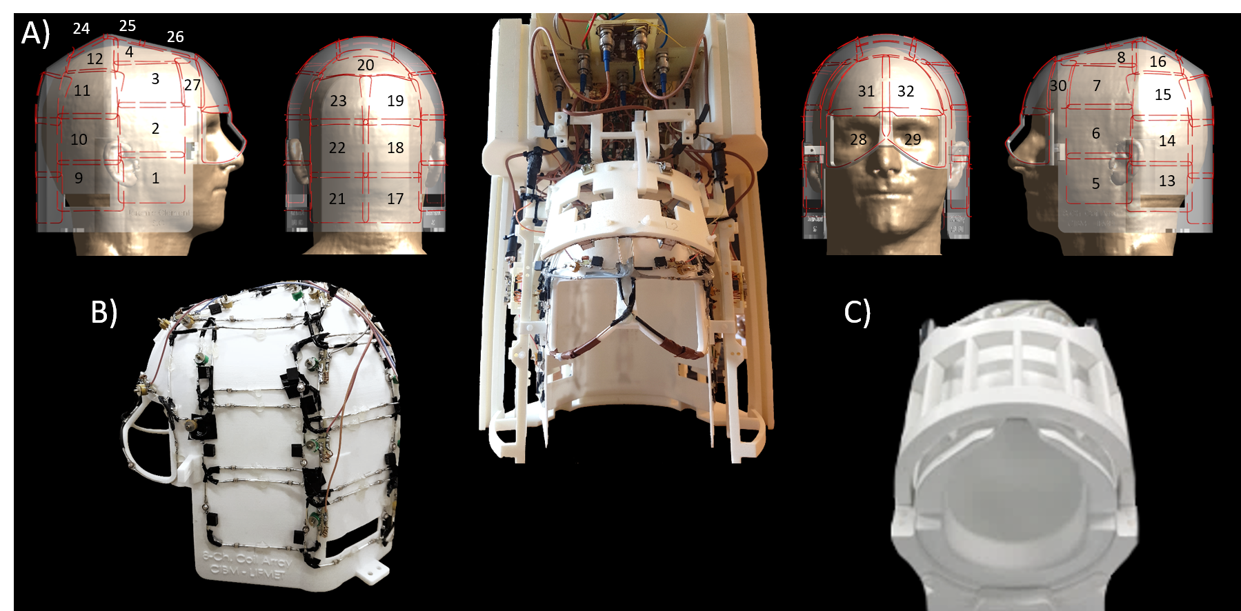
The
transmit coil-array consisted in seven dipoles and two quadrature frontal loops
covering the whole-brain, including cerebellum3. The 32 rectangular loops
were built with silver-platted copper wires, and dimensions ranging from 68x30mm2
to 88x60mm2 (Fig1A-B). They were arranged on a 3D-printed (EOSINT-P395,EOS,Germany)
nylon-helmet (EOS,PA2200) designed to accommodate the human head (AP=222mm,LR=187mm,SI=231mm),
and carefully aligned with the transmit dipoles to avoid transmit-field
cancellations. Receive coil inter-elements decoupling was achieved with
overlapping and low-input impedance preamplifiers (WMM7RP,WantCom,Minnesota,USA).
MRI data was acquired on healthy volunteers using a Magnetom 7T head-gradient MR scanner with an 8x1kW RF-amplifier (Step-2.3,Siemens,Erlangen,Germany) and 32-channel receivers. The worst-case local SAR value was evaluated from the Q-matrix4 on a Duke model5 using FDTD solver (Sim4Life 4.2,ZMT-AG,Switzerland) without including the receivers. Two-dimensional sagittal and transverse fully sampled GRE-data (1x1mm2,slice-thickness=1mm,TR/TE=1000/3.37ms,FA= 48°,matrix=192x192) was acquired with the RF phases optimized according to the slice orientation and areas-of-interest with a PSO method6. Receive noise correlation matrix was computed from a noise-only scan, and the images were reconstructed in SNR units for no-acceleration7. The raw data was therefore under-sampled in post-processing and reconstructed with the SENSE method8. G-factor maps were calculated for acceleration factors of 2,3,4, and 5. RF phases were optimized over the auditory cortices and mid-brain area for the in-house built 8Tx/32Rx dipole coil array, and the B1+-maps were measured for both coils9.
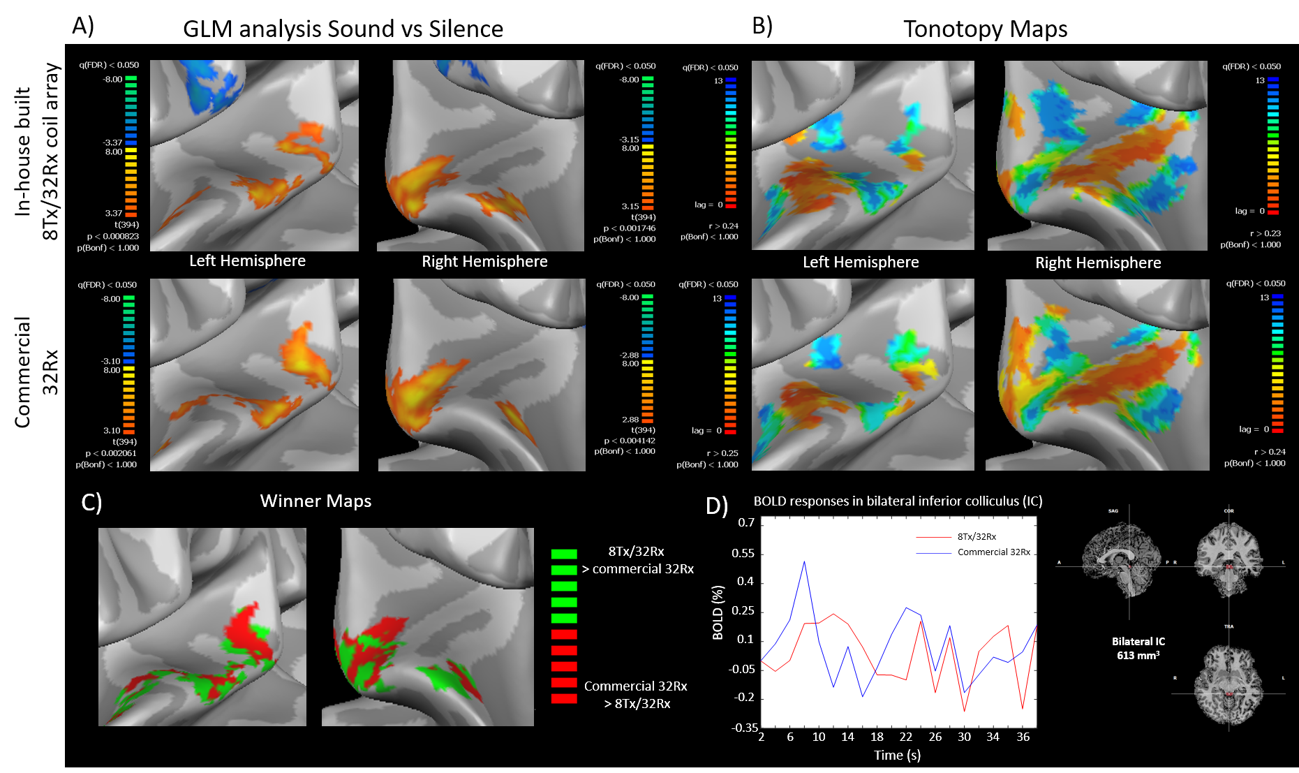
A healthy subject listened passively to two runs of 6 min, in which 10 blocks of pure tones (from 88Hz to 8kHz in half-octave steps) were presented in progression of 2s (14 tones=28s) followed with a “silent”pause of 12s10. Bold signal was acquired for both coils with a 2D-EPI sequence (1.5x1.5x1.5mm3,TR/TE=2000/25ms,FA=90°,GRAPPA=3,43 slices,BW=2252Hz/px,200 volumes). Functional data was analyzed in BrainVoyager 20.6 (Brain Innovation,Maastricht). Resulting cross-correlations, GLM analysis “Sound vs Silence” and winner-maps were projected on segmented inflations of the structural images.
Results
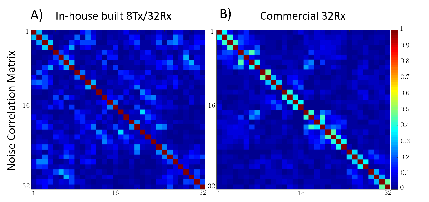
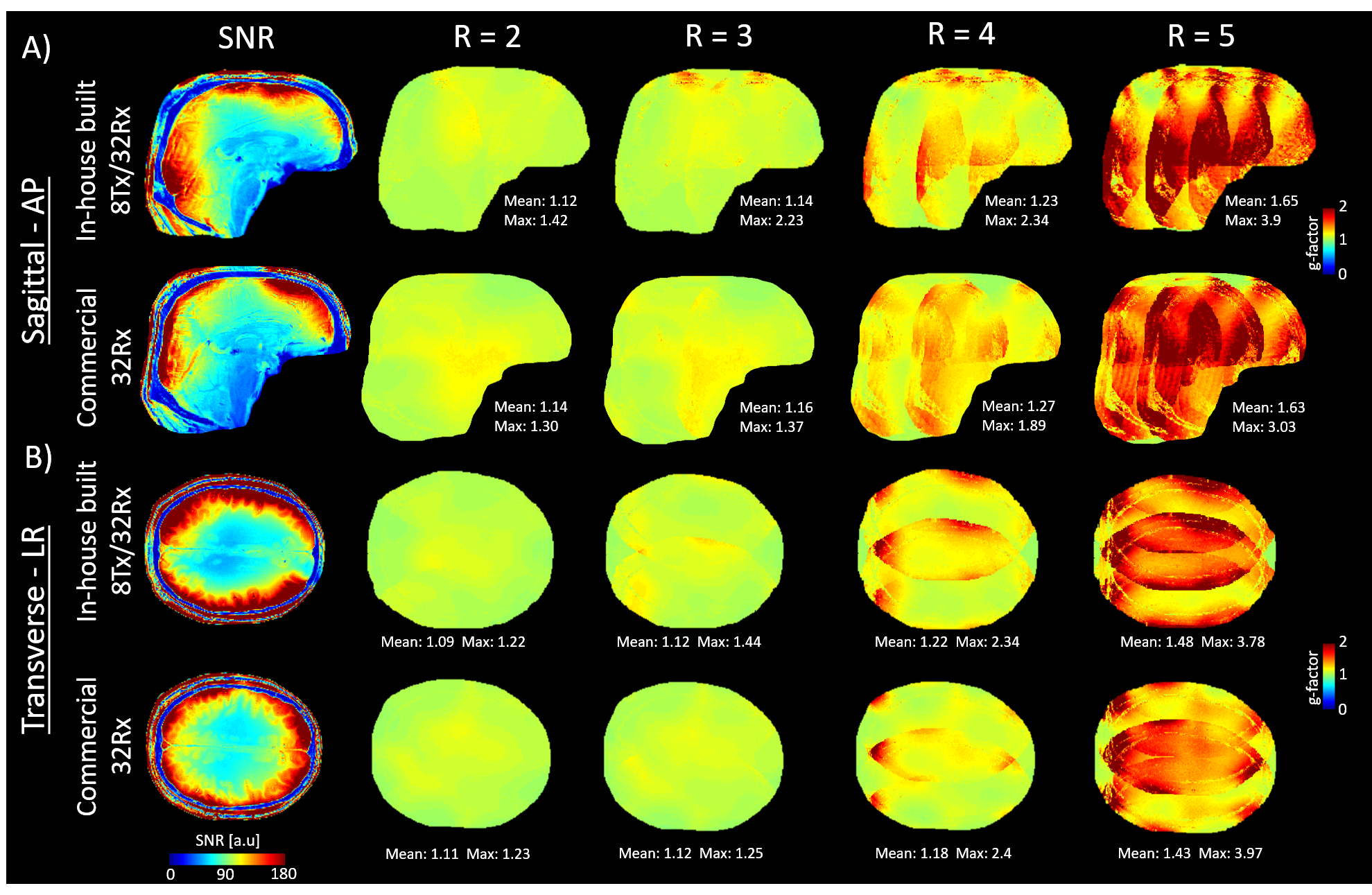
The in-house built 8Tx/32Rx coil array demonstrated a mean and a maximum noise correlation of 6% and 33%, respectively (Fig.2A), while the commercial 32Rx demonstrated a maximum noise correlation of 47%, and a similar mean value (Fig.2B). Both coils produced comparable mean g-factor values for an acceleration up to R=4 (Fig.3B). The highest SNR values were achieved at the periphery of the brain (Fig.3A). Mean SNR values of 115 and 95 were measured over the brain tissues in transverse slice and sagittal slices with the in-house built 8Tx/32Rx coil array. Comparatively, the commercial 32-channel coil demonstrated similar values (111 in transverse and 90 in sagittal).
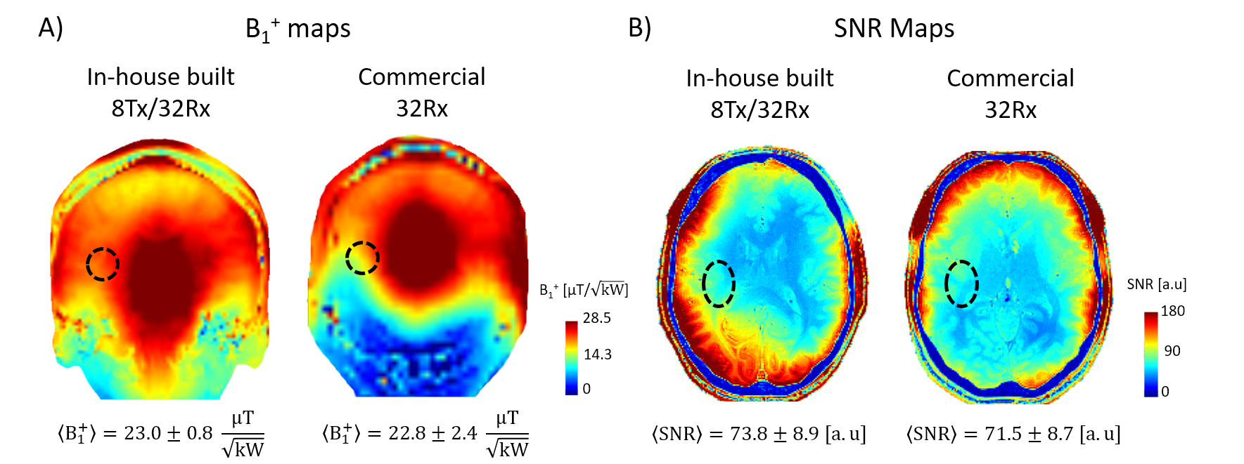
In the auditory cortices, the in-house built 8Tx/32Rx and the commercial 32Rx presented comparable transmit-field efficiencies and SNR levels (Fig.4). Nevertheless, the GLM analysis “Sound vs Silence” revealed smaller clusters with the in-house built 8Tx/32Rx coil array, but with smaller p-values, while the commercial 32Rx demonstrated the opposite pattern (Fig.5A). Tonotopic maps computed for the in-house built 8Tx/32Rx coil array demonstrated better specificity compared to the commercial 32Rx, which showed broader maps despite higher correlation values (Fig.5B). Winner-maps, computed between the two t-maps “Sound vs Silence” showed similar results (Fig.5C). In inferior colliculus (IC), the bold response was less contaminated by noise in the in-house built 8Tx/32Rx coil array compared to the commercial 32Rx (Fig.5D).
Discussions and Conclusion
In this study, significant performances were shown for the combination of a 32-channel receive array with a tight-fitted dipole transmit coil array in terms of SNR and acceleration capabilities. The mean g-factor and SNR values were comparable to the commercial 32Rx. The lower noise correlations between the receivers of the in-house built 8Tx/32Rx may have contributed to improve the robustness of the fMRI results. However, the commercial 32Rx provided larger clusters, which could indicate a better sensitivity.
In bilateral IC, the bold response was slightly better compared to the commercial 32Rx. However, no statistically-significant results could be derived. The hemodynamic response-function being different, the model could be optimized to address more specifically this area. Moreover, a larger number of volunteers could be imaged, and the measurements could target more specifically the IC region by excluding the auditory cortices. Nevertheless, the properties demonstrated in this study, combined to the parallel transmit capabilities make this coil particularly suitable for whole-brain MR studies at 7T.
Acknowledgements
This study was supported by Centre d’Imagerie Biomedicale (CIBM) of the UNIL, UNIGE, HUG, CHUV, EPFL and the Leenaards and Jeantet Foundations.References
[1] Roemer PB et al. The NMR phased array. Magnetic Resonance in Medicine, 16(2), 1990
[2] Clément
JD et al. 31-channel receive coil array
combined with an 8-channel whole-brain dipole transmit array. Proceedings
of the 26th Annual meeting of ISMRM (146), 2018
[3] Clément JD et al. A human cerebral and cerebellar 8-channel transceive RF dipole coil array at 7T. Magnetic Resonance in Medicine, 2018
[4] Ipek O et al. Intersubject local SAR variation for 7T prostate MR imaging with an eight-channel single-side adapted dipole antenna array. Magnetic Resonance in Medicine, 71(4), 2013
[5] Gosselin MC et al. Development of a new generation of high-resolution anatomical models for medical device evaluation: the Virtual Population 3.0. Physics in Medicine and Biology, 59(18), 2014
[6] Clément JD et al. Comparison of passive RF phase shimming methods on the human brain at 7T using particle-swarm optimization. Proceedings of the 33rd Annual meeting of ESMRMB (503), 2015
[7] Kellman P et al. Image reconstruction in SNR units: A general method for SNR measurement. Magnetic Resonance in Medicine, 54(6), 2005
[8] Pruessmann KP et al. SENSE: Sensitivity encoding for fast MRI. Magnetic Resonance in Medicine, 42(5), 1999
[9] Eggenschwiler et al. SA2RAGE: A new sequence for fast B1+-mapping. Magnetic
Resonance in Medicine, 67(6), 2011
[10] Da Costa S et al. Human primary auditory cortex follows the shape of
Heschl’s gyrus. The
Journal of Neuroscience, 31(40), 2011
Figures