0559
Generalize diffusion-MRI-based brain age predictive model using transfer learning1Institute of Medical Device and Imaging, College of Medicine, National Taiwan University, Taipei, Taiwan, 2AcroViz Technology Inc., Taipei, Taiwan, 3Graduate Institute of Brain and Mind Sciences, College of Medicine, National Taiwan University, Taipei, Taiwan, 4Molecular Imaging Center, National Taiwan University, Taipei, Taiwan
Synopsis
Heterogeneity of diffusion MRI data limits the diffusion-MRI-based machine learning model to be generalized to the data acquired at other sites. To generalize the brain age model based on diffusion-MRI-derived features, we used transfer learning techniques to transfer the pre-trained model from the source domain to the target domain with a few tuning data. We found that 75 tuning data with transfer learning framework achieved the acceptable performance, and 150 tuning data achieved the performance comparable to the maximum samples in the target domain. This study provides a practical solution to solve the limitation of diffusion-MRI-based model using transfer learning.
Introduction
Brain age is an emerging imaging biomarker that could be predicted on individuals based on neuroimaging data using machine learning approaches to model trajectories of brain aging[1]. Diffusion MRI (dMRI) is suitable for the investigation of cortical connection and can provide diffusion indices that reflect structural integrity within interconnected networks[2,3]. Using dMRI datasets, dMRI-based brain age is a useful aging biomarker to represent the aging state of white matter[4]. However, the dMRI suffers from between-scanner variations that hinder direct comparisons across different imaging sites[5]. The dMRI-based brain age model is, therefore, limited within the source domain and hard to generalize to the data acquired from other sites. To solve the limitation of dMRI, we aimed to generalize the dMRI-based brain age model using transfer learning (TL) techniques. Specifically, TL applies a brain age model already trained in the source domain to a new target domain by tuning the model parameters with a few data from the target domain.Methods
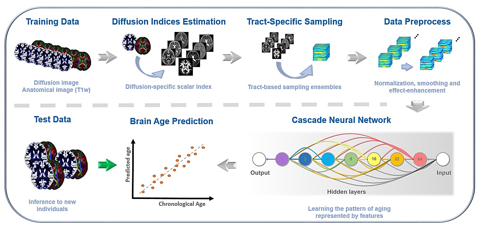
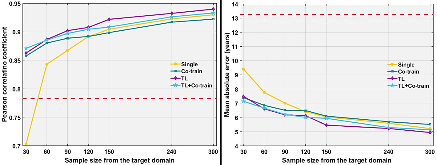
A large cohort as the source domain data used to pre-train a brain age predictive model were obtained from the CamCAN repository[6,7] that consisted of 615 healthy subjects whose age ranged 18-88 years. These data sets, including T1-weighted images and two-shell diffusion tensor images, were split into the training (n=500, called CC-training) and testing (n=115, called CC-testing) sets to develop and evaluate the brain age model, respectively. The data in the target domain were collected from in-house private MRI database that included 300 tuning data at maximum (NTU-tuning) for TL and 100 testing data (NTU-testing) for evaluating the TL performance. The data from the target domain consisted of T1-weighted imaging and diffusion spectrum imaging datasets. To obtain white matter features to predict brain age, the regularized version of diffusion MAP-MRI framework was used to reconstruct diffusion image data into 5 diffusion indices, such as fractional anisotropy[8]. Whole brain tract-specific analysis was conducted to sample the features according to the predefined 76 tracts from each diffusion index[9]. These tract-specific features from the CC-training data were used to create a brain age pre-trained model by cascade neural network (Fig.1). To decide the strategy most effective to adjust the model and the optimal sample size of tuning data, we proposed three transfer strategies to adjust the model so the model predicted brain age in the target domain successfully. The strategies included “co-train” (pool the NTU-tuning and the CC-training data together to train the model), “TL” (re-train the model with the NTU-tuning data and using the parameters of the pre-trained model as the model initial values, no data from the source domain was used) and “TL with co-train” (combine the above two methods). The “single” framework (train the model using the single NTU-tuning data, without TL and/or co-training) was performed as the baseline. The simulated sample size of the tuning data comprised of 30,60,90,120,150,240 and 300 (maximum). The permutation of strategy and sample size would simulate repeatedly for 30 times to estimate the optimal combination. The simulated result was evaluated by applying the transferred model to the NTU-testing data through the statistical metrics including Pearson correlation coefficient and mean absolute error (MAE). After deciding the optimal combination of tuning sample size and strategy, we further improved the performance of transferred model by fine-tuning the hyperparameters and using the advanced optimizer.Results
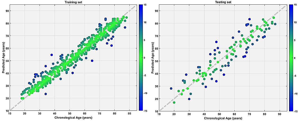
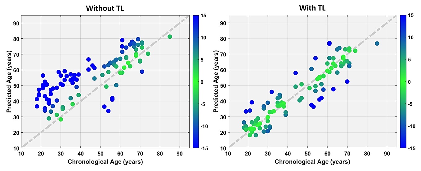
The pre-trained model predicted individuals’ age for both the CC-training and CC-testing sets with satisfactory performance (Fig.2). In the simulation, TL strategy achieved better performance than did the other methods (Fig.3). The performance of transferred model with 150 samples of NTU-tuning data (correlation=0.922, MAE=5.56 years) was comparable to the model trained by maximum 300 NTU-tuning data (correlation=0.931, MAE=5.29 years). If we considered the MAE which was one year more than the standard MAE as the acceptable performance, 75 NTU-tuning data with TL strategy was an acceptable combination (correlation=0.894, MAE=6.26 years) to transfer the pre-trained model. After fine-tuning the transferred model, it attained satisfactory performance as assessed by the NTU-testing data (Correlation=0.916, MAE=5.55 years)(Fig.4).Discussion and Conclusion
We generalized the dMRI-based brain age predictive model to the new data domain using TL. The TL strategy was an effective paradigm to transfer the pre-trained model. We found that 75 image data as the tuning set achieved the acceptable performance compared to the model trained with the 300 datasets in the target domain (3/4 data-saving). Also, the performance of transferred model using 150 tuning data was comparable to the standard performance (1/2 data-saving). This study provides a transfer learning solution to address the problem of generalization in dMRI-based brain age model.Acknowledgements
The CamCAN data collection and sharing for this study was provided by the Cambridge Centre for Ageing and Neuroscience (CamCAN). CamCAN funding was provided by the UK Biotechnology and Biological Sciences Research Council (grant number BB/H008217/1), together with support from the UK Medical Research Council and University of Cambridge, UK.References
[1] Cole, J.H., & Franke, K. (2017) Predicting Age Using Neuroimaging: Innovative Brain Ageing Biomarkers. Trends Neurosci, 40:681-690.
[2] Bennett, I. J., & Madden, D. J. (2014). Disconnected aging: cerebral white matter integrity and age-related differences in cognition. Neuroscience, 276, 187-205.
[3] Alexander, A.L., Hurley, S.A., Samsonov, A.A., Adluru, N., Hosseinbor, A.P., Mossahebi, P., Tromp, D.P., Zakszewski, E., Field, A.S. (2011) Characterization of cerebral white matter propertie using quantitative magnetic resonance imaging stains. Brain connectivity, 1:423-446.
[4] Mwangi, B., Hasan, K.M., Soares, J.C. (2013) Prediction of individual subject's age across the human lifespan using diffusion tensor imaging: a machine learning approach. Neuroimage, 75:58-67.
[5] Mirzaalian, H., Ning, L., Savadjiev, P., Pasternak, O., Bouix, S., Michailovich, O., ... & Flashman, L. A. (2018). Multi-site harmonization of diffusion MRI data in a registration framework. Brain imaging and behavior, 12(1), 284-295.
[6] Taylor, J.R., Williams, N., Cusack, R., Auer, T., Shafto, M.A., Dixon, M., Tyler, L.K., CamCAN, Henson, R.N. (2016). The Cambridge Centre for Ageing and Neuroscience (CamCAN) data repository: Structural and functional MRI, MEG, and cognitive data from a cross-sectional adult lifespan sample. NeuroImage. doi: 10.1016/j.neuroimage.2015.09.018.
[7] Shafto, M.A., Tyler, L.K., Dixon, M., Taylor, J.R., Rowe, J.B., Cusack, R., Calder, A.J., Marslen-Wilson, W.D., Duncan, J., Dalgleish, T., Henson, R.N., Brayne, C., CamCAN, & Matthews, F.E. (2014). The Cambridge Centre for Ageing and Neuroscience (CamCAN) study protocol: a cross-sectional, lifespan, multidisciplinary examination of healthy cognitive ageing. BMC Neurology, 14(204). doi:10.1186/s12883-014-0204-1.
[8] Hsu, Y.C., Tseng, W.Y. (2018) An efficient regularization method for diffusion MAP-MRI estimation. 2018 ISMRM-ESMRMB Joint Annual Meeting.
[9] Chen, Y.J., Lo, Y.C., Hsu, Y.C., Fan, C.C., Hwang, T.J., Liu, C.M., Chien, Y.L., Hsieh, M.H., Liu, C.C., Hwu, H.G., Tseng, W.Y. (2015) Automatic whole brain tract-based analysis using predefined tracts in a diffusion spectrum imaging template and an accurate registration strategy. Hum Brain Mapp, 36:3441-58.
Figures