0552
Multi-Dimensional Diffusion MRI Sampling Scheme: B-tensor Design and Accurate Signal Reconstruction1Research School of Engineering, Australian National University, Canberra, Australia, 2Computer Science department, University of Verona, Verona, Italy, 3Univ Rennes, Inria, CNRS, IRISA, Rennes, France
Synopsis
b-tensor encoding enables the separation of isotropic and anisotropic tensors. However, little consideration has been given as to how to design a b-tensor encoding sampling scheme. In this work, we propose the first 4D basis for representing the diffusion signal acquired with b-tensor encoding. We study the properties of the diffusion signal in this basis to give recommendations for optimally sampling the space of axisymmetric b-tensors. We show, using simulations, that the proposed sampling scheme enables accurate reconstruction of the diffusion signal by expansion in this basis using a clinically feasible number of samples.
Introduction
Multi-dimensional diffusion MRI (MD-dMRI) is a recent diffusion signal modelling and reconstruction framework where the diffusion signal is derived from the diffusion tensor distribution (DTD). Advanced magnetic gradient modulation schemes, resulting in b-tensor encoding, enable separation of isotropic and anisotropic tensors1-3. Often diffusion tensors are assumed to be axisymmetric, so the DTD is 4D4,5. While algorithms have been proposed for recovering the DTD from b-tensor encoded measurements of the diffusion signal4-6, little consideration has been given to the b-tensor sampling scheme. Currently, b-tensors are empirically chosen, often from uniform sampling of b-tensor parameters3, typically resulting in on the order of 1000 samples to recover the full DTD5,6. To our knowledge, no b-tensor sampling scheme has been proposed for optimally sampling the MD-dMRI signal. We present the first 4D basis for representing the MD-dMRI signal. This enables us to study the properties of the signal in this basis to give recommendations for sampling the space of axisymmetric b-tensor, while enabling accurate reconstruction of the MD-dMRI signal. MD-dMRI Signal Basis and b-tensor Sampling Scheme
MD-dMRI Signal Basis and b-tensor Sampling Scheme
$$\frac{S}{S_0}(\mathbf{B})=\int{P(\mathbf{D})\exp(-\mathbf{B}:\mathbf{D})d\mathbf{D}}=\langle\exp(-\mathbf{B}:\mathbf{D})\rangle,$$ is the diffusion signal acquired with b-tensor encoding, where $$$P(\mathbf{D})$$$ is the DTD, $$$\mathbf{D},\mathbf{B}$$$ are second order symmetric positive-definite diffusion and b-tensors respectively, and $$$\mathbf{B}:\mathbf{D}=\sum_i\sum_jb_{ij}D_{ij}$$$. For a discrete set of diffusion tensor populations, $$\frac{S}{S_0}(\mathbf{B})=\sum_{d=1}^{N_{D}}w_{d}\exp(-\mathbf{B}:\mathbf{D}_{d}),$$ where $$$w_{d}$$$ is the proportion of tensor population $$$\mathbf{D}_{d}$$$ and $$$N_{D}$$$ is the number of populations. The axisymmetric diffusion tensor can be parameterised as5,6,
$$\mathbf{D}=\mathbf{R}(\theta,\phi){D_{iso}}\Bigg(\begin{pmatrix}1&0&0\\0&1&0\\0&0&1\end{pmatrix}+D_\Delta\begin{pmatrix}-1&0&0\\0&-1&0\\0&0&2\end{pmatrix}\Bigg)\mathbf{R}(\theta,\phi)^T,$$ where $$$\mathbf{R}$$$ is a rotation operator. The b-tensor can be parametrised in terms of $$$(b_s,b_l,\Theta,\Phi)$$$2,6, $$\mathbf{B}=\mathbf{R}(\Theta,\Phi)\Bigg(\frac{b_{s}}{3}\begin{pmatrix}1&0&0\\0&1&0\\0&0&1\end{pmatrix}+b_{l}\begin{pmatrix}0&0&0\\0&0&0\\0&0&1\end{pmatrix}\Bigg)\mathbf{R}(\Theta,\Phi)^T.$$ Expanding $$$\frac{S}{S_0}(\mathbf{B})$$$, using the above parameterisation, gives a separable expression in $$$b_{s}$$$, and the other three parameters, $$$b_l,\Theta,\Phi$$$2:
$$\frac{S}{S_0}(\mathbf{B})=\sum_{d=1}^{N_{D}}w_{d}\exp(-b_{s}(D_{iso})_d)\exp(-b_{l}(D_{iso})_d)\exp(-2b_{l}(D_{iso})_{d}(D_\Delta)_{d}P_{2}(\cos\beta_d)),$$$$$\cos\beta_d=\cos{\Theta}\cos{\theta_d}+\sin{\Theta}\sin{\theta_d}\cos({\Phi-\phi_d})$$$. This enables $$$\frac{S}{S_0}(\mathbf{B})$$$ to be expanded in a separable and orthogonal 4D basis which is the product of a 1D basis for $$$b_{s}$$$ dimension and a 3D basis for $$$b_{l},\Theta$$$ and $$$\Phi$$$ dimensions. As $$$\frac{S}{S_0}(\mathbf{B})$$$ is a function of negative exponential of $$$b_{s}$$$, we use an exponential modulated with a Laguerre polynomial, which together form an orthogonal basis over $$$b_{s}$$$ dimension7. Similarly, we use the spherical Laguerre basis8, a 3D orthogonal basis with an exponential weighting function in the radial direction, for $$$b_{l},\Theta,\Phi$$$ dimensions, leading to expansion: $$\frac{S}{S_0}(b_{s},b_{l},\Theta,\Phi)=\sum_{p=0}^{P}\sum_{n=0}^{N}\sum_{\ell=0}^{L} \sum_{m=-\ell}^{\ell}c_{pn\ell m}Z_p(b_{s})B_{n\ell m}(b_l,\Theta,\Phi)$$ where $$$B_{n\ell m}(b_l,\Theta,\Phi)=X_n(b_l)Y^m_\ell(\Theta,\Phi)$$$ is the spherical Laguerre basis, with $$$X_n(b_{l})=\sqrt{\frac{n!}{{\zeta_l}^3(n+2)!}}\exp(\frac{-b_{l}}{2\zeta_{l}})L^2_n(\frac{b_l}{\zeta_{l}}),$$$ and $$$Z_p(b_{s}) = \frac{1}{\sqrt{\zeta_s}}\exp(\frac{-b_s}{2\zeta_{s}})L_{p}(\frac{b_{s}}{\zeta_{s}})$$$. $$$Y^m_\ell(\Theta,\Phi)$$$ are spherical harmonics of maximum degree $$$L$$$, $$$\zeta_{l},\zeta_{s}$$$ are scale factors and $$$N,P$$$ are maximum orders for $$$b_{l}$$$ and $$$b_s$$$ bases respectively. Coefficients $$$c_{pn\ell{m}}$$$ are defined by inner product $$$c_{pn\ell{m}}=\langle \frac{S}{S_0}(b_{s},b_{l},\Theta,\Phi),Z_p(b_s)B_{n\ell{m}}(b_{l},\Theta,\Phi)\rangle$$$. Due to the separability of this basis, we can design a separable transform,
$$c_{pn\ell{m}}=\sum_{j=0}^{J}w_jZ_p(b_{s}(j))\sum_{i=0}^{I}w_iX_n(b_{l}(i))\int_{\Theta=0}^{\pi}\int_{\Phi=0}^{2\pi}\frac{S}{S_0}(b_{s}(j),b_{l}(i),\Theta,\Phi)Y^m_\ell(\Theta,\Phi)\sin{\Theta}d\Theta{d}\Phi.$$ with Gauss-Laguerre quadrature9 used to choose the weights $$$w_j,w_i$$$, and sample locations $$$b_{s}(j),b_{l}(i)$$$ for the $$$b_{s}$$$ and $$$b_{l}$$$ dimensions. We use the scheme10,11 to place the samples in the angular dimension. For a band-limited signal, this quadrature enables exact reconstruction and efficient sampling with the number of samples equal to number of coefficients.
Methods
The Magic DIAMOND model is used to study maximum orders needed for expansion of the diffusion signal in the proposed basis and to evaluate reconstruction accuracy of the proposed sampling scheme. Magic DIAMOND12, an extension of the DIAMOND model13,14 for axisymmetric tensor acquisitions, describes the DTD through a mixture of non-central matrix-variate Gamma distributions, where each diffusion compartment is represented through a single distribution with two shape parameters, that set axial and radial heterogeneity of a compartment, and a scale parameter. Compartments are used to represent diffusion molecules hindered and restricted by fascicles, and free water diffusion. We used a range of diffusivities and distribution parameters representative of those in the human brain.Results and Discussion
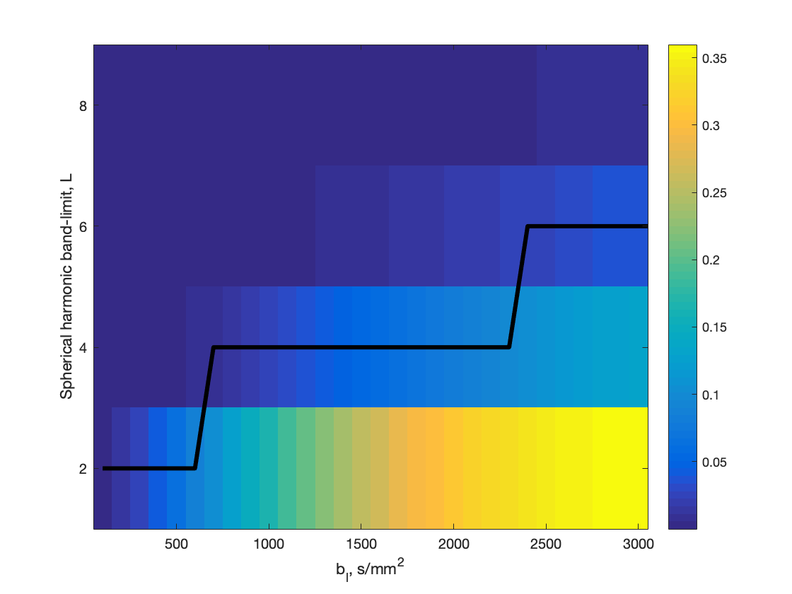
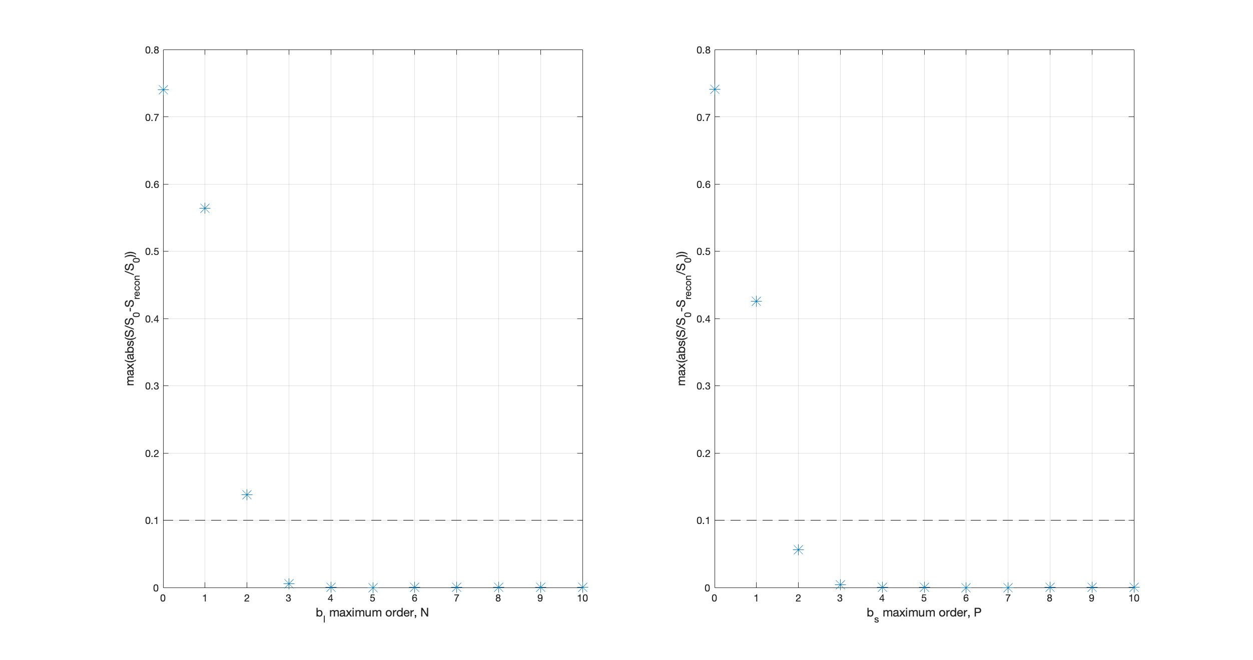
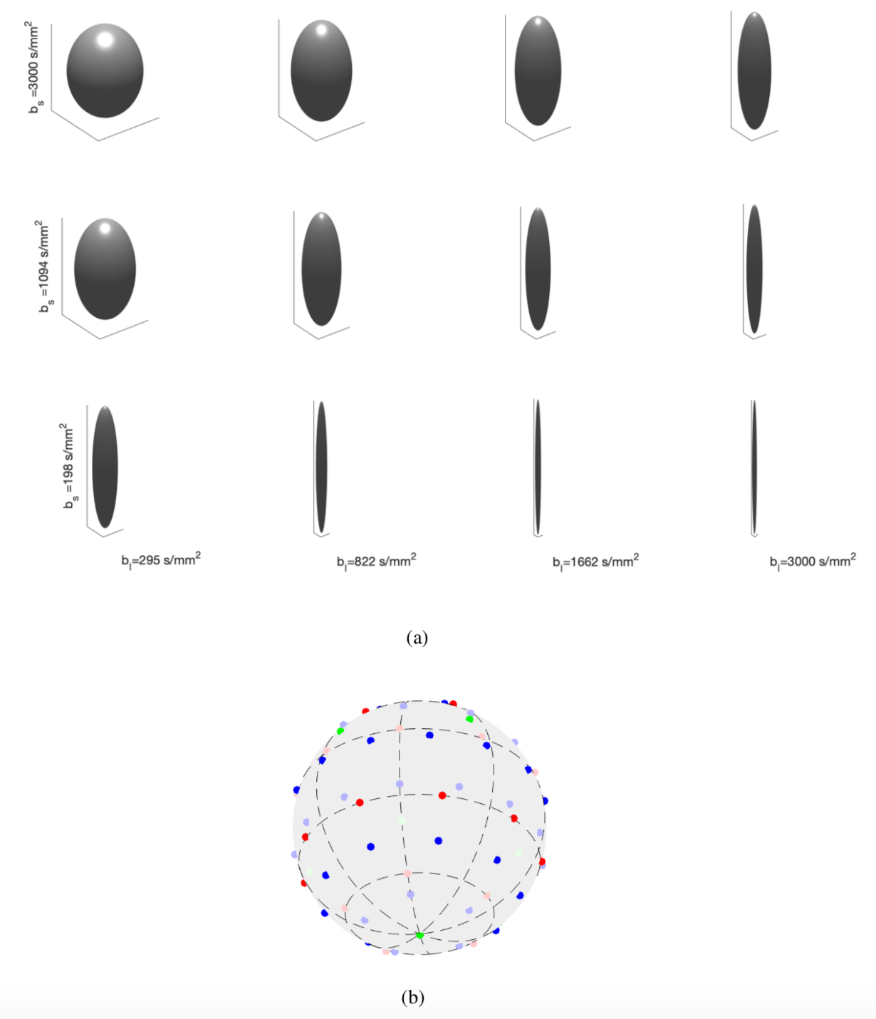
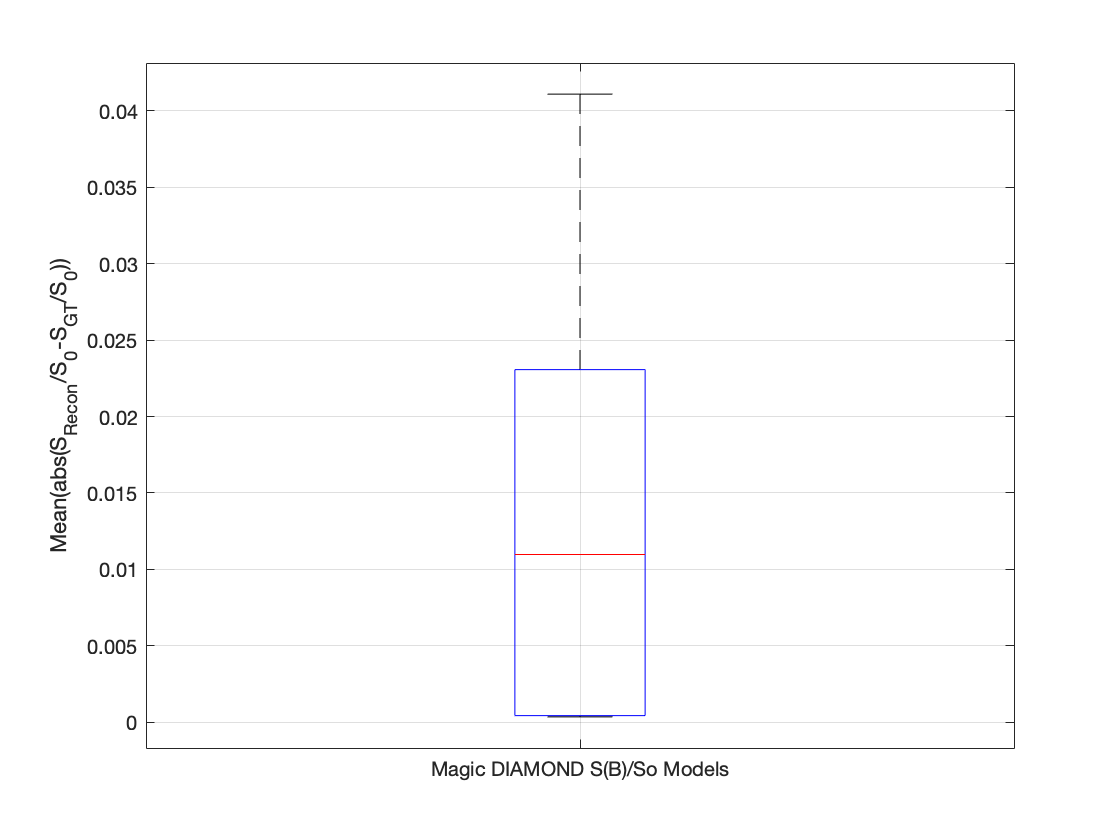
As Magic DIAMOND model is linear, it is sufficient to study each compartment individually; the truncation order of the compartment with the highest truncation order is sufficient to represent all combinations of compartments. Fig.1 shows the spherical harmonic band-limit as a function of $$$b_l$$$. Fig.2 shows the reconstruction error as a function of increasing truncation order for maximum $$$b_l$$$ and $$$b_s$$$ of $$$3000s/mm^2$$$. It was found that $$$N=3,\,P=2,\,L(i)=[2,4,4,6]$$$ were sufficient for reconstruction error smaller than $$$S/S_0=0.1$$$3 for all compartments, resulting in the sampling scheme (Fig.3) with 192 samples, the same order as several state-of-the-art sampling schemes in q-space15. Fig.4 shows the mean reconstruction error evaluated at 1080 uniformly placed measurements for 640 realisations of Magic DIAMOND.Conclusion
We present the first 4D basis and reconstruction algorithm for representing and reconstructing the axisymmetric b-tensor encoded diffusion signal. We study the properties of the signal to inform how many and which b-tensors should be used, thereby providing sampling recommendations. Although preliminary, the high reconstruction accuracy achieved on Magic DIAMOND model is promising for interpolation of b-tensors encoded measurements to enable algorithms for recovering the DTD to achieve higher accuracy and/or reduce the number of measurements.Acknowledgements
Alice Bates was supported by the Australian Research Council’s Discovery Projects funding scheme (Project no. DP170101897).
Alessandro Daducci is supported by the Rita Levi Montalcini, MIUR for the recruitment of young researchers.
References
1. D. Topgaard, Chapter 7 NMR methods for studying microscopic diffusion anisotropy, in Diffusion NMR of Confined Systems: Fluid Transport in Porous Solids and Heterogeneous Materials. The Royal Society of Chemistry, 226–259 (2017).
2. D. Topgaard, Multidimensional diffusion MRI, J. Magn. Reson. 275, 98–113 (2017).
3. C.-F. Westin et al., Q-space trajectory imaging for multidimensional diffusion MRI of the human brain, NeuroImage 135, 345–362 (2016).
4. J. P. de Almeida Martins and D. Topgaard, Multidimensional correlation of nuclear relaxation rates and diffusion tensors for model-free investigations of heterogeneous anisotropic porous materials, Scientific Reports 8 (2018).
5. S. Eriksson et al., NMR diffusion-encoding with axial symmetry and variable anisotropy: Distinguishing between prolate and oblate microscopic diffusion tensors with unknown orientation distribution, J Chem Phys. 142, 104201 (2015).
6. J. P. de Almeida Martins and D. Topgaard, Two-dimensional correlation of isotropic and directional diffusion using NMR, Phys. Rev. Lett. 116, 087601 (2016).
7. R. H. Fick et al., Non-parametric graphnet-regularized representation of dMRI in space and time, Med. Image Anal. 43, 37–53 (2018).
8. B. Leistedt and J. D. McEwen, Exact wavelets on the ball, IEEE Trans. Signal Process. 60, 6257–6269 (2012).
9. M. Abramowitz and I. A. Stegun, Handbook of mathematical functions with formulas, graphs, and mathematical tables, ser. National Bureau of Standards Applied Mathematics Series. Dover, New York: U.S. Government Printing Office 55 (1964).
10. A. P. Bates et al., An optimal dimensionality sampling scheme on the sphere with accurate and efficient spherical harmonic transform for diffusion MRI, IEEE Signal Process. Lett. 23, 15–19 (2016).
11. A. P. Bates et al., An optimal dimensionality multi-shell sampling scheme with accurate and efficient transforms for diffusion MRI, in Proc. IEEE Int. Symp. Biomed. Imaging, ISBI, Melbourne, Australia, 770–773 (2017).
12. A. Reymbaut and et al., The “magic DIAMOND” method: probing brain microstructure by combining b-tensor encoding and advanced diffusion compartment imaging, in Proc. Int. Soc. Magn. Reson. Med, Paris, France (2018).
13. B. Scherrer et al., Characterizing brain tissue by assessment of the distribution of anisotropic microstructural environments in diffusion-compartment imaging (DIAMOND), Magn. Reson. Med. 76, 963–977 (2015).
14. B. Scherrer and et al., Decoupling axial and radial tissue heterogeneity in diffusion compartment imaging, in Springer International Publishing, Cham, 440–452 (2017).
15. S. N. Sotiropoulos et al., Advances in diffusion MRI acquisition and processing in the Human Connectome Project, NeuroImage 80, 125–143 (2013).
Figures