0551
Disentangling diffusion-weighted SSFP: ADC estimates in terms of an effective diffusion time1Wellcome Centre for Integrative Neuroimaging, FMRIB, Nuffield Department of Clinical Neurosciences, University of Oxford, Oxford, United Kingdom, 2Department of Radiology, University of Chicago, Chicago, IL, United States, 3Clinical Neurology, Nuffield Department of Clinical Neurosciences, University of Oxford, Oxford, United Kingdom, 4Biomedical Image Computing and Analytics, University of Pennsylvania, Philadelphia, PA, United States
Synopsis
Diffusion-weighted SSFP (dwSSFP) is a high-SNR-efficiency diffusion imaging method. Unlike conventional diffusion measurements, the dwSSFP signal reflects a range of diffusion times because the signal is recycled over multiple excitations. This complicates interpretation and leads to an ill-defined b-value. We present a framework to describe dwSSFP-derived ADC estimates in terms of an effective diffusion time. To achieve this, we require dwSSFP measurements at two flip-angles. Experimental results are presented in a whole, postmortem brain at 7T. This enables us to simultaneously addresses flip-angle inhomogeneity at 7T and provide ADC estimates that are more comparable to conventional diffusion MRI.
Introduction
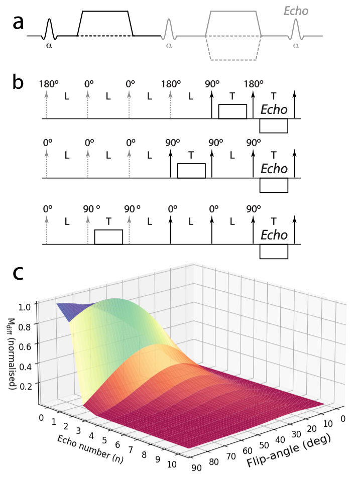
Diffusion-weighted steady-state free precession (dwSSFP) is a highly efficient diffusion-weighted imaging method. This efficiency results from a steady-state signal that recycles magnetisation over multiple excitations (i.e. there are multiple signal-forming coherence pathways). As a result of this signal-forming mechanism, the signal reflects a range of diffusion times, each with its own b-value. Moreover, each echo pathway has distinct T1 and T2 weighting, and the relative contribution of these pathways depends on flip-angle and TR, leading to a complicated signal dependence1 (Figure 1). dwSSFP has demonstrated higher SNR efficiency than spin echoes for imaging fixed, post-mortem tissue with very short T22. Further SNR benefits have been gained at 7T3, but require addressing flip-angle (B1) inhomogeneity. Previous work at 7T acquired dwSSFP images at two flip-angles, demonstrating improved homogeneity of principal diffusion direction (PDD) estimates4. However, that work encountered the additional challenge that the weighting of diffusion times is also flip-angle dependent5. Here, we turn this challenge into an opportunity: this dependence means that multiple flip-angle dwSSFP measurements can in theory be extrapolated to a single effective diffusion time, $$$\Delta_{eff}$$$, making dwSSFP measurements more comparable to conventional diffusion measurements. We present a framework to obtain ADC estimates as a function of $$$\Delta_{eff}$$$ from multiple flip-angle dwSSFP data. Experimental validation is performed in a whole, postmortem brain.
Theory
The “two transverse approximation” of dwSSFP signal considers only coherence pathways where the magnetisation is in the transverse plane for two TR periods1,6, as a summation of pathways corresponding to one spin- and many stimulated-echoes: $$M_{\text{ADC}}=\frac{M_{0}(1-E_{1})E_{1}E_{2}^{2}\sin\alpha}{2\left(1-E_{1}\cos\alpha\right)}\left(\frac{1-\cos\alpha}{E_{1}}\cdot A_{\text{ADC}}+\sin^{2}\alpha\sum_{n=1}^{\infty} \left[(E_{1}\cos\alpha)^{n-1}\cdot A_{\text{ADC}}^{n+1}\right]\right)\;\;\;[1]$$ where $$$E_{1}=e^{-TR/T_{1}}$$$, $$$E_{2}=e^{-TR/T2}$$$, $$$A_{ADC}=e^{-q^{2}\cdot ADC\cdot TR}$$$, $$$q$$$ is the area under the diffusion gradient, $$$\alpha$$$ is the flip-angle and $$$n$$$ is the number of TRs between the transverse periods for a given stimulated-echo6.
The diffusion time of the signal is well
defined for each pathway (spin-echo: $$$\Delta=TR$$$, stimulated-echo: $$$\Delta=(n+1)\cdot TR$$$). We define $$$\Delta_{eff}$$$ as the weighted-mean of these
diffusion times, with each echo weighted by its relative signal contribution, obtaining
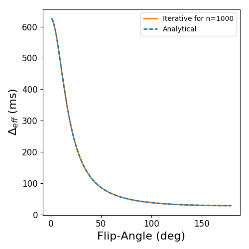
(Figure 2): $$\Delta_{\text{eff}}=\frac{(1+2E_{1}-E_{1}^2\cos\alpha)}{(1+E_{1})(1-E_{1}\cos\alpha)}\cdot\text{TR}\;\;\;[2]$$ The dependence of calculated diffusivity on
diffusion time reflects non-gaussian diffusion, which can be captured with a Gamma
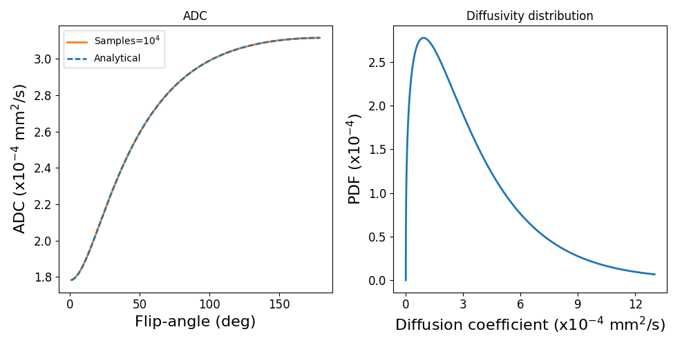
distribution of diffusion coefficients7. For a given distribution,
we calculate the dwSSFP signal and translate this into a single ADC. For a Gamma
pdf of mean $$$\mu$$$ and standard deviation $$$\sigma$$$, this gives (Figure 3): $$\text{ADC}=-\frac{1}{q^{2}\text{TR}}\cdot\ln{\left[\frac{-(S\cdot E_{1}\cos\alpha+1)+[(S\cdot E_{1}\cos\alpha+1)^{2}+4E_{1}\cdot S]^{\frac{1}{2}}}{2E_{1}}\right]}\;\;\;[3]$$ where: $$ S=\left(\frac{\mu}{\mu+q^{2}\cdot\text{TR}\cdot\sigma^2}\right)^{\frac{\mu^{2}}{\sigma^{2}}}+ (1+\cos\alpha)\cdot E_{1}\cdot\left(\frac{\mu}{q^{2}\cdot\text{TR}\cdot\sigma^2}\right)^{\frac{\mu^{2}}{\sigma^{2}}}\cdot\Phi\left(E_{1}\cos\alpha,\frac{\mu^{2}}{\sigma^{2}},2+\frac{\mu}{q^{2}\cdot\text{TR}\cdot\sigma^2}\right)\;\;\;[4]$$
and $$$\Phi$$$ is the Lerch transcendent. As Eqs. [2] and [3] both depend on flip-angle, we can use measurements at multiple flip-angles to calculate the ADC for a target $$$\Delta_{eff}$$$.
Method
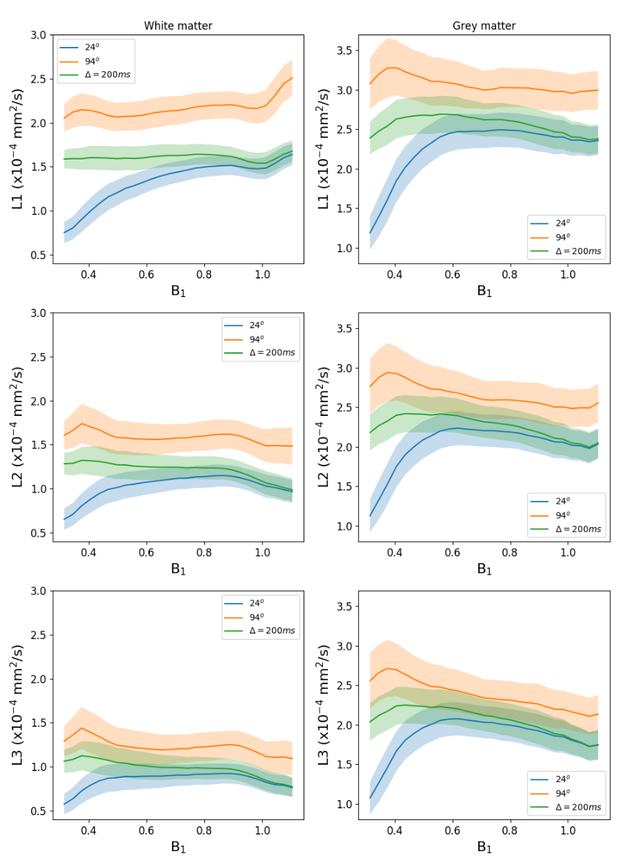
A fixed postmortem brain was scanned at 7T using a protocol described previously8. Briefly, the dwSSFP acquisition used flip-angles 24o and 94o, 120 directions per flip-angle, q=300/cm, resolution=0.85mm3, along with T1, T2 and B1 mapping. Diffusion-tensor (DT) estimates were calculated using the full dwSSFP signal model1,9, defining unique eigenvalues ($$$L_{1,2,3}$$$) at each flip-angle, but shared eigenvectors ($$$\overrightarrow{V}_{1,2,3}$$$). $$$L_{1,2,3}$$$ estimates at each flip-angle were fit to Eq. [3] to determine $$$\mu$$$ and $$$\sigma$$$: $$\min_{\mu,\sigma}||ADC_{\alpha_{low},\alpha_{high}}(\mu,\sigma)-L_{\alpha_{low},\alpha_{high}}||_2^2+||\mu-L_{\alpha_{high}}||_2^2\;\;\;[5]$$ where regularisation ensured that $$$\mu$$$ remains on the order of $$$L_{1,2,3}$$$.
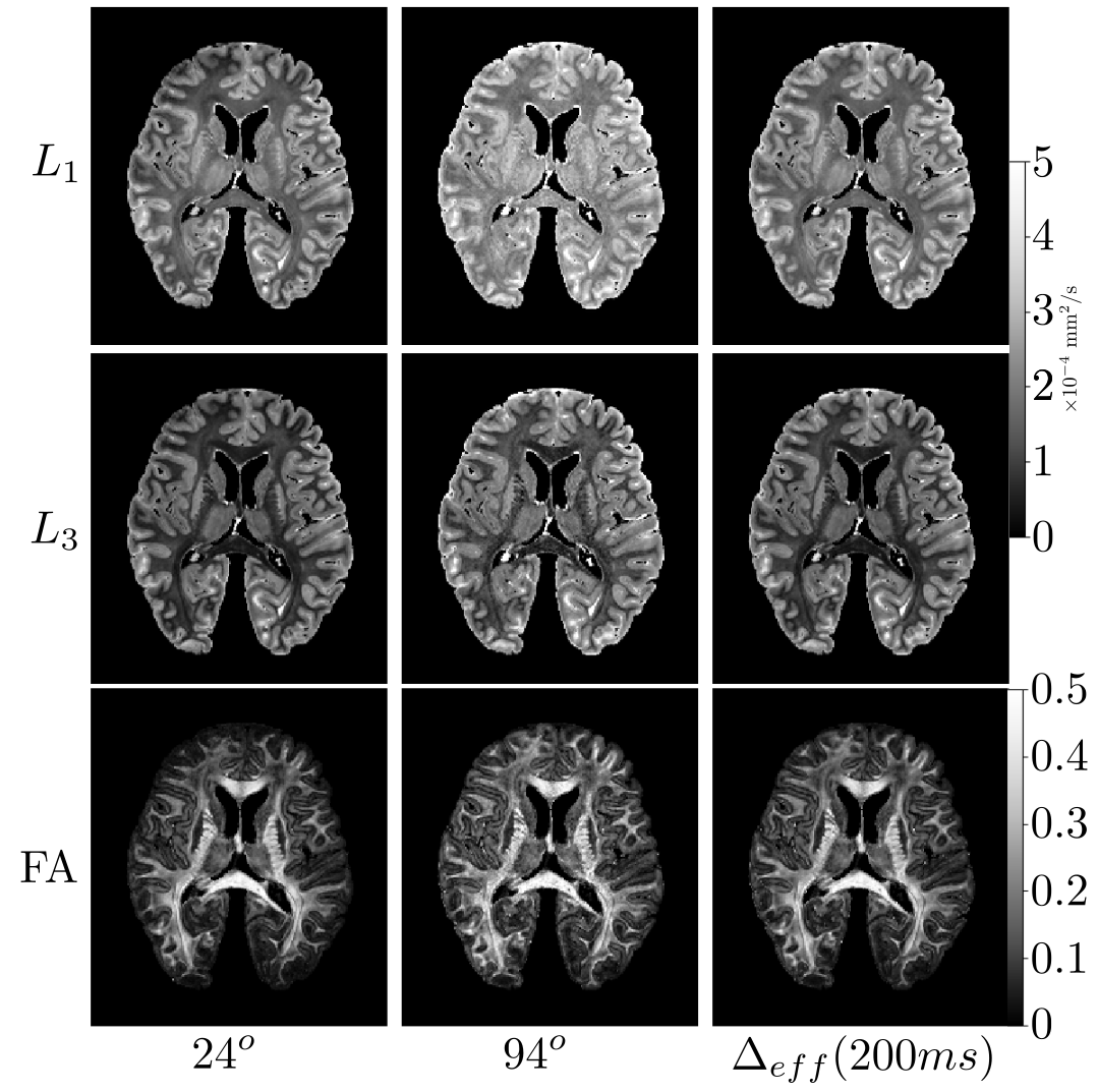
The $$$\mu$$$ and $$$\sigma$$$ maps were subsequently input into Eq. [3], where we substituted $$$\cos\alpha$$$ with an expression in terms of $$$\Delta_{eff}$$$ (rearranging Eq. [2]) to obtain $$$L_{1,2,3}$$$ estimates for $$$\Delta_{eff}$$$=200ms over the entire brain. $$$\Delta_{eff}$$$=200ms was selected due to the low diffusivity of postmortem tissue. Furthermore, it corresponds to $$$\approx$$$26o in our sample, an SNR-efficient flip-angle in dwSSFP.
Results and discussion
Figure 4 reveals how $$$L_{1,2,3}$$$ varies with B1 in the
postmortem brain. The reconstructed $$$\Delta_{eff}$$$=200ms maps (corresponding to $$$\approx$$$26o flip-angle) give good
agreement to the 24o dataset at high B1 whilst maintaining
a flatter distribution, highlighting the removal of B1-induced
ADC variability in our reconstruction. At 94o, we observe higher $$$L_{1,2,3}$$$ estimates in B1 matched areas, in agreement with simulation (Figure
3).
However, little B1-influence is observed at 94o, suggesting a reduced ADC-sensitivity at high flip-angles (Figure
3).
The dwSSFP signal is SNR-efficient at low flip-angles1 (Figure 1). Figure 5 reveals that by extrapolating our dual flip-angle datasets to $$$\Delta_{eff}$$$=200ms, we are able to simultaneously preserve the SNR of the low flip-angle dataset whilst removing the influence of B1, generating results in terms of a single effective diffusion time over the entire brain.
Conclusion
The framework presented here generates high-SNR dwSSFP ADC estimates in terms of a single effective diffusion time, removing the influence of B1. This methodology could be extended to other diffusion models, distributions, or to derive ADC estimates in terms of $$$\Delta_{eff}$$$ for a single-orientation, multiple flip-angle dwSSFP dataset, making dwSSFP more comparable to conventional diffusion measurements.Acknowledgements
No acknowledgement found.References
1Buxton, Richard B. "The diffusion sensitivity of fast steady‐state free precession imaging." Magnetic resonance in medicine 29.2 (1993): 235-243.
2Miller, Karla L., et al. "Diffusion tractography of post-mortem human brains: optimization and comparison of spin echo and steady-state free precession techniques." Neuroimage 59.3 (2012): 2284-2297.
3Foxley, Sean, et al. "Improving diffusion-weighted imaging of post-mortem human brains: SSFP at 7 T." Neuroimage 102 (2014): 579-589.
4Foxley, Sean, et al. “Correcting for B1 inhomogeneities in post-mortem DWSSFP human brain data at 7T using multiple flip-angles.” 22nd Proc. Intl. Soc. Mag. Reson. Med., 2014: #4438.
5Jbabdi, Saad, et al. “Modelling multiple flip-angle diffusion weighted SSFP data.” 23rd Proc. Intl. Soc. Mag. Reson. Med., 2015: #2928.
6McNab, Jennifer A., and Karla L. Miller. "Sensitivity of diffusion weighted steady state free precession to anisotropic diffusion." Magnetic Resonance in Medicine 60.2 (2008): 405-413.
7Jbabdi, Saad, et al. "Model‐based analysis of multishell diffusion MR data for tractography: How to get over fitting problems." Magnetic Resonance in Medicine 68.6 (2012): 1846-1855.
8Pallebage-Gamarallage, Menuka, et al. "Dissecting the pathobiology of altered MRI signal in amyotrophic lateral sclerosis: A post mortem whole brain sampling strategy for the integration of ultra-high-field MRI and quantitative neuropathology." BMC neuroscience 19.1 (2018): 11.
9Hernandez-Fernandez Moises, et al. Using GPUs to accelerate computational diffusion MRI: From microstructure estimation to tractography and connectomes. BioRxiv. 2018
10McNab, Jennifer A., and Karla L. Miller. "Steady‐state diffusion‐weighted imaging: theory, acquisition and analysis." NMR in Biomedicine 23.7 (2010): 781-793.
11Jenkinson, Mark, et al. "Fsl." Neuroimage
62.2 (2012): 782-790.
Figures