0544
Imaging injectable liposomal hydrogels using CEST MRI for applications in the brainXiongqi Han1, Jianpan Huang1, Peng Xiao1, Joseph Ho Chi Lai1, and Kannie Wai Yan Chan1,2,3
1City University of Hong Kong, Hong Kong, China, 2Department of Radiology and Radiological Science, The Johns Hopkins University School of Medicine, Baltimore, MD, United States, 3F.M. Kirby Research Center for Functional Brain Imaging, Kennedy Krieger Research Institute, Baltimore, MD, United States
Synopsis
Nanocarrier-loaded hydrogel shows good prospects in biomedical application. However, there is lack of noninvasive methods for
Introduction
Hydrogel has gained considerable attention in recent years, especially with the incorporation of nanocarriers, such as liposomes, which is the first FDA approved nanocarrier for drug delivery1. It offers unique properties such as minimizing burst release and site-specific therapy. Alginate and HAMC are two kinds of most widely used hydrogels in biomedical realm. However, there is lack of noninvasive methods for real time monitoring hydrogel and drug release to guide therapy. Chemical exchange saturation transfer (CEST) could be used as a versatile imaging for hydrogel compositions, degradation and the encapsulated cells viabilities2-5. In this study, we developed drug-loaded CEST liposomal (LipoCEST) injectable hydrogels. The resultant liposomal hydrogel showed porous structure with mechanical property comparable to native brain tissue. In addition to the CEST contrast of our model drug at 5 ppm, we also observed the nuclear overhauser enhancement (NOE, -3.5 ppm) from the phospholipid bilayers in our LipoCEST hydrogel formulations.Methods
Liposomes were prepared by thin film hydration method1, 6, 7, composed of DPPC, cholesterol and DSPE-PEG2000 in a molar ratio of 10:8:1. Briefly, the lipid mixture was dried on a rotary evaporator to form a homogeneous thin film layer. Afterwards, 1 mL barbituric acid (BA) solution (25 mg/mL, pH 7.2) was added to hydrate under 65 oC for 1 h. The resulting mixture was sonicated and extruded through 400 nm polycarbonate filters. Liposomes was then mixed with either alginate or HAMC to form liposomal hydrogels. Phantoms were imaged on a horizontal bore 3T preclinical Bruker MRI system at 37 oC with a 38 mm volume coil. The B0 field was shimmed and a modified rapid acquisition with relaxation enhancement (RARE) sequence was used to generate the Z-spectrum. Images ware acquired with the following parameters: Slice thickness=1 mm, field of view (FOV) =2020 mm, image size = 6464, RARE factor = 32, repetition time/echo time (TR/TE) = 6000/4.7 ms, -7 to + 7 ppm, 0.2 ppm steps.Results
BA liposomes showed good loading capacity (up to 76.8%) with size about 200 nm. It generated both CEST and NOE contrast at 5.0 ppm (BA, drug) and -3.4 ppm (liposomes) with the highest intensity up to 8.46% and 4.20% acquired at B1 = 0.4 μT and Tsat = 3000 ms. All hydrogels showed storage modulus in the range of 100-300 Pa at 10 Hz. After the addition of liposomes into hydrogels, G’ was slight decreased (5.6~22.6%). Both kinds of hydrogels showed interconnected microporous structures with similar pore size about 750 and 10 μm2 in alginate and HAMC, respectively. Moreover, the resultant LipoCEST hydrogel showed contrasts of 1.82±0.09%/1.47±0.12% for alginate and 2.16±0.11%/2.25±0.17% for HAMC at 5 ppm/-3.4 ppm, respectively. Most interestingly, we also observed the CEST signals at 1 ppm and -1.4 ppm in HAMC hydrogels.Discussion
We have systemically investigated the effects of liposomes on rheological properties and porosities of the resultant hydrogels. The goal is to develop injectable liposomal hydrogels for imaging-guided therapy in brain. Both CEST and NOE contrasts of liposomes were proportional to drug loading and liposomes number, respectively. Moreover, there were peaks at 1.0 ppm and -1.4 ppm for HAMC hydrogel, which could be ascribed to the hydroxyl groups of HA and methoxy protons of methylcellulose, respectively. These multiple CEST readouts could be favorable for monitoring drug, nanocarrier and hydrogel composition independently and simultaneously in hydrogel matrix. After addition of liposomes, G’ was slightly decreased, but all in the range of 10-300 Pa, which is comparable to the native brain tissue 8. Besides, both types of hydrogel showed porous structure, with large pore size of alginate (>50 μm) facilitating nutrient exchange and cell migration9 and the smaller pore size (<20 μm) of HAMC favoring drug retention.Conclusions
We have developed injectable hydrogels with unique CEST contrasts and mechanical properties comparable to the brain. This multiparametric imaging platform shows good prospects in imaging-guided therapy in vivo for monitoring drug, nanocarrier and hydrogel matrix simultaneously and independently.Acknowledgements
This study was supported by CityU: P9610362; P7200516; P6000612;P7004859; RGC: GRF-9042620; NSFC: 81871409-H1808.References
1. Torchilin, Vladimir P. Nature reviews Drug discovery 4.2 (2005): 145. 2. Liang, Y. et al. Biomaterials 2015:42:144-150. 3. Jin, T. et al. Biomaterials 2017:113:176-190. 4.Shazeeb, M. S. et al. Biomaterials 2018:178:326-338. 5.Chan, K. W. et al. Nat Mater 2013:12:268-275. 6.Chan, K. W. et al. WIREs Nanomed Nanobiotechnol 2013:6:111-124. 7.Chan, K. W. et al. J Control Release 2014:180:51-59. 8. M.J. Rowland et al. / Biomaterials 2018:179:199-208. 9. Majumder, Poulami, et al. Angew. Chem. Int. Ed. 2018, 57, 1-6Figures
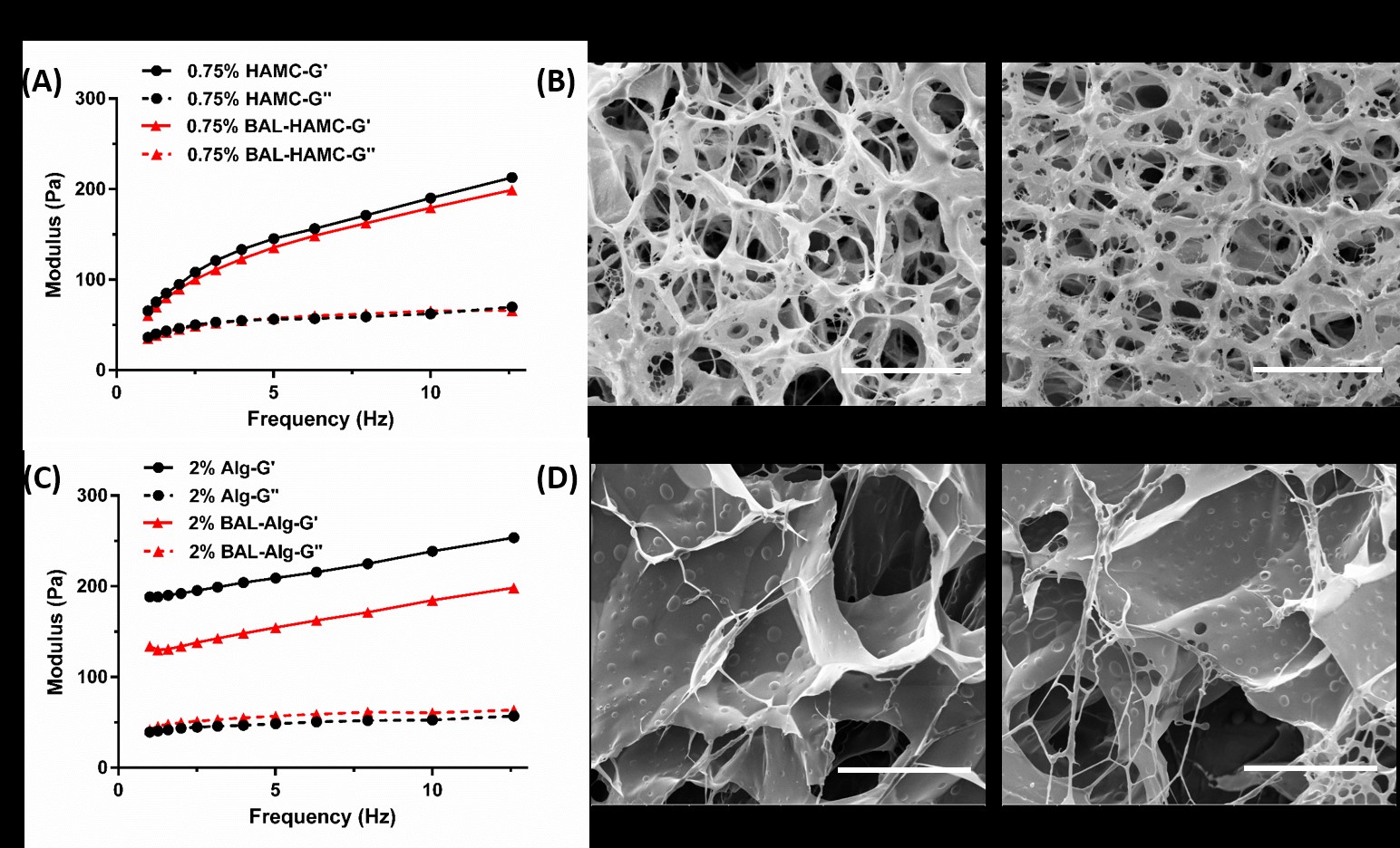
Fig.1
Rheological properties and representative SEM images of liposomal hydrogels.
(A) and (B) were frequency sweep results and SEM images of HAMC hydrogel with
or without liposomes, scale bar=50 μm; (C) and (D) were frequency sweep results
and SEM images of alginate hydrogel with or without liposomes, scale bar=100
μm. BA-liposomes (BAL) concentrations were 1014 particle/mL.
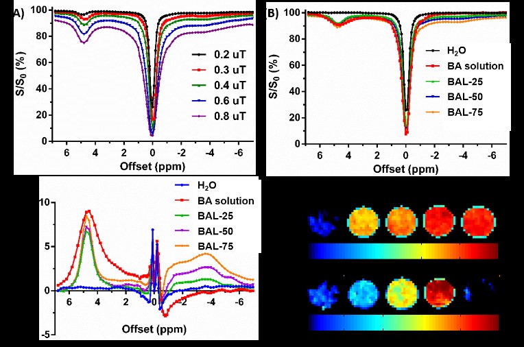
Fig. 2 CEST
properties of BA-liposomes (n=3). (A) Z-spectra of BA liposomes with 75 mg/mL
lipids under various B1 fields. (B), (C) and (D) were Z-spectra,
CEST contrast (%) and the representative maps (at 5.0 and -3.4 ppm) of BA
liposomes with different concentrations.
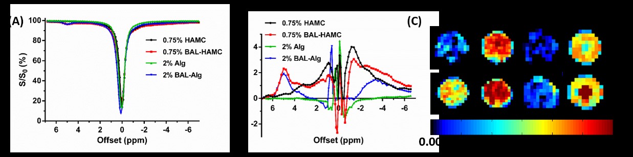
Fig.3
CEST
properties of BA-liposomes incorporated alginate and HAMC hydrogel (n=3).
(A) Z-spectra and (B) CEST contrast (%) of liposomal hydrogels. (C) Corresponding
CEST parametric maps at 5.0 and -3.4 ppm.