0529
Quantitative MRI with spatiotemporal normalization can detect longitudinal changes in primary lateral sclerosis cervical spinal cord1Physics and Astronomy, University of British Columbia, Vancouver, BC, Canada, 2International Collaboration on Repair Discoveries, University of British Columbia, Vancouver, BC, Canada, 3Institute of Psychiatry, Psychology & Neuroscience, King's College, London, United Kingdom, 4Radiology, University of British Columbia, Vancouver, BC, Canada, 5Kinesiology, University of British Columbia, Vancouver, BC, Canada, 6Medicine, University of British Columbia, Vancouver, BC, Canada
Synopsis
Analysis of longitudinal quantitative MRI data can be confounded by a variety of factors. We use spatiotemporal normalization and a symmetric diffeomorphic normalization algorithm to compare quantitative MRI metrics in a patient-specific halfway space. Utilizing these methods, myelin water imaging and diffusion tensor imaging were able to detect changes in primary lateral sclerosis (PLS) spinal cord similar to those found in the faster-progressing amyotrophic lateral sclerosis (ALS). Future studies could employ spatiotemporal normalization with more subjects and imaging timepoints to investigate the use of longitudinal MWI and DTI as a biomarker for motor neuron disease progression.
Background
Primary lateral sclerosis (PLS) is a motor neuron disease that causes degeneration of upper motor neurons which carry motor information down the spinal cord. MRI techniques sensitive to changes in myelin have been used to detect myelin loss in PLS brain1-3, but not spinal cord. Myelin water imaging (MWI) is a quantitative MRI technique that measures the myelin water fraction (MWF), a metric with high specificity to myelin4,5. The 3D multi-echo gradient and spin echo (GRASE) sequence has recently been implemented for rapid, high-resolution spinal cord MWI6. Fractional anisotropy (FA) and radial diffusivity (RD) are well established diffusion tensor imaging (DTI) metrics, sensitive to myelin but more generally related to microstructural tissue integrity. Quantitative MRI results from these techniques can be confounded by a variety of factors during analysis, such as bias or noise from cross-sectional template creation methods7, similarity metrics introducing circularity bias8, or poor anatomical alignment of images in common space. These issues are amplified in longitudinal studies, where results can easily be obscured by biased or underperforming methods.
This study demonstrates the use of spatiotemporal normalization for analyzing longitudinal quantitative spinal cord MRI data. We also provide pilot data for future studies of primary lateral sclerosis.
Subjects and Imaging
Subjects: 2 volunteers with PLS (P01: 44 year-old female, P02: 64 year-old female) and 6 healthy controls (4 females, 2 males, mean=48yrs, σ=11yrs, range 30-62yrs). PLS scans were repeated after 10 (P01) and 11 (P02) months.
Data acquisition: Imaging was performed on a 3T scanner (Achieva, Philips Medical Systems, Best, The Netherlands) with a 6-channel spine coil. Anatomical images included a sagittal T2 weighted image and a high-resolution multi-echo gradient echo (mFFE) sequence with strong white/gray matter contrast for inter-subject registrations. Quantitative MRI included GRASE rapid spinal cord MWI6 and 32-direction DTI. Axial scans were centered at the level of the C2/C3 disk (Figure 1). Acquisition details are provided in Table 1.
Data processing: GRASE data was processed using a regularized non-negative least-squares fitting algorithm with stimulated echo correction9,10 and MWF was calculated as the fractional signal with T2<40ms11. The Spinal Cord Toolbox12 software was used to motion correct DTI data before calculating FA and RD metrics, and for spinal cord segmentation.
Analysis Methods
The Advanced Normalization Tools (ANTs) software was used for all registrations13. Intra-subject/intra-visit normalization of GRASE (echo 16) and DTI (mean diffusion weighted) data to mFFE space allowed metric maps to be transformed with their corresponding mFFE image. A combination of rigid, affine, and greedy symmetric diffeomorphic transformation algorithms was optimized for anatomical alignment of internal cord structure during inter-subject/inter-visit mFFE normalizations. For each PLS subject, mFFE images were normalized to create a spatiotemporal halfway space (subject-specific template without bias towards any timepoint14). Similarly, control data was normalized to create a template space with mean and standard deviation metric maps. Voxel-wise metric difference maps were calculated in each PLS subject halfway space, then normalized to control template space. Finally, longitudinal Z score maps were calculated as $$$ Z= \frac{Metric[PLS\ Visit\ 2]-Metric[PLS\ Visit\ 1]}{\sigma_{Metric}[Controls]} $$$ with a coefficient of variation threshold at 0.7 . This process is outlined in Figure 2.Results
Figure 1 shows representative slices of MWF, FA, and RD maps in their native image space for P01 and P02 at baseline and follow-up. Figure 3 shows longitudinal MWF, FA, and RD difference Z score maps for P01 and P02. Areas of negative Z-scores indicating decreasing MWF (Figure 3 top) and FA (Figure 3 middle) over 10-11 months were apparent for both P01 and P02, though more extensive for P01. Positive Z-scores indicating increasing RD (Figure 3 bottom) were also observed, particularly for P02.Discussion
PLS presents with axonal degeneration in the motor cortex of the brain, which would cause Wallerian degeneration along the distal portion of the axon, leading to subsequent degradation of the myelin sheath. This should be visible as demyelination of tracts extending from the motor cortex of the brain to the lower motor neurons in the spinal cord. Our results show areas of decreasing MWF over time, consistent with demyelination. Longitudinally decreasing FA and increasing RD also suggest demyelination (though with less specificity than MWF).Conclusions
A spatiotemporal normalization method for MWI and DTI data analysis can detect longitudinal changes in quantitative MRI metrics of PLS spinal cord over less than a year. Future studies can apply these analysis methods with more subjects and an arbitrary number of imaging timepoints to investigate the use of longitudinal MWI and DTI as a biomarker for motor neuron disease progression.Acknowledgements
We thank all of the participants in this study, without whom our research would not be possible, and the UBC MRI Centre and its MRI technologists for their steadfast support of our work.References
1. Kolind, S., et al., Myelin imaging in amyotrophic and primary lateral sclerosis. Amyotrophic Lateral Sclerosis and Frontotemporal Degeneration, 2013. 14(7-8): p. 562-573.
2. Ulug, A.M., et al., Diffusion tensor imaging in the diagnosis of primary lateral sclerosis. Journal of Magnetic Resonance Imaging, 2004. 19(1): p. 34-39.
3. Muller, H.P., et al., Identical patterns of cortico-efferent tract involvement in primary lateral sclerosis and amyotrophic lateral sclerosis: A tract of interest-based MRI study. Neuroimage Clin, 2018. 18: p. 762-769.
4. MacKay, A., et al., In vivo visualization of myelin water in brain by magnetic resonance. Magn Reson Med, 1994. 31(6): p. 673-7.
5. Laule, C., et al., Magnetic resonance imaging of myelin. Neurotherapeutics, 2007. 4(3): p. 460-84.
6. Ljungberg, E., et al., Rapid myelin water imaging in human cervical spinal cord. Magn Reson Med, 2016.
7. Reuter, M. and B. Fischl, Avoiding asymmetry-induced bias in longitudinal image processing. Neuroimage, 2011. 57(1): p. 19-21.
8. Tustison, N.J., et al., Logical circularity in voxel-based analysis: Normalization strategy may induce statistical bias. Human Brain Mapping, 2014. 35(3): p. 745-759.
9. Prasloski, T., et al., Applications of stimulated echo correction to multicomponent T2 analysis. Magn Reson Med, 2012. 67(6): p. 1803-14.
10. Yoo, Y.J., et al., Fast Computation of Myelin Maps From MRI T-2 Relaxation Data Using Multicore CPU and Graphics Card Parallelization. Journal of Magnetic Resonance Imaging, 2015. 41(3): p. 700-707.
11. Laule, C., et al., Two-year study of cervical cord volume and myelin water in primary progressive multiple sclerosis. Mult Scler, 2010. 16(6): p. 670-7.
12. De Leener, B., et al., SCT: Spinal Cord Toolbox, an open-source software for processing spinal cord MRI data. Neuroimage, 2017. 145(Pt A): p. 24-43.
13. Avants, B.B., et al., A reproducible evaluation of ANTs similarity metric performance in brain image registration. Neuroimage, 2011. 54(3): p. 2033-2044.
14. Reuter, M., et al., Within-subject template estimation for unbiased longitudinal image analysis. Neuroimage, 2012. 61(4): p. 1402-1418.
Figures
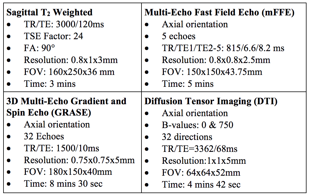
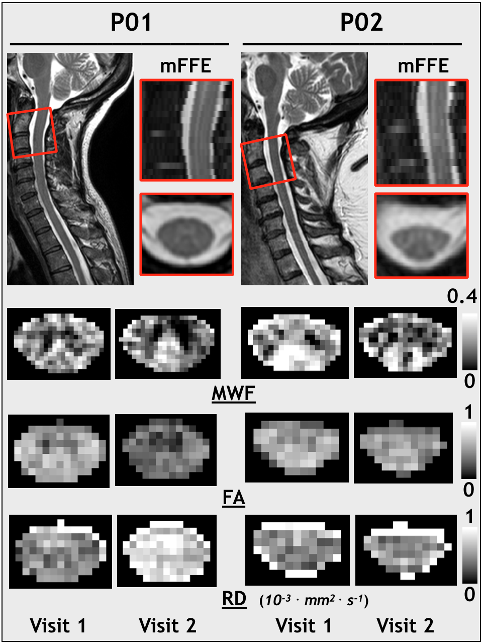
Figure 1: Anatomical and quantitative MRI results in their native image space.
Visit 1 sagittal T2 weighted images are show with the anatomical mFFE image field of view superimposed. Representative slices of the myelin water fraction (MWF), fractional anisotropy (FA) and radial diffusivity (RD) maps are presented for Visit 1 and Visit 2 of both PLS patients.
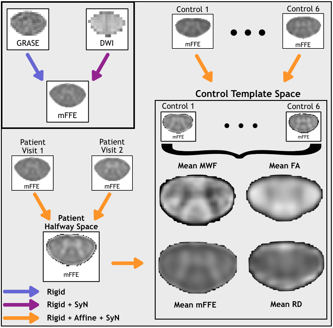
Figure 2: Workflow of analysis methods.
Rigid, affine, and symmetric diffeomorphic normalization (SyN) transformation algorithms are used to create a spatiotemporal halfway space with longitudinal data for each patient and a study-specific template with healthy control data. SyN normalizations determine the minimal deformations required to align anatomical mFFE image cords and internal structures (gray matter). Intra-subject normalization of MWI and DTI data to mFFE space allows creation of healthy control mean and standard deviation maps and patient Z score maps in template space for myelin water fraction (MWF), fractional anisotropy (FA) and radial diffusivity (RD).
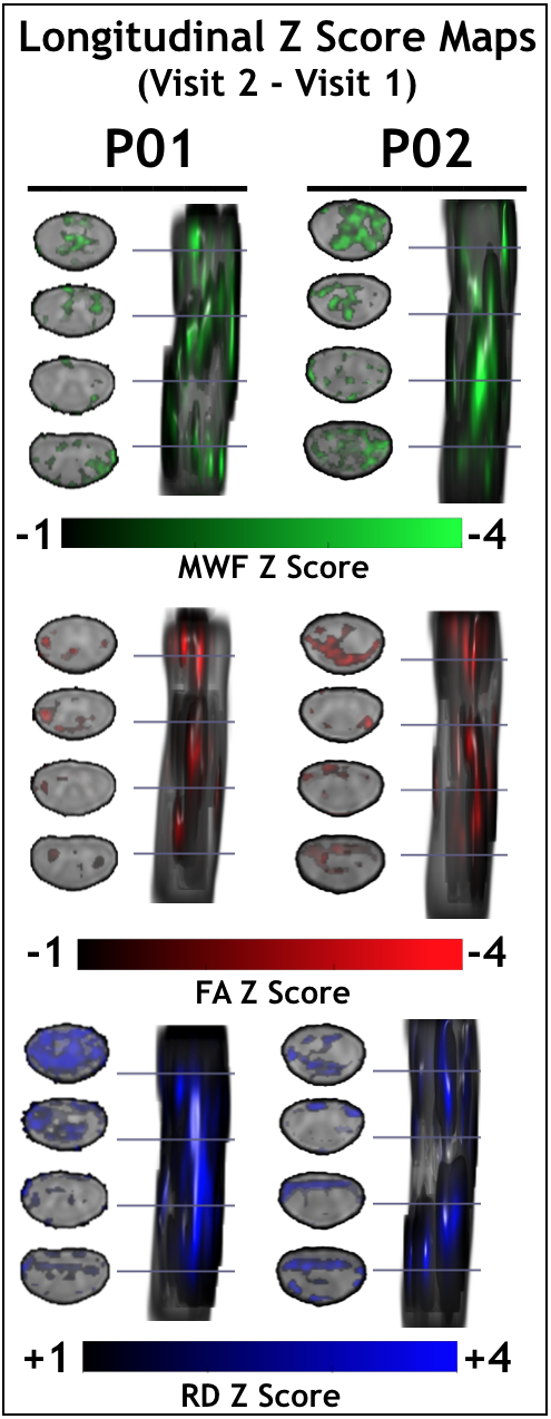
Figure 3: Longitudinal Z scores maps.
The change in myelin water fraction (MWF), fractional anisotropy (FA) and radial diffusivity (RD) metrics between Visit 1 and Visit 2 is calculated in the spatiotemporal halfway space for both PLS patients. The resulting difference maps are transformed to template space and divided by healthy control standard deviation maps to produce longitudinal Z score maps. A threshold was applied at 0.7 for the coefficient of variation (healthy control standard deviation divided by mean).