0515
Towards absolute quantification of brain metabolites using Electronic REference To access In vivo Concentrations (ERETIC) for MR spectroscopic imaging (MRSI)1Dept. of Radiology, MGH, A. A. Martinos Center for Biomedical Imaging, Charlestown, MA, United States, 2Harvard Medical School, Boston, MA, United States, 3Mittelhessen University of Applied Science, Giessen, Germany, 4Siemens Healthcare, Erlangen, Germany, 5Siemens Medical Solutions USA, Charlestown, MA, United States
Synopsis
Absolute quantification of metabolite concentration from MRSI data requires a reference signal of known concentration. An external synthetic electronic reference signal method (Electronic REference To access In vivo Concentrations – ERETIC) has shown great promise for absolute quantification and calibration. However, ERETIC based absolute quantification is challenging for MR spectroscopic imaging (MRSI) data and here we set on investigating strategies and performance of ERETIC in combination with MRSI and multi-channel receive arrays.
INTRODUCTION
For 1H-MRSI the water signal is typically used as internal reference assuming a known concentration. However, in disease conditions water content varies due to inflammation, edema and cell death. The external synthetic electronic reference (Electronic REference To access In vivo Concentrations – ERETIC1-2 has shown great promise to avoid these biases while mitigating coil loading differences from phantom replacement calibrations. The absolute quantification of metabolite can be achieved by injecting a calibrated ERETIC reference signal using a micro-coil coupled inductively with the receive RF coil during acquisition of MR signal2. A particular challenge with the use of ERETIC in MRSI experiments arise from the fact that ERETIC signal does not see the localization gradients of the pulse sequence3 . In addition, ERETIC has not been yet demonstrated with multi-channel receive array coils. In this work we used a custom build ERETIC hardware and receive array coil and investigated localization strategies for ERETIC signal in MRSI experiments.METHOD
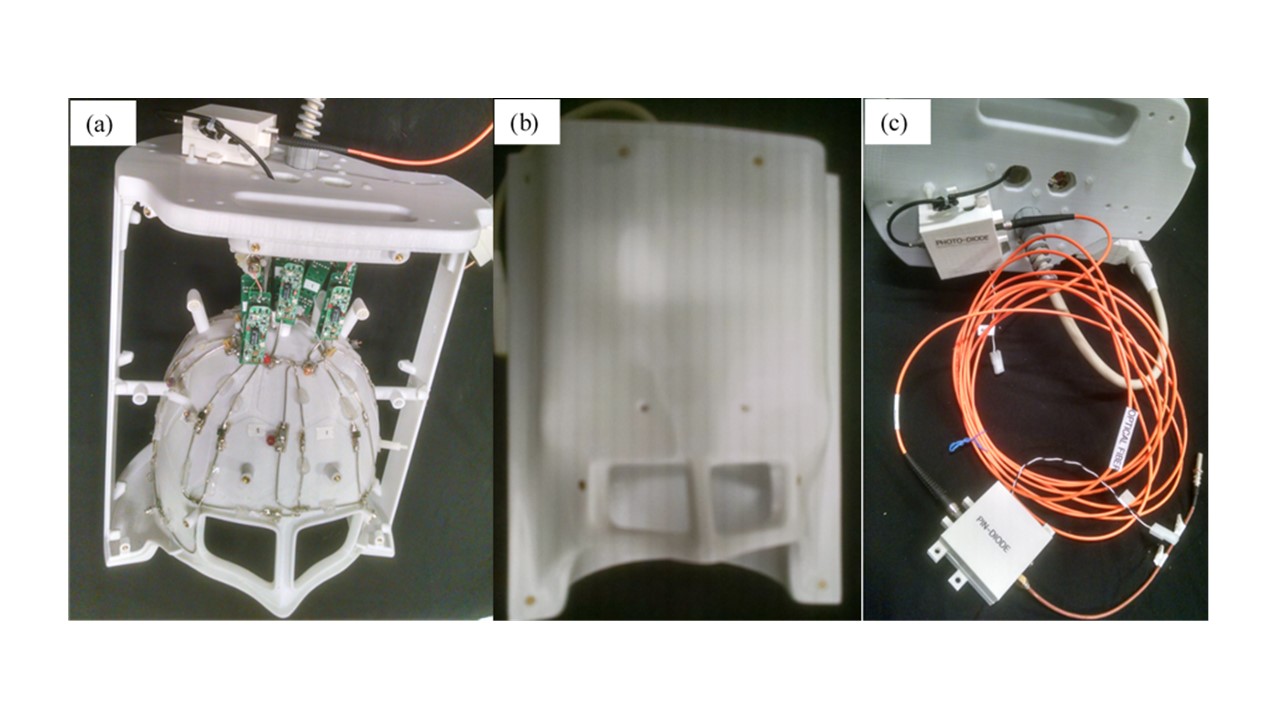
We build an 8-channel receive array RF coil (trapezoidal shaped elemental coil: anterior two coils of dimension: 11 x 11 x11 x 6 cm and 6 lateral and posterior coils of dimension: 17 x 11 x 17 x 6 cm) on the 3D printed head frame designed using Rhinoceros cadsoftware as shown in Fig. 1(a-b) for the whole brain imaging. A low power ERETIC signal was synthesized from TTX port of the scanner (3T Tim Trio, Siemens) and optical signal transmission line was used to send ERETIC from TTX to 8-channel receive array as previously proposed4 in order to eliminate the parasitic coupling and variability of ERETIC signal with RF coax cables. An ERETIC micro-coil of was placed at an equidistance from the apex of the RF coils, or a distributed ensemble of ERETIC coils (diameter 3 mm and 3 turns) was used for the uniform excitation of the array coil by ERERTIC pulse. Fig.1 depicts the photograph of (a) an 8 channel receive only human brain dedicated RF coil with trapezoidal elements, (b) the coil with cover, and (c) RF over fiber link consisting of a PIN diode, optical fiber, and photo diode for the transmission of ERETIC pulse. The ERETIC signal was programmed inside the MRSI pulse sequence and was controlled by the host computer of the Siemens scanner. A 2D LASER-MRSI5 sequence (TR/TE=1500/45 ms) with FOV=220x220, matrix 22x22, 20 mm thickness was used for metabolite localization, An ERETIC FID (8 Hz Lorentzian line at -0.5 ppm offset from DSS, 6oo ms duration) was transmitted during acquisition of the MRSI signal. A basis set was simulated with the ERETIC signal included and data were fitted with LCModel6 . The method was tested in two phantoms and two healthy volunteers.RESULTS
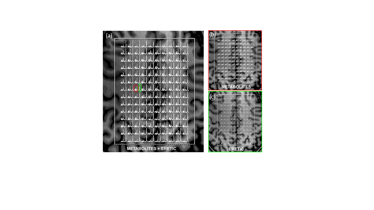
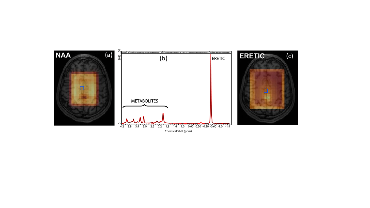
Our aim was to encode ERETIC signal in all the MRSI voxels to provide a reference for all brain regions. Previous demonstrations showed ERETIC encoded only in the center voxel of the MRSI data3. Although ERETIC signal does not sense the gradient localization of the pulse sequence, the amplitude and phase of the ERETIC can be altered as to encode a certain k-space pattern. The MRSI spectral grid with in vivo brain metabolites and ERETIC signal obtained from one volunteer is shown in Fig. 2. In Fig 2a, the entire spectral range of metabolites + ERETIC (4.2 to -1.5 ppm) is shown throughout the volume of interest, while Fig2b shows the metabolite spectral range (4.2 to 0.8 ppm), and Fig 2c shows the ERETIC spectral range (0.5 to -1.5 ppm). Encoding of the ERETIC signal can be noted in all the MRSI voxels with comparable signal amplitude. Fig. 3 reflects the LCModel fitting, with a map of N-Acetyl-aspartate (NAA) and ERETIC signal. Fig. 3b represents the LCModel fitting of metabolites and ERETIC signal obtained from a representative voxel (blue outlined) in NAA map in Fig 3a or ERETIC signal map in Fig. 3c.DISCUSSION
These preliminary data indicate that integration of ERETIC with clinical scanners, receive arrays and different spatial encoding strategies are possible and promising for absolute quantification of MRSI data. Further investigation and demonstration in patients with brain tumors is underway.CONCLUSION
Combination of ERETIC and receive arrays for MRSI with multivoxel localization of ERETIC is possible and might be used for absolute quantification.Acknowledgements
This work is supported by NIH (1R01CA211080-02).References
[1] Heinzer-Schweizer, S., N. De Zanche, M. Pavan, G. Mens, U. Sturzenegger, A. Henning, and P. Boesiger. 2010. 'In-vivo assessment of tissue metabolite levels using 1H MRS and the Electric REference To access In vivo Concentrations (ERETIC) method', NMR Biomed, 23: 406-13.
[2] Marro KI, Lee D, Shankland EG et al. Synthetic signal injection using inductive coupling. J Magn Reson. 2008; 194(1): 67–75.
[3] Zoelch, N., A. Hock, S. Heinzer-Schweizer, N. Avdievitch, and A. Henning. 2017. 'Accurate determination of brain metabolite concentrations using ERETIC as external reference', NMR Biomed, 30.
[4] Heinzer-Schweizera S, Zanchea ND, Pavana M et al. In-vivo assessment of tissue metabolite levels using 1H MRS and the Electric REference To access In vivo Concentrations (ERETIC) method. NMR Biomed. 2010; 23: 406–413.
[5] Andronesi, O. C., S. Ramadan, E. M. Ratai, D. Jennings, C. E. Mountford, and A. G. Sorensen. 2010. 'Spectroscopic imaging with improved gradient modulated constant adiabaticity pulses on high-field clinical scanners', Journal of Magnetic Resonance, 203: 283-93.
[6] Provencher, S. W. 2001. 'Automatic quantitation of localized in vivo H-1 spectra with LCModel', NMR Biomed, 14: 260-64.
Figures