0493
Deuterated water labeling followed by deuterium MRI for visualization of tumors in vivo1Experimental Transplantation and Immunology Branch (ETIB), National Cancer Institute (NCI), National Institutes of Health (NIH), Bethesda, MD, United States, 2Radiation Biology Branch, NCI, NIH, Bethesda, MD, United States, 3Laboratory of Functional and Molecular Imaging, National Institute of Neurological Disorders and Stroke (NINDS), NIH, Bethesda, MD, United States, 4ETIB, NCI, NIH, Bethesda, MD, United States, 5Radiation Biology Branch (RBB), NCI, NIH, Bethesda, MD, United States, 6Urologic Oncology Branch, NCI, NIH, Bethesda, MD, United States, 7RBB, NCI, NIH, Bethesda, MD, United States
Synopsis
In vivo DNA labeling with deuterated water (2H2O) has been used for cell kinetics research and more recently to image rapidly proliferating immune cells in the context of graft-versus-host disease. Using a custom dual-resonance coil (1H-2H) we demonstrate that this approach can be applied to the in vivo detection of tumors via MRI in a xenograft tumor mouse model. Therefore, this novel imaging technique could serve as a sensitive, safe, and non-radioactive method of tumor detection with significant impact on the field of oncology.
Introduction
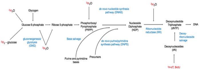
In vivo deuterated water (2H2O) labeling has been used in cell kinetics research because deuterium in body water incorporates into newly synthesized DNA bases via de novo nucleotide synthesis1, Figure 1. Thus, we developed a novel imaging approach using in vivo deuterated water labeling followed by deuterium magnetic resonance imaging (dMRI) for visualization of rapidly proliferating cells2. Our group has previously demonstrated that this approach could be used to image dysregulated immunity, such as graft-versus-host disease, which is characterized by infiltration of target organs by rapidly proliferating immune cells. Since deuterium enrichment serves as a proxy for cell proliferation, we hypothesized that dMRI could also be used for in vivo tumor imaging, in a non-radioactive and clinically relevant manner.Methods
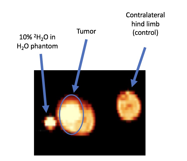
We tested our imaging approach in a xenograft tumor model. One hind limb of each athymic nude mouse (Foxn1nu) was injected with 10 million cultured HT-29, human colon adenocarcinoma, cells, Figure 2. The contralateral hind limb served as a control and did not receive a tumor injection. 2H2O labeling to 8% total body water (TBW) was initiated at the time of tumor injection, per previously described protocol2-4. Following 7 and 14 days of in vivo 2H2O labeling (and 7 and 14 days of in vivo tumor growth, respectively), mice underwent anatomical and deuterium MRI, under anesthesia. Both hind limbs were placed into a custom elliptical dual-resonance coil (1H-2H). A 3-mm diameter phantom containing 10% 2H2O in H2O was placed adjacent to the limbs and was included with each scan and served as a reference. Imaging was performed on 9.4 and 11.7 Tesla (T) magnets. CSI imaging was performed using the following parameters: 64 x 64 matrix, FOV = 32 x 32 mm, slice thickness = 3 mm, 3 slices, TR = 397 ms, total scan time = 27 minutes, fid acquisition time = 127.8 ms, 512 points, bw = 4000 Hz, fid acquisition starts 2.3 ms after rf pulse. The anatomical imaging was performed using the following parameters: spin-echo sequence, TR/TE = 500/8 ms, with the same geometry as CSI, but with matrix size of 128 x 128. All imaging data were analyzed using custom code written in Python. A urine sample was collected for each mouse immediately following dMRI to confirm TBW enrichment per previously published protocol2,4. Following the final imaging session, the mice were euthanized, and bulk tumor and a section of muscle from the contralateral limb were excised for gas chromatography-tandem mass spectrometry (GC-MS/MS) DNA deuterium enrichment analysis2-4.Results
Anatomical and CSI images were collected for each mouse. Following 7 and 14 days of in vivo 2H2O labeling and in vivo tumor growth, tumors appeared to have greater 2H signal, CSI signal normalized to the phantom (nCSI), compared to tissue surrounding tumor as well as contralateral control hind limb, using either a 9.4 or 11.7T magnet, Figure 3.Discussion
Using our approach of deuterated water labeling to 8% TBW we demonstrate higher in vivo deuterium signal in tumor versus unaffected tissue. Deuterated water labeling to 8% TBW is safe, non-toxic, and non-radioactive1; combining such labeling with dMRI allows sensitive detection of tumors in vivo.Conclusion
Our novel imaging approach is likely to have a significant impact on the field of oncology by allowing safe, non-radioactive, and sensitive in vivo detection of tumors. Clinical translation of this imaging approach is planned via a clinical protocol to image patients receiving care at the Clinical Cancer Center (CCR) of the National Cancer Institute (NCI).Acknowledgements
The authors acknowledge Dr. Stephen Dodd, Functional and Molecular Metabolism Section, National Institute of Neurological Disorders and Stroke (NINDS), and Dr. Martin Lizak, In Vivo NMR Center, NINDS for facilitating imaging experiments on the 11.7T and 9.4T magnets, respectively. Our work was supported by National Cancer Institute (NCI), National Institutes of Health (NIH) intramural funding.References
1. Busch, R., Neese, R.A., Awada, M., Hayes, G.M. & Hellerstein, M.K. Measurement of cell proliferation by heavy water labeling. Nat Protoc 2, 3045-3057 (2007).
2. Buxbaum, N.P., et al. In vivo kinetics and nonradioactive imaging of rapidly proliferating cells in graft-versus-host disease. JCI Insight 2(2017).
3. Farthing, D.E., et al. Sensitive GC-MS/MS method to measure deuterium labeled deoxyadenosine in DNA from limited mouse cell populations. Anal Chem 85, 4613-4620 (2013).
4. Farthing, D.E., et al. Comparing DNA enrichment of proliferating cells following administration of different stable isotopes of heavy water. Sci Rep 7, 4043 (2017).
Figures