0482
Human Brain 31P NMR Spectra at 7T: UDP-Glucose Assignment Revisited1Advanced Imaging Research Center, UT Southwestern Medical Center, Dallas, TX, United States, 2Department of Radiology, UT Southwestern Medical Center, Dallas, TX, United States, 3Department of Chemistry, University of Texas at Dallas, Richardson, TX, United States, 4VA North Texas Health Care System, Dallas, TX, United States
Synopsis
In a typical human brain 31P NMR spectrum, a small peak is often present at ~-9.7 ppm, near the α-ATP and NAD+/NADH signals. This 31P resonance, accounting for ~1/30th of the brain α-ATP signal, has been considered to be a doublet and assigned to UDPG. Here we present strong evidence to show that the -9.7 ppm signal is in fact a finely structured quartet. This finding has direct impact on current 31P NMR method for evaluation of brain redox, which requires UDPG correction. It may also have implication on our interpretation of brain energy metabolism based on 31P NMR data.
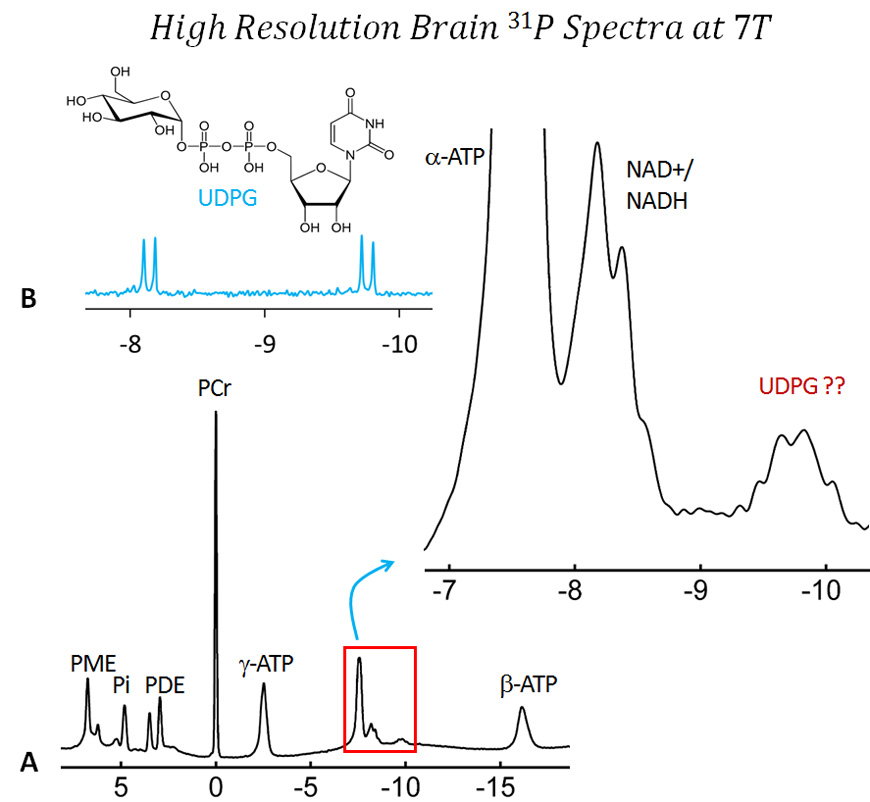
INTRODUCTION In a typical 31P MR spectrum acquired from human brain, a small peak often appears at ~-9.7 ppm (referenced to PCr at 0 ppm), in the upfield region near α-ATP (-7.6 ppm) and NAD+/NADH (-8.3) signals. This 31P resonance, accounting for about 1/30th of the brain α-ATP signal, has been assigned to UDPG, given the match between its chemical shift and the findings from studies of UDPG in solution and historical assignment of a similar signal found in plant cells.1-7 The assignment of the -9.7 ppm signal to UDPG in human brain is however difficult to confirm in vivo. Provided that the UDPG assignment is correct, then one would expect, as a part of weakly coupled AB spin system from UDPG pyrophosphate moiety, a doublet for the -9.7 ppm signal. Another doublet, which is 1.6 ppm downfield, falls in the region where NAD signal resides and becomes unresolvable due to overlap with the α-ATP signal.
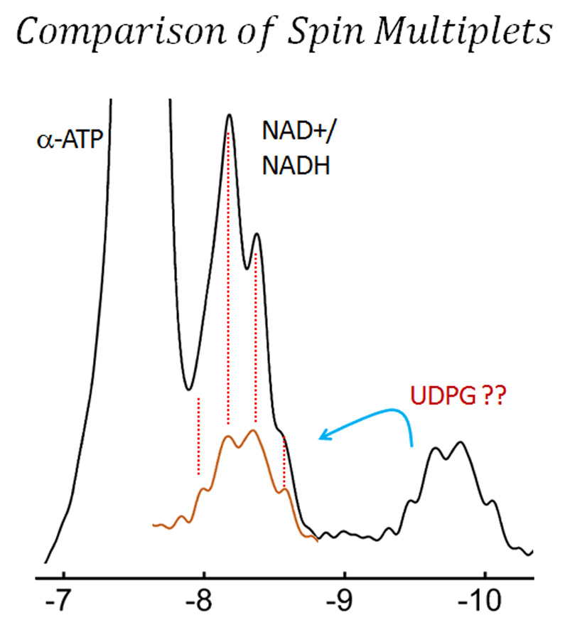
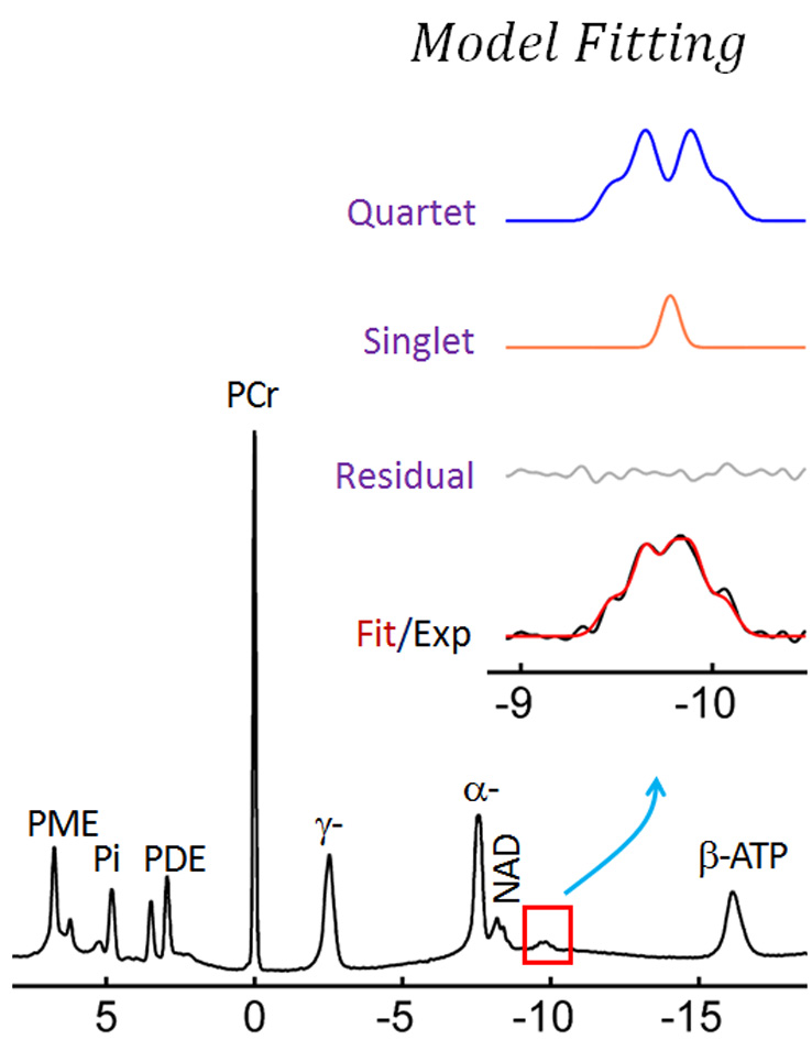
However, there is strong evidence, as will be revealed in this study by high-resolution NMR spectra acquired from human brain, that the -9.7 ppm signal cannot be reasonably characterized by a simple doublet as expected for UDPG. Instead, this signal appears to be a finely-structured quartet, suggesting that the traditional UDPG assignment needs to be revisited. This finding has direct impact on the current method for evaluation of brain redox state using NAD+/NADH ratio, which requires UDPG correction. This finding may also have potential implication in our interpretation of brain energy metabolism based on 31P NMR data.
Acknowledgements
This work was supported by NIH grant P41 EB-015908 and internal UTSW-AIRC grants FY18_IA0001 and FY18_IA0009.References
1. de Graaf RA, De Feyter HM, Brown PB, Nixon TW, Rothman DL, Behar KL. Detection of cerebral NAD+ in humans at 7T. Magn Reson Med. 2017;78(3):828-835.
2. Kim SY, Cohen BM, Chen X, Lukas SE, Shinn AK, Yuksel AC, Li T, Du F, Öngür D. Redox Dysregulation in Schizophrenia Revealed by in vivo NAD+/NADH Measurement. Schizophr Bull. 2017;43(1):197-204. 3. Chouinard VA, Kim SY, Valeri L, Yuksel C, Ryan KP, Chouinard G, Cohen BM, Du F, Öngür D. Brain bioenergetics and redox state measured by 31P magnetic resonance spectroscopy in unaffected siblings of patients with psychotic disorders. Schizophr Res. 2017;187:11-16.
4. Ren J, Sherry AD, Malloy CR. 31P-MRS of healthy human brain: ATP synthesis, metabolite concentrations, pH, and T1 relaxation times. NMR Biomed. 2015;28(11):1455-62.
5. Xin L, Ipek Ö, Beaumont M, Shevlyakova M, Christinat N, Masoodi M, Greenberg N, Gruetter R, Cuenoud B. Nutritional Ketosis Increases NAD+/NADH Ratio in Healthy Human Brain: An in Vivo Study by 31P-MRS. Front Nutr. 2018;5:62.
6. Lei H, Zhu XH, Zhang XL, Ugurbil K, Chen W. In vivo 31P magnetic resonance spectroscopy of human brain at 7 T: an initial experience. Magn Reson Med. 2003;49(2):199-205.
7. Roby C, Martin JB, Bligny R, Douce R. Biochemical changes during sucrose deprivation in higher plant cells. Phosphorus-31 nuclear magnetic resonance studies. J Biol Chem. 1987;262(11):5000-7.
8. Gout E, Rébeillé F, Douce R, Bligny R. Interplay of Mg2+, ADP, and ATP in the cytosol and mitochondria: unravelling the role of Mg2+ in cell respiration. Proc Natl Acad Sci U S A. 2014;111(43):E4560-7.
Figures