0471
Application of a k-Space Interpolating Artificial Neural Network to In-Plane Accelerated Simultaneous Multislice Imaging for Motion Monitoring1Radiation Oncology, Medical College of Wisconsin, Milwaukee, WI, United States, 2Radiology, Medical College of Wisconsin, Milwaukee, WI, United States
Synopsis
A scan-specific deep learning approach to Cartesian k-space interpolation was extended to in-plane accelerated simultaneous multislice imaging. This method yields images more accurate and less noisy reconstructions than conventional SMS parallel imaging algorithms, particularly as acceleration factors approach the number of receive coils. Furthermore, the models trained in one particular motion state are applicable to test data from a different motion state. This suggests that this method will be useful for cine imaging.
Introduction
Precisely delivering radiation to an abdominal or thoracic tumor is difficult due to substantial complex motion that can occur within each treatment fraction. The advent of MR-guided radiation therapy (MR-gRT) allows for unprecedented visualization of the target and surrounding tissues during treatment delivery. Gating and tracking based on real-time cine imaging are two motion management strategies achievable on integrated MR-RT treatment machines. Increasing slice coverage while maintaining high frame rates is one approach that may improve the precision of tracking algorithms in MR-gRT. The goal of this study was to extend the parallel imaging-like scan-specific 3-layer convolutional neural network known as RAKI[1] to unalias simultaneously excited (SMS) slices and perform subsequent phase-encoding line interpolation.Methods
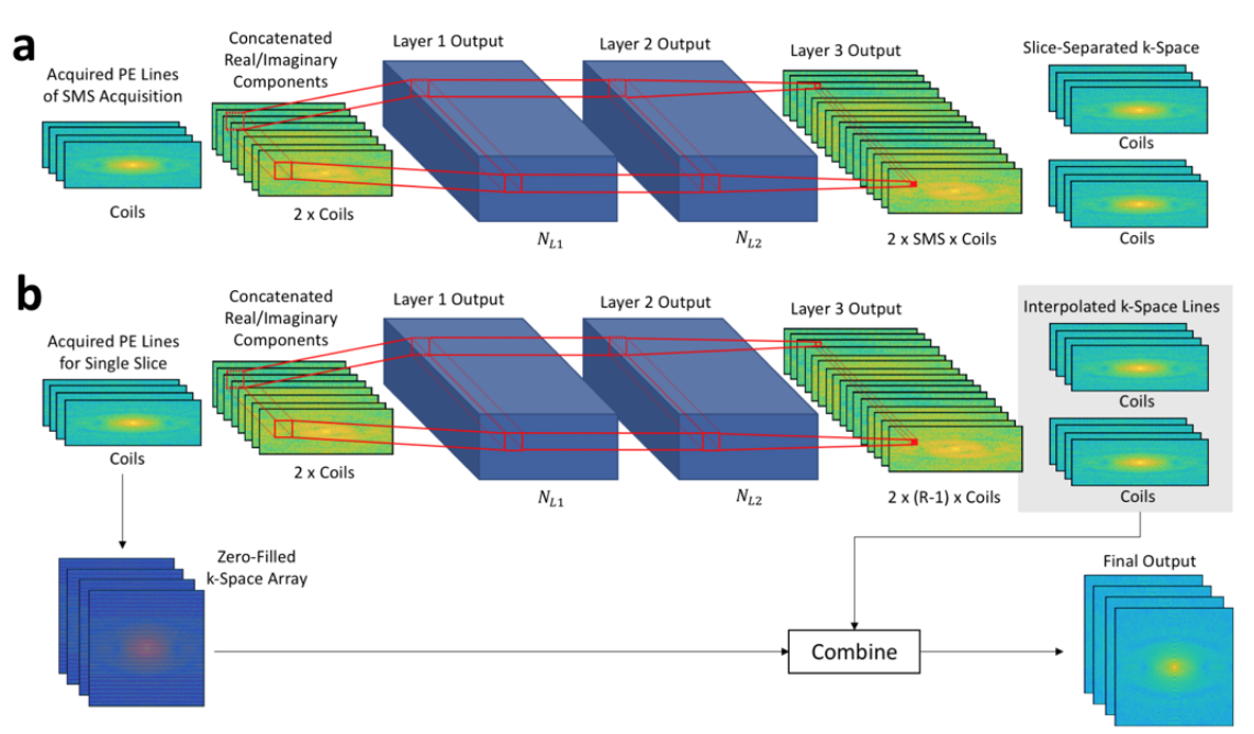
Separate slice-interpolating and phase-encode line interpolating networks were trained using the architectures shown in Figure 1. The models were trained using fully sampled data from individual slices. The real and imaginary components of the measured k-space are concatenated along the coil (“channel”) dimension. The first layer convolves the undersampled measured k-space with filters of size N1,PE x N1,RO x (2xNc) x NL1. Nc represents the number of receive coils. The second layer convolves the output of layer one with convolution filters of size N1,PE x N1,RO x NL1 x NL2 followed by a ReLU operation. Finally, layer three convolves the output of layer two with filters of size N1,PE x N1,RO x NL2 x NOUT. Values for NL1 and NL2 were both set to 128. For slice- and in-plane-interpolation, NOUT was set to SMS*2*Nc and (R-1)*2*Nc, respectively.
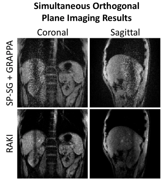
A retrospective undersampling experiment was performed on a fully-sampled respiratory-correlated 4D bSSFP dataset acquired on an Elekta 1.5T MR-Linac with eight coils (Nc=8). Two averages were acquired; one was used for training the models while the other was used for testing. RAKI was compared to split slice-GRAPPA (SP-SG)[2] with subsequent in-plane GRAPPA[3] for SMS and in-plane acceleration factors up to SMS and R=4 for 100 Monte Carlo iterations. Structural similarity metrics[4] (SSIM) were calculated to quantitatively compare RAKI to SP-SG+GRAPPA. To test robustness to respiratory motion, RAKI was trained for SMS=2 x R=3 acceleration on end-inspiratory data and applied to end-expiratory data. Finally, a simultaneous orthogonal plane imaging (SOPI)[5] dataset was also acquired on a Siemens 3T Verio with Nc=21 and SMS=2 orthogonal slices with R=3 in-plane acceleration. RAKI was compared qualitatively to SP-SG+GRAPPA. For SOPI, the calibration data were acquired with separate, low-resolution (32 phase encoding lines) scans for each slice.
Results
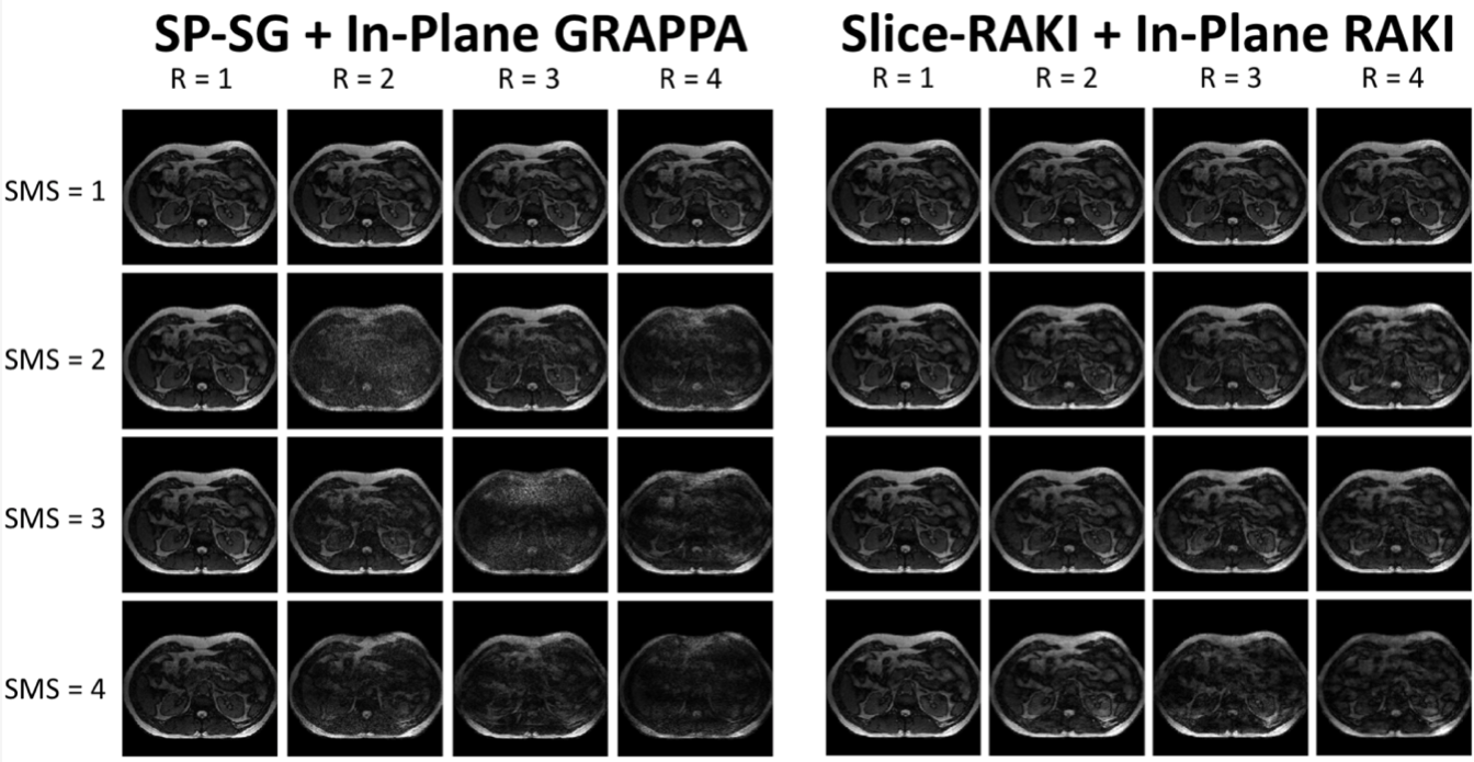
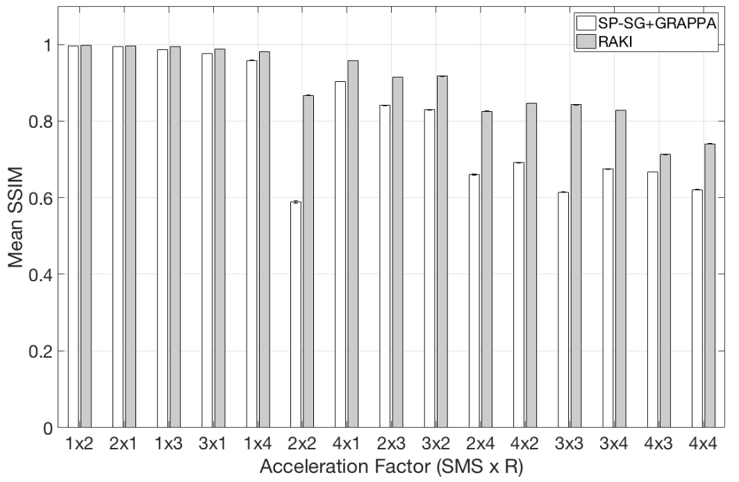
A single SMS slice for all tested acceleration factors is shown in Figure 2. No differences in image quality were observed between parallel imaging and RAKI images when either the SMS or R acceleration factors are equal to one. However, as both SMS and R increase, the SP-SG+GRAPPA begins to fail as evidenced by incomplete unaliasing and noise enhancement. The mean SSIM values shown in Figure 3 from RAKI are significantly higher than those from parallel imaging for all tested acceleration values.
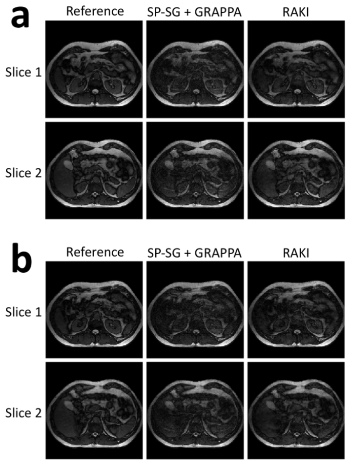
A closer look at the SMS=2 + R=3 acceleration factor is shown in Figure 4a. Here, the benefits of RAKI for removing artifacts and minimizing noise enhancement are clearly demonstrated. In Figure 4b, the robustness of RAKI to respiratory motion can be seen. The artifacts when applying expiratory-trained models to inspiratory data are still more benign than those introduced by conventional parallel imaging.
The SOPI results are shown in Figure 5. SP-SG+GRAPPA left severe noise enhancement near the intersection of the orthogonal slice planes. These images would have limited utility since the anatomy of interest lies at the intersection of the slices. RAKI was able to greatly reduce the amount of noise in the reconstructed SOPI images.
Discussion
RAKI was extended to interpolate in-plane accelerated SMS data and was successful in providing reconstructed in-plane accelerated images with similar or better quality than conventional parallel imaging methods. Visual noise enhancement and residual aliasing artifacts are minimized with the use of RAKI.Conclusion
This initial implementation of RAKI for in-plane accelerated simultaneous multislice imaging has been shown to be useful for improving reconstructed image quality. Following a more rapid implementation on multiple GPUs, SMS RAKI will be useful for providing real-time SMS imaging for motion monitoring in MR-gRT and potentially other applications.Acknowledgements
No acknowledgement found.References
[1] Akçakaya M, Moeller S, Weingärtner S, Uğurbil K. Scan-specific robust artificial-neural-networks for k-space interpolation (RAKI) reconstruction: Database-free deep learning for fast imaging. Magn Reson Med 2018. doi:10.1002/mrm.27420.
[2] Cauley SF, Polimeni JR, Bhat H, Wald LL, Setsompop K. Interslice leakage artifact reduction technique for simultaneous multislice acquisitions. Magn Reson Med 2014;72:93–102. doi:10.1002/mrm.24898.
[3] Griswold MA, Jakob PM, Heidemann RM, Nittka M, Jellus V, Wang J, et al. Generalized autocalibrating partially parallel acquisitions (GRAPPA). Magn Reson Med 2002;47:1202–10. doi:10.1002/mrm.10171.
[4] Wang Z, Bovik AC, Sheikh HR, Simoncelli EP. Image Quality Assessment: From Error Visibility to Structural Similarity. IEEE Trans Image Process 2004;13:600–12. doi:10.1109/TIP.2003.819861.
[5] Mickevicius NJ, Paulson ES. Simultaneous orthogonal plane imaging. Magn Reson Med 2017;78:1700–10. doi:10.1002/mrm.26555.
Figures