0461
Natively Fat-Suppressed Fast Interrupted Steady State (FISS) for 5D Whole Heart Imaging at 1.5 and 3T1Radiology, UNIL-CHUV, Lausanne, Switzerland, 2Advanced Clinical Imaging Technology, Siemens Healthcare AG, Lausanne, Switzerland, 3Center for Biomedical Imaging, Lausanne, Switzerland, 4Department of Radiology, NorthShore University HealthSystem, Evanston, IL, United States, 5The University of Chicago Pritzker School of Medicine, Chicago, IL, United States, 6Northwestern University Feinberg School of Medicine, Chicago, IL, United States
Synopsis
Fast interrupted steady state (FISS) sequences provide bSSFP signal contrast and concomitant fat signal suppression. In this work, 3D radial FISS was implemented as part of a respiratory self-gated free-breathing cardiac and respiratory motion-resolved 5D imaging framework. Its capabilities for fat suppression were tested and characterized at both 1.5 and 3T. Combined with a XD-GRASP reconstruction, FISS offers a versatile alternative for motion-resolved fat suppressed high-resolution whole-heart anatomical and functional cine imaging with a scan time as low as 8 minutes.
Introduction
Fast interrupted steady state (FISS) sequences retain the high signal-to-noise ratio (SNR) of balanced steady state free precession (bSSFP) sequences while simultaneously suppressing fat.1,2 Fat suppression is important for high vessel conspicuity in coronary MRA. However, high resolution imaging using continuous radial phyllotaxis acquisitions that can be reconstructed in arbitrary dimensions (Free-running framework) often suffers from sub-optimal fat suppression. In this context, a natively fat-suppressed sequence may reduce scan time compared to the use of water excitation and avoid sequence pauses with periodically applied fat saturation.3 The aim was to develop an optimized FISS sequence into the Free-running framework for fat-suppressed motion-resolved whole-heart imaging at 1.5 and 3T.Methods
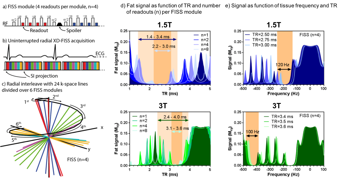
Fat suppression in FISS depends on the repetition time (TR) of the readouts, and the number of readouts per FISS module (n). Bloch equation simulations were performed to determine the effect of n on the TR needed for fat suppression at 1.5 and 3T, and on fat suppression bandwidth. Simulation parameters: T1/T2 1600/180ms, 1200 readouts, RF duration 0.3ms, RF excitation angle 40°, tissue frequencies ranging from -500 to 100Hz in steps of 5Hz.
A FISS acquisition scheme (Fig.1a) was implemented in a prototype 3D radial sequence following a phyllotaxis trajectory.4 Each radial interleave was fixed to 24 segments that can be divided over different amounts of FISS modules to enable 1,2,3,4,6,8,12, or 24 readouts per FISS module (Fig.1b-c).
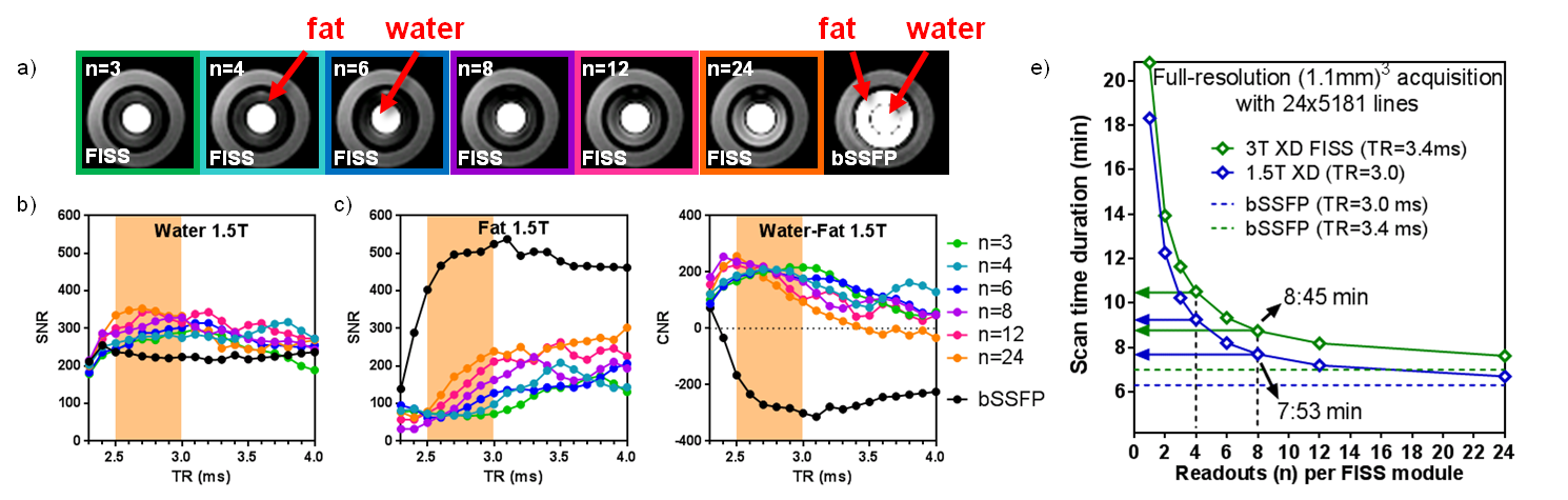
Phantom experiments were performed to quantify fat suppression with FISS and bSSFP, using TRs ranging from 2.3ms to 4.0ms, and varying n from 3 to 24. FISS experiments were performed in eight volunteers at 1.5 or 3T (MAGNETOM Aera and Prisma, Siemens Healthcare, Erlangen, Germany) using a TR found in simulations and phantom experiments. Experiments were performed with n=4 or n=8. The 3D data were sorted into a 5D motion-resolved dataset.3,5 The respiratory signal was extracted from a superior-inferior (SI) projection (Fig.1b), and cardiac motion from a recorded ECG signal. Images were reconstructed using XD-GRASP.5 A single respiratory and cardiac motion state was extracted for coronary analysis.
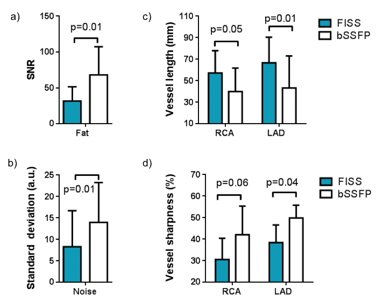
SNR and contrast to noise ratio (CNR) of fat were quantified in phantoms and non-motion-resolved volunteer data. Standard deviation of the noise was used as measure of streaking artifacts. Vessel sharpness and length of the RCA and LAD were obtained using SoapBubble.6 Paired Student’s t tests were used to compare bSSFP and FISS results with p<0.05 considered statistically significant.
Results
For whole-heart free-running FISS, the TR needed for fat suppression at 1.5T is in the range of 1.4ms to 3.4ms (n=1) or 2.2ms to 3.0ms (n=8) (Fig.2d, top). At 3T, the needed TR varies from 2.4ms to 4.0ms (n=1) or 3.1ms to 3.6ms (n=8) (Fig.2d, bottom). The fat suppression bandwidth that can be achieved is in the order of 120 Hz at 1.5 T (Fig.2e, top), and about 100 Hz at 3T.
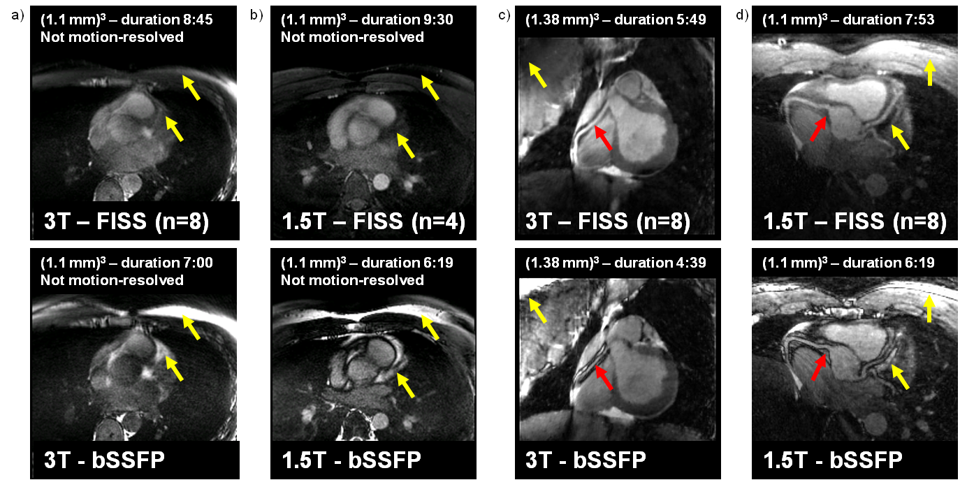
In phantoms, a fat suppression decrease was observed with increasing n (Fig.2a). For a TR between 2.5 and 3.0 ms, up to 8 readouts per FISS module achieves fat suppression and high water-fat contrast at 1.5T (Fig.2b-d). Since scan time is dependent on TR and n, a high resolution (1.1mm isotropic) whole-heart XD-FISS acquisition can be achieved in 7:53min at 1.5T (n=8, TR=3.0ms) and 8:45min at 3T (n=8, TR=3.4ms)(Fig.2e). Corresponding bSSFP acquisitions (not fat-suppressing) were 6:19min and 7:00min respectively.
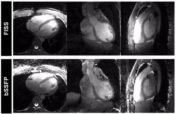
Whole-heart free-running FISS distinguishes clearly the different cardiac and respiratory motion states in volunteers (Fig.3, animation). Fat signals are visibly suppressed comparing FISS and bSSFP in non-motion-resolved images (Fig. 4ab) as well as in the coronary reformats (Fig.4cd, yellow arrows). SNR of chest fat was significantly decreased (Fig.5a) as well as background noise (Fig.5b) comparing FISS with bSSFP. Detectable vessel length was significantly increased in FISS compared with bSSFP (Fig.5c). Vessel sharpness was significantly higher in bSSFP (Fig.5d).
Discussion
Whole-heart free-running FISS demonstrated the feasibility for natively fat-suppressed structural and functional cardiac imaging. Increasing n reduces scan time, but narrows the range of available TRs and fat suppression bandwidth. FISS achieved similarly high signal levels compared to bSSFP while significantly reducing fat signal, as previously shown.1,2 Increased vessel sharpness in bSSFP was attributed to dephasing artifacts causing black borders at water fat interfaces (Fig.4, red arrows). FISS increases the scan time compared with bSSFP, but to a lesser extent than water excitation pulses that may lengthen the bSSFP TR and introduce banding artifacts.Conclusion
FISS, optimized and integrated with cardiac and motion-resolved 5D imaging, offers a versatile natively fat-suppressed imaging approach to measure cardiac structure and function with scan times of less than 8 minutes.Acknowledgements
No acknowledgement found.References
1) I Koktzoglou, RR Edelman. Radial fast interrupted steady-state (FISS) magnetic resonance imaging. Magn Reson Med. 2018 Apr;79(4):2077-2086. doi: 10.1002/mrm.26881
2) RR Edelman, A Serhal, A Pursnani, J Pang, I Koktzoglou. Cardiovascular cine imaging and flow evaluation using Fast Interrupted Steady-State (FISS) magnetic resonance. Journal of Cardiovascular Magnetic Resonance. 2018. doi: 10.1186/s12968-018-0433-3
3) L Feng, S Coppo, D Piccini, J Yerly, RP Lim, PG Masci, M Stuber, DK Sodickson, R Otazo. 5D Whole-heart sparse MRI. Magn Reson Med. 2018 Feb;79(2):826-838. doi: 10.1002/mrm.26745
4) D Piccini, A Littmann, S Nielles-Vallespin, MO Zenge. Spiral phyllotaxis: the natural way to construct a 3D radial trajectory in MRI. Magn Reson Med. 2011 Oct;66(4):1049-56. doi: 10.1002/mrm.22898
5) L Feng, L Axel, H Chandarana, KT Block, DK Sodicson, R Otazo. XD-GRASP: Golden-angle radial MRI with reconstruction of extra motion-state dimensions using compressed sensing. Magn Reson Med. 2016 Feb;75(2):775-88. doi: 10.1002/mrm.25665
6) A Etienne, RM Botnar, AM Van Muiswinkel, P Boesiger, WJ Manning, M Stuber. "Soap-Bubble" visualization and quantitative analysis of 3D coronary magnetic resonance angiograms. Magn Reson Med. 2002 Oct;48(4):658-66.
Figures