0442
Echo Planar Imaging of the Human Brain with 100 mT/m Gradients1Institute for Biomedical Engineering, ETH Zurich and University of Zurich, Zurich, Switzerland
Synopsis
First results obtained with Echo Planar Imaging using a gradient insert producing 100 mT/m with 1200 T/m/s slew rate are reported. High-resolution single-shot images of human head with a very low level of distortion and blur were obtained without the need of high parallel acceleration. However, at these extreme gradient strengths, high-order fields produced by eddy-currents require special measures to avoid spatially modulated “Nyquist ghosts”. Our strategy consisted of multi-position monitoring of eddy-current fields, fitting these results with 5th-order spherical harmonics, and including this model in the signal encoding matrix inverted during reconstruction.
Introduction
Echo-Planar Imaging (EPI)1 requires high and fast-switching gradients to produce high-resolution images without distortions and blur. We report first results obtained with EPI using a recently developed insert coil generating 100 or 200 mT/m maximum gradient dependent on amplifier configuration2. The key elements of this design are the limited spatial range of the gradient field, which allows its full usage for human head and limbs without peripheral nerve stimulation, and a low inductance allowing the slew rate of 1200 T/m/s with the 100 mT/m configuration. As shown in Fig.1, the slew rate growing in concert with the gradient amplitude is essential for running EPI at low echo spacing, which reduces distortions, and with a possibly flattened readout waveform, which improves the SNR3. It appears that using EPI with this unprecedented gradient strength requires special means to deal with the “Nyquist ghost” caused by high-order eddy currents. The original solution presented here is based on dynamic field monitoring4.Methods
All experiments were carried out on a 3T Achieva scanner (Philips Healthcare, The Netherlands) with the gradient insert mentioned above connected to the system’s standard amplifiers. A custom-made transmit-receive 8-channel array5 was used. Field monitoring was performed with a 16-probes field camera (Skope MR Technologies, Switzerland) in the empty RF coil using identical sequence parameters and geometry as later used for head scanning. Each sequence was monitored with 11 positions of the camera, which allowed the gradient- and eddy-current-generated fields to be fitted with up to 10th order spherical harmonics. The manufacturer’s implementation of gradient-echo EPI was used with minor modifications allowing convenient triggering of the field camera. Transverse images of a water phantom and of the head of a healthy male volunteer (participating in the study in agreement with the institution’s ethics policy) ware measured with resolution ranging from 1.5 to 1.0 mm in-plane and undersampling (acceleration) factors from 1 to 2. The reconstruction involved a regularized least-squares inversion of the high-order encoding matrix6 including gradient- and sensitivity encoding, B0 map, as well as the eddy-current-related phase terms measured with the camera. Concomitant field contributions were also included based on transverse field maps derived from the coil design. The high-order reconstruction procedure, highly time-consuming in its full form, was significantly accelerated by Fourier-transforming each echo and making 1-dimensional algebraic inversions in the hybrid space, as recently proposed for de-ghosting and unwarping of multiband EPI7,8.Results
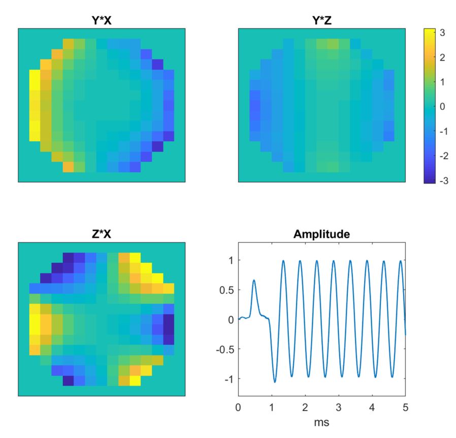
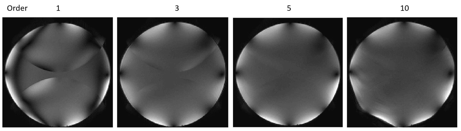
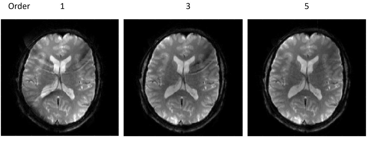
Principal component analysis of the field monitoring data shows the presence of a strong spatially nonlinear field pattern that oscillates along with the readout gradient (Fig. 2) and explains the presence of a 2D-modulated ghost in the “classically” reconstructed image, i.e. with off-resonance demodulation and trajectory-based gridding (Fig. 3). With increasing correction order, the ghost is reduced, however, still not sufficiently with the 3rd order correction, for which a single position of the field camera would be sufficient. Starting from 5th order model, containing 36 basis functions and requiring at least three camera positions for a well-conditioned fit, the ghost was reduced to an acceptable level, making it hardly visible in-vivo (Fig. 4). Using higher orders did not appear necessary. Field monitoring also revealed a slight smoothing of the readout shape by the limited bandwidth of the amplifier, which slightly reduced the effective resolution (e.g. 1.6 instead of 1.5 mm).
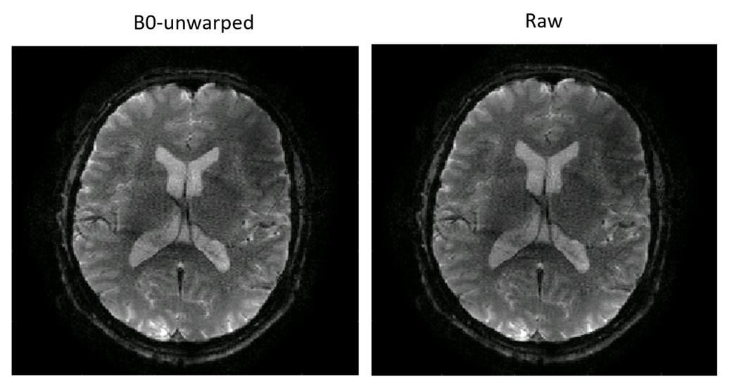
Echo-planar images of the human head acquired at 3T with the gradient insert show a very low level of distortion, even at a resolution of 1mm (relatively high for single-shot EPI) and a moderate parallel acceleration. This is demonstrated by comparison of the same slice reconstructed with and without including the field map-related phase in the encoding matrix (i.e. with and without unwarping) (Fig. 5).
Discussion and Conclusions
It is highly beneficial for the quality of EPI – especially
at high resolution – to use gradients as high 100 mT/m provided the slew rate
is sufficient to take advantage of such gradient amplitudes in practice.
However, since the eddy-currents cannot be completely avoided, and their
high-order field components grow with the gradient amplitude, non-standard methods
of Nyquist ghost suppression become necessary. Classic two-dimensional ghost
corrections9,10 can be shown to fail with oscillating field patterns
observed here due to ill-conditioning. The solution could be based on measuring
the correction patterns by monopolar-echo reconstructions and incorporating
them in the final SENSE inversion11 in a manner similar to MUSE12.
The approach presented here, although requiring additional equipment, has the
advantage of accommodating any kind of eddy current perturbations, not only
those perfectly following the oscillation of the readout gradient.
Acknowledgements
No acknowledgement found.References
- P. Mansfield, J Phys C. 10 (1977)
- M. Weiger et al. Magn Reson Med 2018 https://doi.org/10.1002/mrm.26954
- J Pipe and JL Duerk, Magn Reson Med (1995) 34, 170-178.
- C Barmet et al, Magn Reson Med (2008) 60, 187-197.
- M Roessler et al. ISMRM 2019, submitted
- B Wilm et al., Magn Reson Med (2011) 65 1690-1701.
- K Zhu et al, IEEE Trans Med Imaging (2016) 99 DOI 10.1109/TMI.2016.2531635
- B Zahneisen et al, Neuroimage (2017) 153, 97-108
- F Hennel, MAGMA (1999) 9: 134. https://doi.org/10.1007/BF02594610
- N Chen, Magn Reson Med (2004) 51, 1247-1253.
- VB Xie et al, Magn Reson Med (2017) DOI 10.1002/mrm.26710
- N Chen, Neuroimage (2013) 72, 41-47
Figures