0439
Development of a Local Pituitary Coil for Assessment of Pituitary Microadenomas1Radiological Sciences, University of California, Los Angeles, Los Angeles, CA, United States, 2Electrical Engineering, University of California, Los Angeles, Los Angeles, CA, United States, 3Radiology and Imaging sciences, University of Utah, Salt Lake City, UT, United States, 4Neurosurgery, University of California, Los Angeles, Los Angeles, CA, United States, 5California NanoSystems Institute, University of California, Los Angeles, Los Angeles, CA, United States
Synopsis
Pituitary microadenomas are difficult to detect due to their small size, and the surgeon is sometimes forced to
Introduction
Pituitary adenomas, commonly found in one out of five adults, are the leading cause of Cushing’s syndrome, a potentially debilitating endocrinological disorder with symptoms ranging from generalized weakness to severe depression and psychosis. Most patients with Cushing's disease have small pituitary adenomas, called pituitary microadenomas. The pituitary microadenomas are difficult to detect due to their small size, and the surgeon is sometimes forced to multiply slice to explore the pituitary gland – at considerable risk of damaging this essential master hormone gland. In this study, we design and evaluate a local pituitary coil that allows placing it in direct proximity to the pituitary gland. The local pituitary coil is intended to increase signal-to-noise ratio (SNR) that can provide sufficient spatial resolution to detect or characterize the pituitary microadenomas. The feasibility of the increased SNR at around 1cm depth from the coil is tested using the prototype design of the local pituitary coil in a 3T MRI scanner.Methods
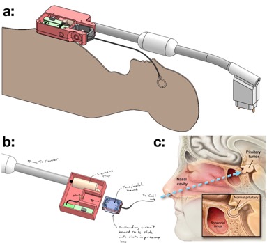
A minimally-invasive endonasal, endoscopic approach is commonly used to surgically remove the pituitary microadenoma via the nasal passageway. The overall concept is that surgeons will situate and secure the local pituitary coil against the pituitary gland (Fig. 1), and the patient will then undergo an intraoperative MRI exam, immediately followed by completion of the surgery with removal of the pituitary microadenoma. The local pituitary coil will be positioned surgically, aided by a novel deployment design necessary that can operate within the small confines of the nasal pathway and sinus cavity harboring the pituitary gland (Fig 2).
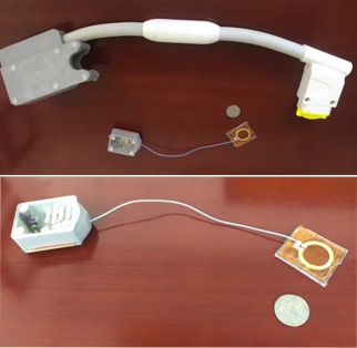
Coil Construction: The prototype local coil is constructed for a 3T MRI scanner (Prisma, Siemens Healthcare). The local coil with a 2cm diameter loop made from a single contiguous copper trace (3.5mm in width and 17.8μm in thickness) is attached to a coax cable, 1mm diameter and 20cm length, such that there are no electrical components on the coil. All tune and match components are on the opposite end of the cable (Fig 3), away from the loop of the coil [1]. The remote tune circuit also included active decoupling of the loop during the transmit portion of the pulse sequence. The 3D printed housing encloses a Siemens in-line cable trap and one preamp. The built-in docking port relays RX signal from the coil remote tune/match box to the preamp via an MCX connector. The remote tune/match box remains undocked for ease of surgical deployment, to be docked later for MR scanning (Fig 3).
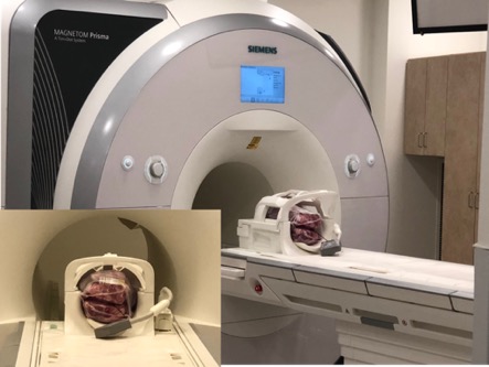
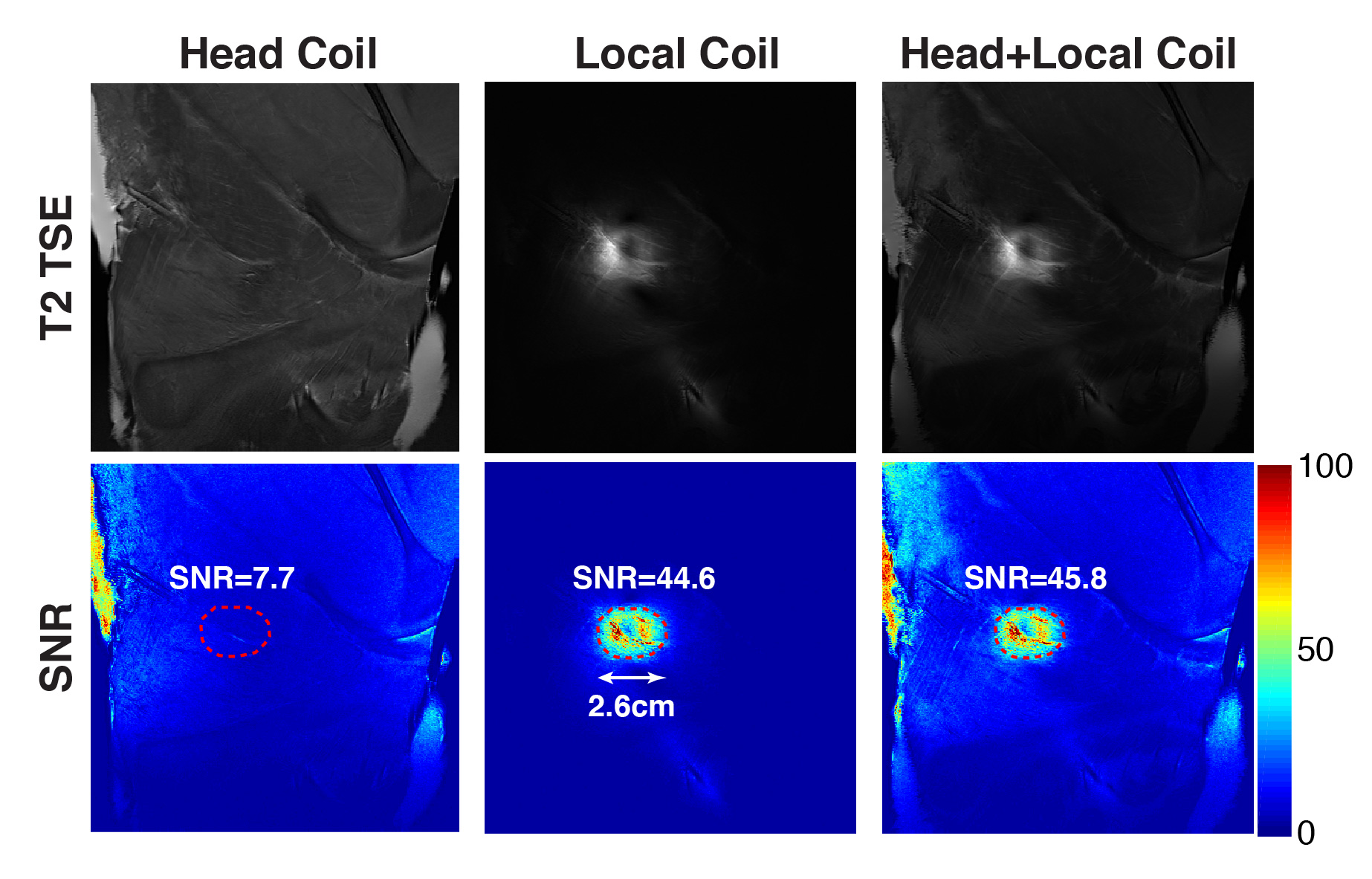
Evaluation: The prototype local coil is inserted between two ex-vivo meat slabs, a similar size of the head, and both head and local coils are connected to the 3T MRI scanner to test SNR, as shown in Fig 4. Three sets of coils are separately evaluated, including a commercial head coil (Head/Neck 20-channel coil, Siemens Healthcare), local pituitary coil, and a combination of two. The coronal T2-Turbo Spin Echo (TSE) sequence (TE/TR=89/4000ms, ETL=17, slice thickness=2mm, resolution=0.5 x 0.5mm2, FOV = 19cm, and #slices = 13) was repeated 48 times to calculate pixel-by-pixel SNR maps. The pixel-wise mean and standard deviation were computed across different measurements. The coronal slice 1cm away from the coil was selected, and the region of interest (ROI) was manually defined with an area of around 5cm2. (a width of approximately 2.6cm).
Results and Discussion
The SNR comparison of three sets of coils is shown in Fig 5. The SNRs within ROI are 7.7, 44.6 and 45.8 for the head coil, the local coil and the combination of two, respectively, at around 1cm depth from the coil. The prototype coil has increased SNR about a factor of six while the SNR difference between the local coil and the combination of two coils was subtle. This shows that the local coil alone has sufficient coverage and SNR improvement for the whole pituitary microadenoma as the typical size of pituitary microadenoma is 1-1.5cm in depth and width. In addition, the local coil can potentially improve motion sensitivity as it can suppress signal artifacts caused by the patient motion. Furthermore, image reconstruction algorithms, such as compressed sensing, in a combination of the local coil will enable translation of the increased SNR into high-resolution pituitary imaging in a 3T MRI system.Conclusion
We developed a local pituitary coil that allows to place it to the bone surface below the pituitary, while conventional MRI coils cannot access the pituitary directly due to their size. The prototype local coil provides an increased SNR with a factor of six with sufficient coverage and SNR for the pituitary microadenoma.Acknowledgements
This study was supported by the DGSOM Seed Grant Program Award (The Spitzer Grant Research Program).References
1. Hadley et all. Remote Tuning and matching of a non-resonant wire loop. Proc. Intl. Soc. Mag. Reson. Med. 26 (2018) pg. 1721Figures