0429
Fast Bound Pool Fraction mapping via steady-state MT saturation using single-shot EPI1Queen Square MS Centre, Department of Neuroinflammation, UCL Queen Square Institute of Neurology, Faculty of Brain Sciences, UCL, London, United Kingdom, 2Philips UK, Guilford, United Kingdom, 3Centre for Medical Imaging Computing, Department of Computer Science, UCL, London, United Kingdom, 4Centre for Medical Imaging Computing, Department of Medical Physics and Biomedical Engineering, UCL, London, United Kingdom, 5Universitat Oberta de Catalunya, Barcelona, Spain, 6Department of Brain and Behavioural Sciences, University of Pavia, Pavia, Italy
Synopsis
The bound pool fraction (BPF) is a quantitative parameter that reflects macromolecular tissue fraction, and has shown sensitivity to myelin content in human white matter. BPF mapping is still largely unexploited for characterizing white matter disease in vivo due to the long MRI protocols needed for its accurate and precise computation. In this work, we develop a new method that allows fast unbiased BPF estimation, suitable for clinical applications.
Introduction
The bound pool fraction (BPF) is a key biophysical parameter for quantifying the magnetization transfer (MT) effect, as it describes the fraction of macromolecular protons undergoing chemical exchange and cross-relaxation with protons in mobile water molecules. The BPF has been associated with tissue macromolecular content, and has shown correlation with myelin content in the central nervous system1, hence the interest in developing methods to robustly extract this parameter in vivo.
BPF mapping for clinical applications remains challenging given the complexity of the two-pool model2 used for its estimation. Existing fast methods rely on fixing unknown model parameters to population average values3,4, which may introduce bias when deviating from the healthy condition.
Here, we develop a new approach for fast BPF
mapping. Hard constraints adopted in previous methods are relaxed by
using approximations on the two-pool model that can be invoked under:
(i)
steady-state conditions, and (ii)
“fast-exchange”
regime conditions. A single-shot spin-echo (ssh-SE-) EPI sequence is
adapted to accommodate (i)
and (ii),
giving an acquisition time of under 10 minutes.
Methods
In the fast-exchange regime, bound and free protons exchange magnetization with a time scale much shorter than spin-lattice relaxation5. This allows the steady-state signal under off-resonance saturation to be expressed as6:
$$\frac{M_{ss}(Δ,θ)}{M_0}=\frac{1-(δ_BBPF\,e^{-\frac{PRT}{T_1}})}{1-(1-δ_BBPF)\,e^{-\frac{PRT}{T_1}}} \,\,\,\,\,\,\,\,\,\,\,\,\,\,\,\,\,\,\,\,\,\,\,\,[1]$$
assuming that: (i) pulse repetition time (PRT) is long compared to transfer time (i.e. PRT>120ms7); (ii) off-resonance saturation does not affect the free pool (i.e. offset frequency Δ>2kHz); (iii) bound pool saturation (expressed by δB in Equation 1) takes place instantaneously (i.e. no exchange or relaxation during off-resonance saturation). Mss, as a function of Δ and saturation flip angle θ, can be fitted to extract BPF and bound pool T2 (T2B), given an external measure of T1.
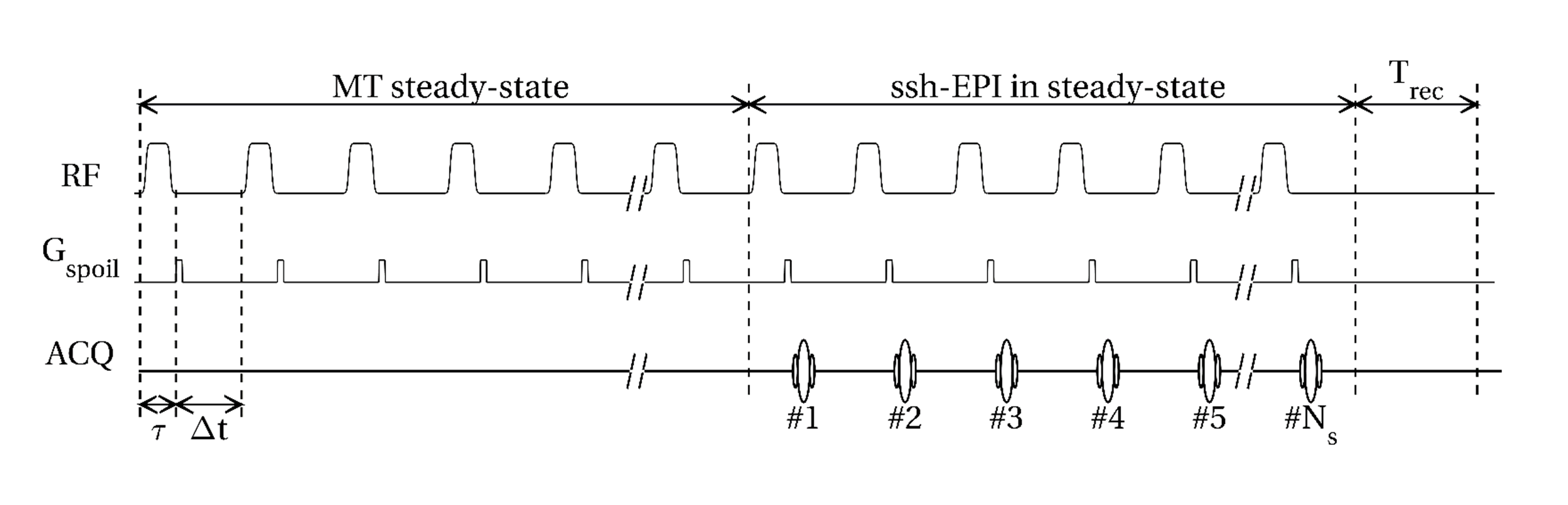
Time-efficient sampling of the steady-state is achieved using the sequence shown in Figure 1. The pulsed steady-state is attained with an initial period of saturation, and maintained during the acquisition of Ns slices with ssh-SE-EPI readouts. Short recovery times between sequence repetitions are allowed, as the steady-state established by the subsequent preparation is independent from the magnetization initial state.
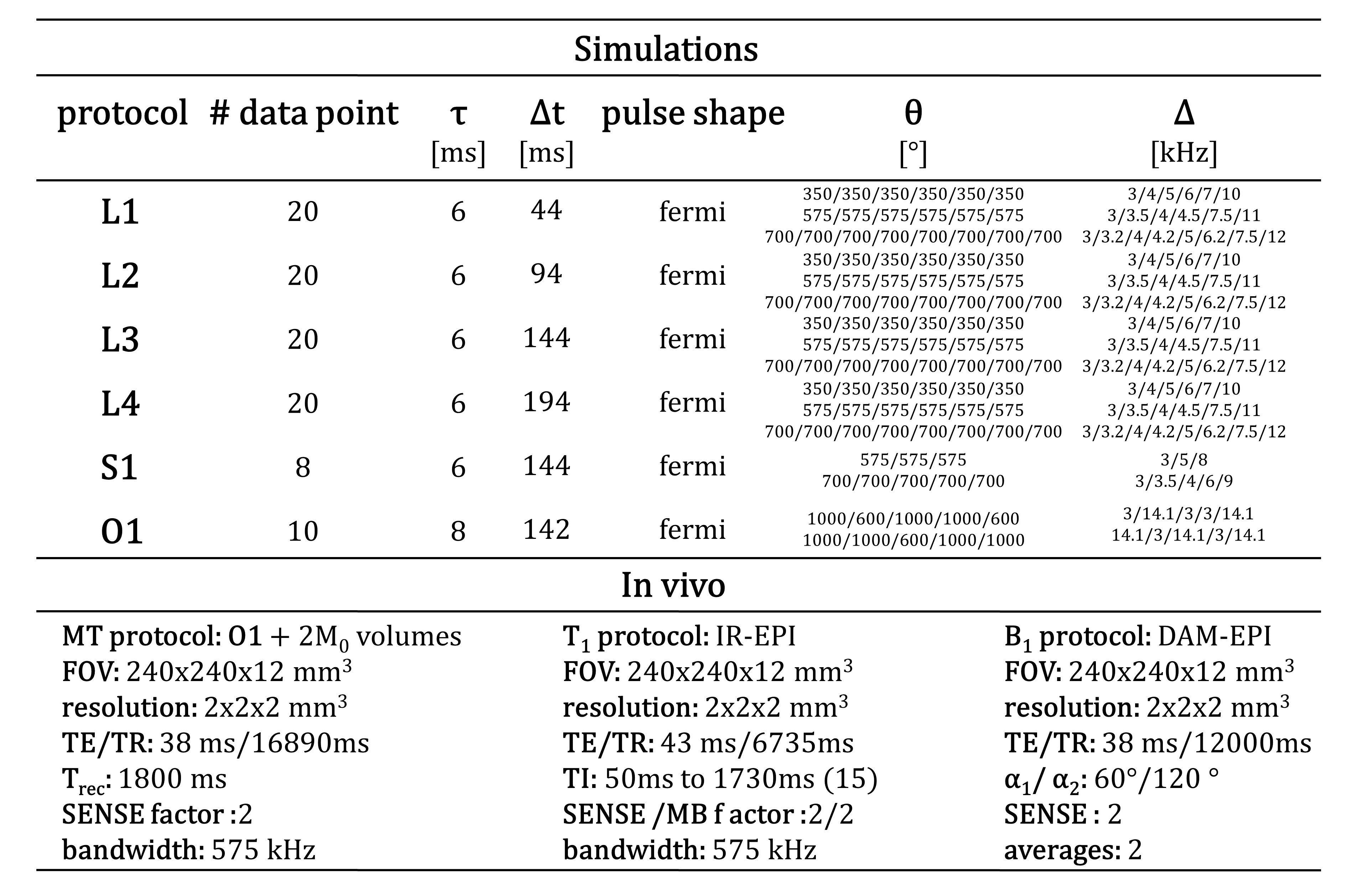
The effect of sequence parameters and number of data points is investigated through simulations. Full two-pool model equations are used to generate steady-state signals for physiologically plausible value of tissue parameters, then fitted by Equation 1. Error on parameter estimates is evaluated for protocols of Figure 2. In vivo acquisition is performed using the optimized protocol O1. A FOV of 224x224x120mm3 at 2mm3 isotropic resolution is acquired with ssh-SE-EPI readouts for: (i) MT steady-state; (ii) Inversion Recovery (IR) for T1 mapping; and (iii) Double-Angle Method (DAM) for B1 mapping8. Total protocol duration: 8min 44sec. A 3D-T1-weighted scan is added to allow regional characterization of the BPF. Six healthy subjects are scanned on a 3T Philips Ingenia CX MRI.
Results
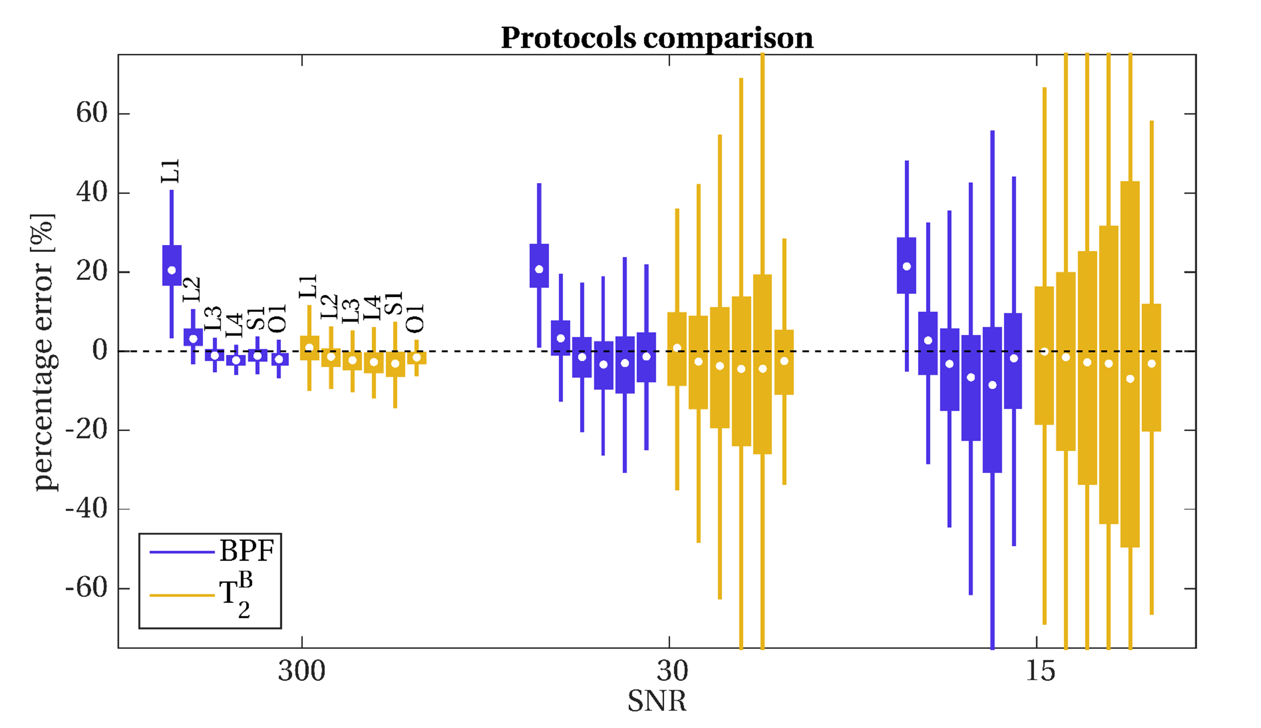
The effect of sequence parameters and number of data points on parameters estimation is shown in Figure 3. The use of long PRT>100ms is necessary to ensure the validity of model approximations. Shorter PRTs in fact produce large bias on BPF, even at high SNR. The BPF is estimated more reliably than T2B, however precision and accuracy of both parameters deteriorate at low SNR~15, regardless the number of data point used. Protocol optimization reduces parameters errors enabling the sampling of less data points.
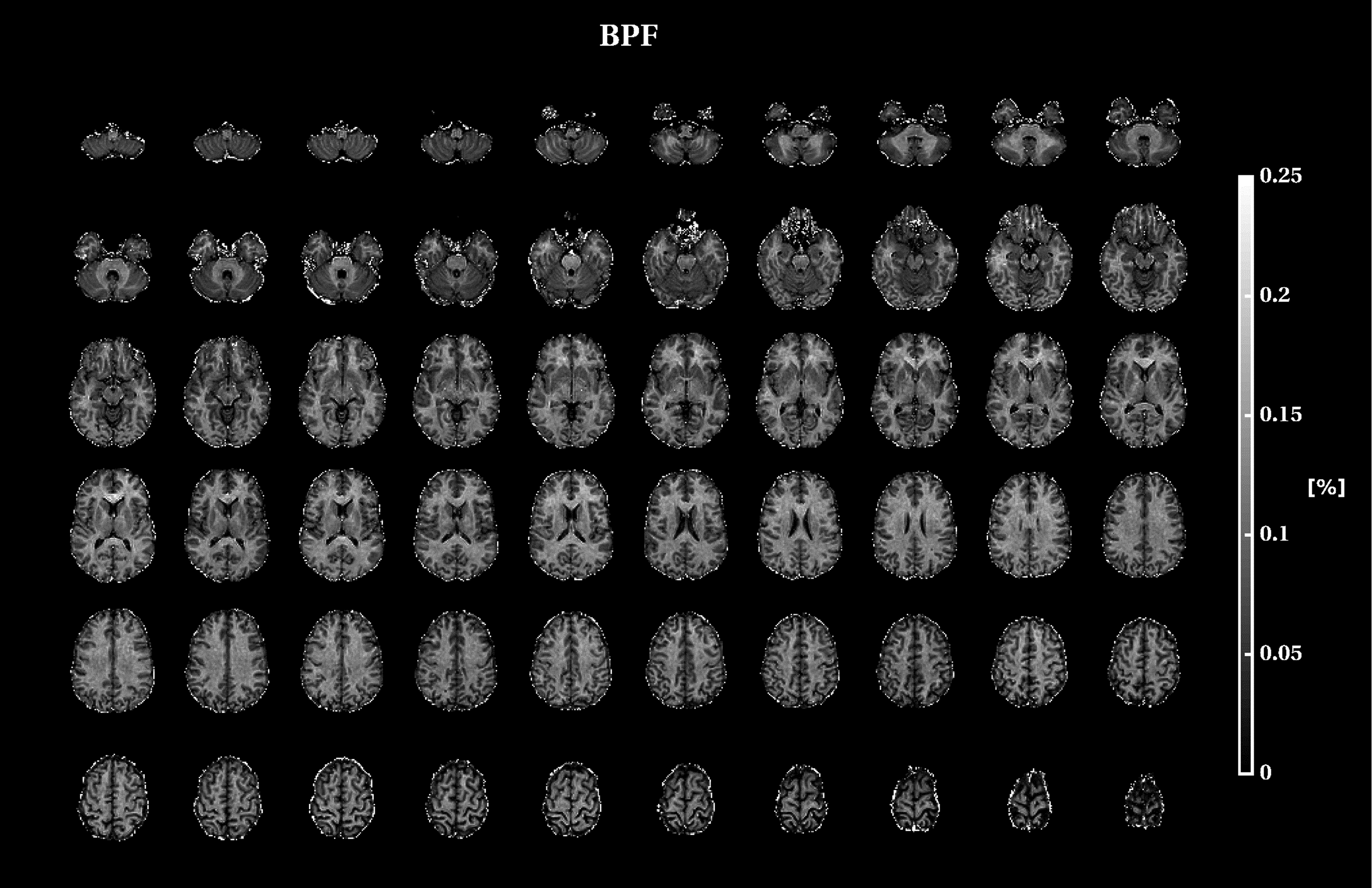
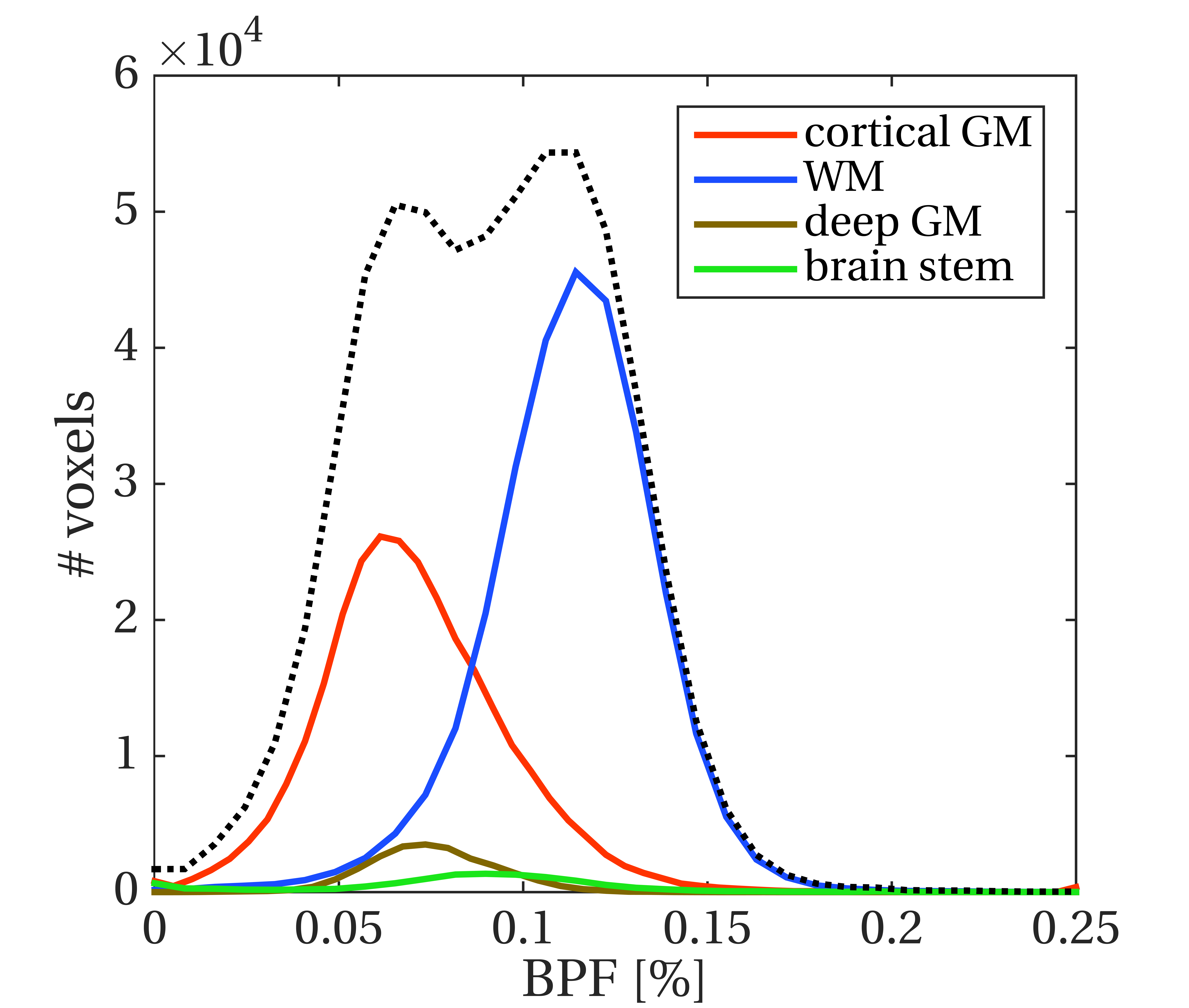
In vivo BPF maps depict the expected contrast, as shown in Figure 4 for a representative subject. Average values in WM and GM are in agreement with literature values3, with population median WM/GM BPF of 0.114/0.068. BPF distributions from all subjects pooled together are shown in Figure 5, displaying similar patterns of previous studies9.
Discussion
The method developed efficiently exploits the fast-exchange regime approximation for the steady-state MT, where off-resonance saturation is applied at long PRT, by acquiring the entire k-space between saturation pulses. This produces an MT-weighted volume per TR~15-20seconds. The interference of a multi-slice readout on the MT steady-state is reduced by avoiding fat suppression pulses and adopting an interleaved slice order in the acquisition. However, further investigation is required to quantify any residual effect, as well as to assess the impact of different pulse shape and/or duration.
The negligible BPF
bias, the lack of
explicit constraints on model parameters, and the short scan time
needed are promising factors for the translation of the method to
clinical applications.
Conclusions
A new, fast approach to map a key parameter of the quantitative MT two-pool model has been developed and applied in a cohort of healthy volunteers. The approach has the potential to be applied in a patient population in clinical studies.Acknowledgements
UK Multiple Sclerosis Society.
Spinal Research (UK), Wings for Life (Austria) and Craig H. Neilsen
Foundation (USA) for INSPIRED.
Engineering and Physical Sciences Research Council (EPSRC
EP/R006032/1, M020533/1, G007748, I027084, M020533, N018702).
Department of Health's National
Institute for Health Research, Biomedical Research Centres (BRC R&D
03/10/RAG0449). Guarantors of Brain post‐doctoral non-clinical
fellowships. This
project has received funding from the European Union’s Horizon 2020
research and innovation programme under grant agreement No. 634541.
References
[1] Schmierer, Klaus, et al. "Quantitative magnetization transfer imaging in postmortem multiple sclerosis brain." Journal of Magnetic Resonance Imaging 26.1 (2007): 41-51; [2] Henkelman, R. Mark, et al. "Quantitative interpretation of magnetization transfer." Magnetic resonance in medicine 29.6 (1993): 759-766; [3] Yarnykh, Vasily L. "Time‐efficient, high‐resolution, whole brain three‐dimensional macromolecular proton fraction mapping." Magnetic resonance in medicine 75.5 (2016): 2100-2106; [4] Dortch, Richard D., et al. "Optimization of selective inversion recovery magnetization transfer imaging for macromolecular content mapping in the human brain." Magnetic resonance in medicine (2018); [5] Helms, Gunther. "Interaction of exchange and differential relaxation in the saturation recovery behavior of the binary spin‐bath model for magnetization transfer." Concepts in Magnetic Resonance Part A 28.4 (2006): 291-298; [6] Helms, Gunther, and Andreas Piringer. "Simultaneous measurement of saturation and relaxation in human brain by repetitive magnetization transfer pulses." NMR in Biomedicine 18.1 (2005): 44-50; [7] Soellinger, M., et al. "Fast bound pool fraction mapping using stimulated echoes." Magnetic resonance in medicine 66.3 (2011): 717-724; [8] Stollberger, Rudolf, and Paul Wach. "Imaging of the active B1 field in vivo." Magnetic Resonance in Medicine 35.2 (1996): 246-251; [9] Mchinda, Samira, et al. "Whole brain inhomogeneous magnetization transfer (ihMT) imaging: Sensitivity enhancement within a steady‐state gradient echo sequence." Magnetic resonance in medicine 79.5 (2018): 2607-2619; [10] Alexander, Daniel C. "A general framework for experiment design in diffusion MRI and its application in measuring direct tissue‐microstructure features." Magnetic Resonance in Medicine 60.2 (2008): 439-448; [11] Cardoso, M. Jorge, et al. "Geodesic information flows: spatially-variant graphs and their application to segmentation and fusion." IEEE transactions on medical imaging 34.9 (2015): 1976-1988.
Figures