0426
The influence of temperature on two ex vivo myelin specific imaging protocols: Inhomogeneous Magnetization Transfer and Myelin Water Imaging1UBC MRI Research Centre, Vancouver, BC, Canada, 2International Collaboration on Repair Discoveries (ICORD), Vancouver, BC, Canada, 3Physics & Astronomy, University of British Columbia, Vancouver, BC, Canada, 4Department of Radiology, University of British Columbia, Vancouver, BC, Canada, 5MR Clinical Science, Philips Healthcare Canada, Markam, ON, Canada, 6Department of Pathology & Laboratory Medicine, University of British Columbia, Vancouver, BC, Canada
Synopsis
The impact of temperature on inhomogeneous magnetization transfer (ihMT) measurements in formalin-fixed human brain was studied. White matter (WM) ihMT signal increased with temperature when a T1D-filter was applied, supporting the hypothesis that components with long T1D (>1ms) are more sensitive to temperature variations. Grey matter ihMT did not vary with temperature. This suggests a difference in T1D values between WM and GM, and confirms that several T1D components contribute to myelin ihMT. Myelin water fraction values decreased with increasing temperature, possibly due to faster exchange between water compartments. Temperature is an important factor to consider in order to characterize the microstructure.
Introduction
Inhomogeneous magnetization transfer (ihMT) is a new MR method developed to be specific to tissues with a long dipolar relaxation time (T1D) such as myelin1–3. IhMT was validated as a myelin biomarker4 and shown to be sensitive to myelin concentration changes in vivo, as demonstrated by a mouse model of demyelination5. However, some studies show ihMT is also sensitive to factors other than myelin concentration, including temperature3,6, making ex vivo studies more challenging. In addition, the presaturation scheme of the ihMT sequence can also be adjusted to reveal or filter structures based on their T1D values7, thereby making ihMT more or less specific to myelin. We investigated the impact of temperature on the ihMT signal from different T1D components found in ex vivo human cortical brain samples, and we compared these measurements with myelin water fraction (MWF) determined using a multi-echo spin echo sequence at the same temperatures.Method
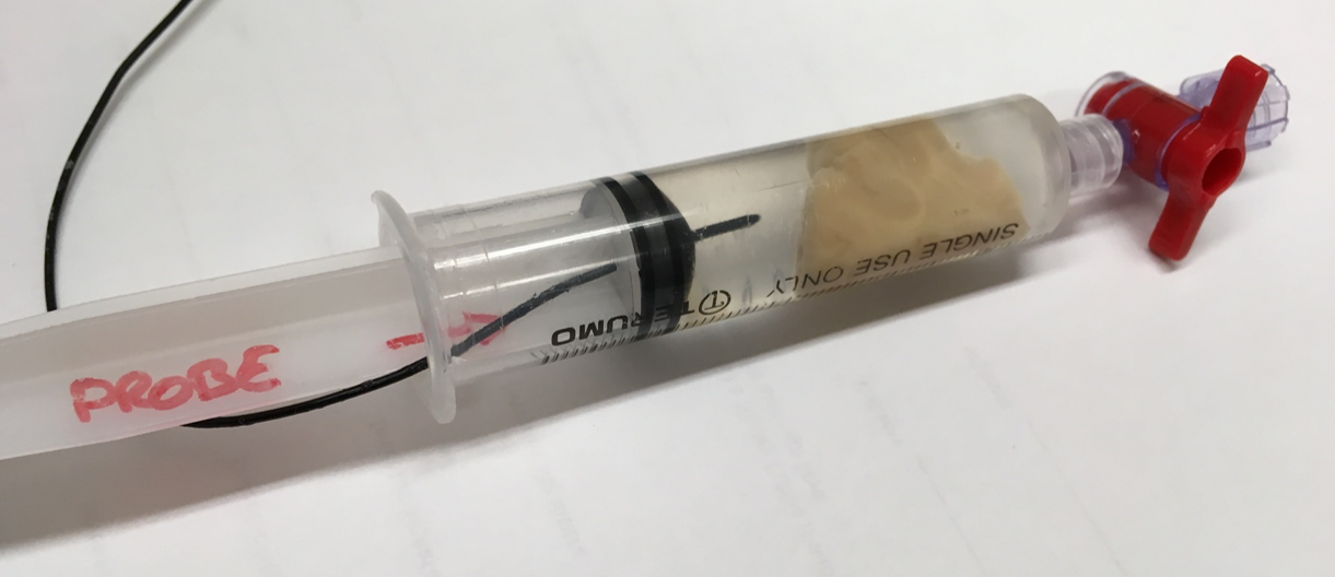
Experimental setup: MR experiments were conducted on a 7T Bruker Biospec system (Bruker Biospin, Ettlingen, Germany) with a quadrature volume coil (inner diameter = 35mm). Two formalin-fixed samples of human cortical brain were placed in a tube of 4% formalin (Figure 1). A temperature probe inserted into the tube controlled an external air flow heating system, enabling accurate temperate control during data acquisition at 20, 25, 30, 33 and 37°C.
Imaging
protocol:
[1] ihMT (FLASH scans exploiting the boost
approach8,9, frequency
offset = 8kHz, total deposited energy B1rms=6.7µT, 3
levels of T1D-filtering7: i) No
T1D-filtering (allowing the detection of all structures with a
non-zero T1D): dual saturation using two 1ms cosine-modulated pulses,
TR=50ms; ii) Weak T1D-filtering
(more specific to structures with long T1D): 12 frequency-alternated
saturation pulses with short pulse repetition times (1.3ms), TR=100ms; iii) Strong T1D-filtering
(very specific to structures with long T1D): 12
frequency-alternated saturation pulses with long pulse repetition times (3.3ms),
TR=100ms; total scan time=45min).
[2] Myelin water imaging (multi-echo spin echo, 64 echoes, TR/TE = 1720/6.753ms,
echo spacing=TE, scan time=30min).
All scans had FOV=28x20mm², matrix=112x80 (250μm in-plane resolution), slice thickness=1mm.
Data processing: IhMT ratio (ihMTR) maps were created using an in-house Matlab program with ihMTR=(Spos+Sneg–2Sdual)/S0. MWF maps were generated by fitting the T2 decay curve using NNLS10 with the MWF defined as the fraction of the total signal with T2 < 20ms. Regions of interest in white matter (WM) and gray matter (GM) were selected to determine mean ihMTR and MWF.
Results and Discussion
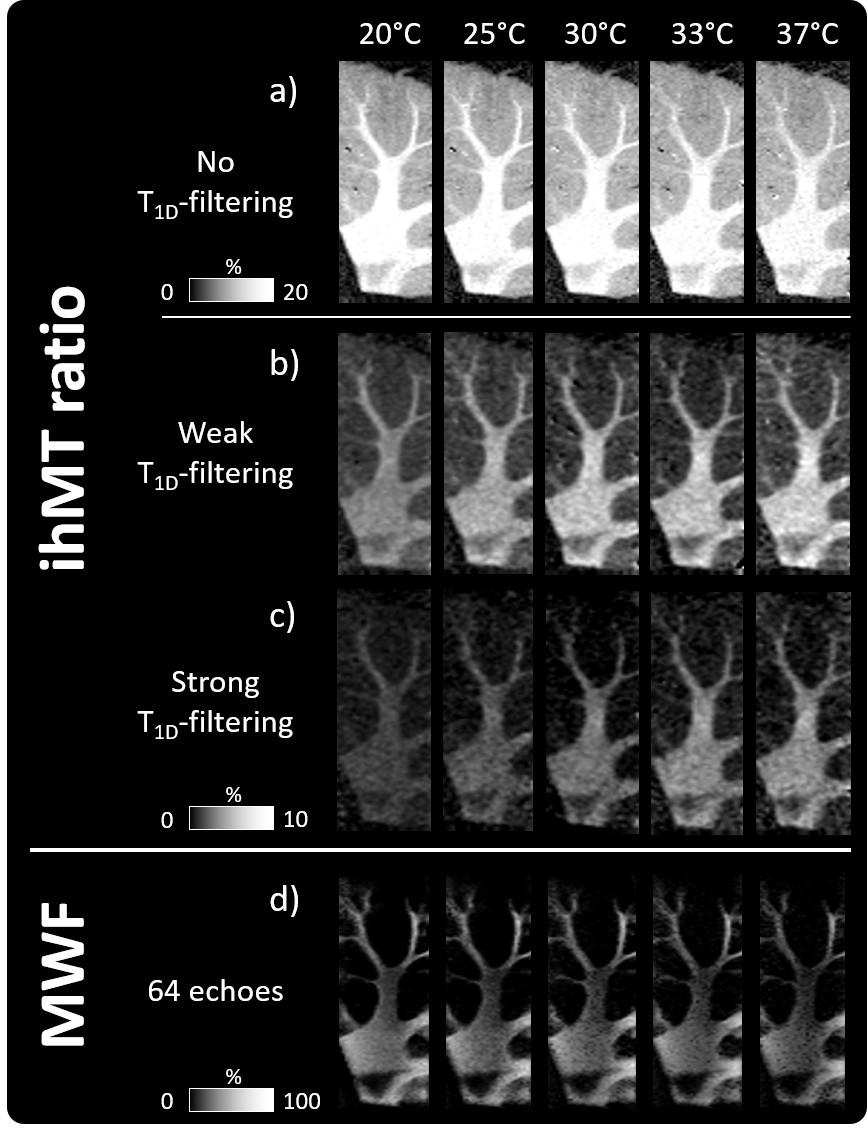
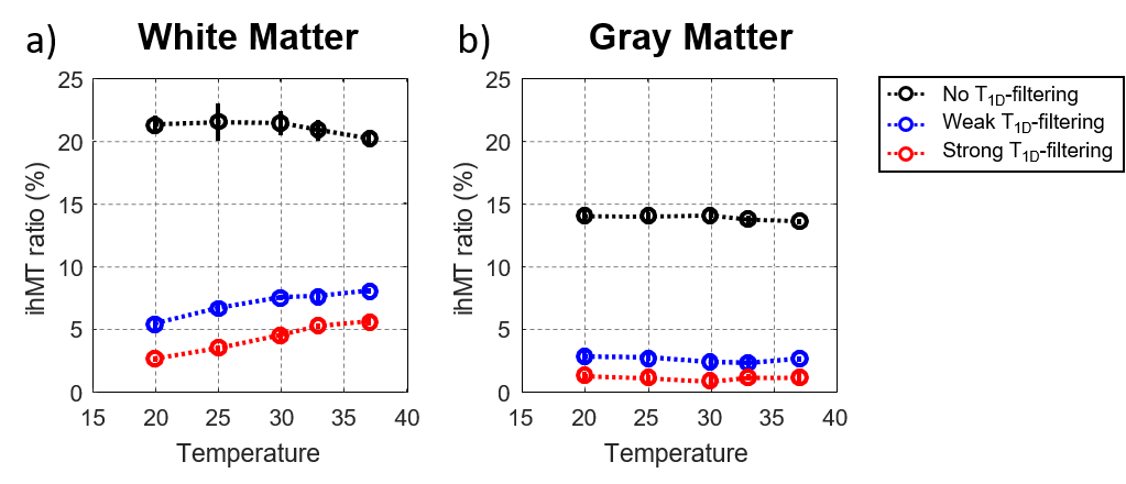
Representative ihMTR and MWF images at various temperatures are shown in Figure 2. Mean ihMTR and MWF in WM and GM are plotted in Figures 3 and 4. WM ihMTR exhibited sensitivity to temperature variation (Fig. 3a). In particular, ihMTR signal increased with temperature when a T1D-filter was applied, whether weak (blue curve) or strong (red curve). The use of cosine-modulated pulses (no T1D-filtering, black curve) enhanced the ihMTR signal intensity by 4x compared to the T1D-filtered signal, due to the inclusion of the short T1D components (arbitrarily defined in this work as T1D < 1ms). IhMTR with no T1D-filtering, which reflects the average of all the T1D components, did not change significantly within the range of temperatures tested, supporting the hypothesis that long T1D components (defined as T1D > 1ms) are more sensitive to temperature variations than short T1D component. Irrespective of the saturation scheme applied, GM ihMTR did not vary with temperature (Fig. 3b), leading to a WM/GM contrast enhancement in the weak and strong T1D-filtering configurations at higher temperature. This phenomenon suggests a difference of T1D values between WM and GM in these experimental conditions. Our results confirmed the hypothesis that myelin ihMT signal is driven by several T1D components4, which exhibit different behaviors with varying temperature.
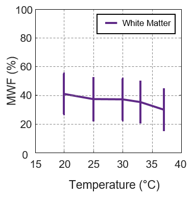
MWF was also influenced by temperature, with WM MWF decreases observed at higher temperatures (Figure 4). This slight trend can be explained by an increase in exchange between the myelin water and intra/extracellular water pools. Complementary prior work has shown that fixative increases the MWF11,12, but our study provides evidence that sample temperature may also explain MWF differences between in vivo and ex vivo studies at room temperature.
Conclusion
Temperature is an important factor interacting with myelin signal, as measured by ihMT and MWF protocols, which has to be monitored and controlled or which can be used to characterize the microstructure.Acknowledgements
We thank the MS patients and families for tissue donation. Wayne Moore and Cornelia Laule hold operating grants from the MS Society of CanadaReferences
1. Varma G, Girard OM, Prevost VH, Grant AK, Duhamel G, Alsop DC. Interpretation of magnetization transfer from inhomogeneously broadened lines (ihMT) in tissues as a dipolar order effect within motion restricted molecules. J. Magn. Reson. 2015;260:67–76 doi: 10.1016/j.jmr.2015.08.024.
2. Girard OM, Prevost VH, Varma G, Cozzone PJ, Alsop DC, Duhamel G. Magnetization transfer from inhomogeneously broadened lines (ihMT): Experimental optimization of saturation parameters for human brain imaging at 1.5 Tesla: Optimizing Saturation Parameters for ihMT Brain Imaging at 1.5T. Magn. Reson. Med. 2015;73:2111–2121 doi: 10.1002/mrm.25330.
3. Swanson SD, Malyarenko DI, Fabiilli ML, Welsh RC, Nielsen J-F, Srinivasan A. Molecular, dynamic, and structural origin of inhomogeneous magnetization transfer in lipid membranes: Origin of ihMT Contrast. Magn. Reson. Med. 2017;77:1318–1328 doi: 10.1002/mrm.26210.
4. Prevost VH, Girard OM, Cayre M, et al. Validation of inhomogeneous Magnetization Transfer (ihMT) as a myelin biomarker. ISMRM Congr. 2017.
5. Prevost VH, Cayre M, Carvalho V, et al. Inhomogeneous Magnetization Transfer (ihMT) sensitivity to myelin impairments in cuprizone mouse model. ISMRM Congr. 2018.
6. Carvalho V, Girard OM, Prevost VH, et al. Dipolar relaxation time (T1D) mapping to assess myelin in vivo. ISMRM Congr. 2018.
7. Prevost VH, Girard OM, Mchinda S, Varma G, Alsop DC, Duhamel G. Optimization of inhomogeneous magnetization transfer (ihMT) MRI contrast for preclinical studies using dipolar relaxation time ( T 1D ) filtering. NMR Biomed. 2017:e3706 doi: 10.1002/nbm.3706.
8. Mchinda S, Varma G, Prevost VH, et al. Whole brain inhomogeneous magnetization transfer (ihMT) imaging: Sensitivity enhancement within a steady-state gradient echo sequence: Whole Brain Inhomogeneous Magnetization Transfer (ihMT). Magn. Reson. Med. 2018;79:2607–2619 doi: 10.1002/mrm.26907.
9. Varma G, Girard OM, Mchinda S, et al. Low duty-cycle pulsed irradiation reduces magnetization transfer and increases the inhomogeneous magnetization transfer effect. J. Magn. Reson. 2018;296:60–71 doi: 10.1016/j.jmr.2018.08.004.
10. Whittall KP, MacKay AL. Quantitative interpretation of NMR relaxation data. J. Magn. Reson. 1969 1989;84:134–152 doi: 10.1016/0022-2364(89)90011-5.
11. Chen HS-M, Holmes N, Liu J, Tetzlaff W, Kozlowski P. Validating myelin water imaging with transmission electron microscopy in a rat spinal cord injury model. NeuroImage 2017;153:122–130 doi: 10.1016/j.neuroimage.2017.03.065.
12. Seifert AC, Umphlett M, Fowkes M, Junqian X. Formalin Tissue Fixation Biases Myelin Density Measurement by Quantitative Magnetization Transfer and Myelin Water Imaging. ISMRM Congr. 2018.
Figures