0418
Detecting articular cartilage and meniscus deformations under mechanical load using magnetization transfer ultrashort echo time (MT-UTE) modeling: ex vivo study1Radiology, University of california, San Diego, San Diego, CA, United States, 2Radiology Service, VA San Diego Healthcare System, San Diego, CA, United States
Synopsis
Ultrashort echo time (UTE) MRI can be used for quantitative assessment of compressive tissues in knee joint such as articular cartilage (AC) and meniscus. In osteoarthritis, the mechanical properties of AC and menisci are altered prior to gross morphological changes. In this study, magnetization transfer UTE imaging (UTE-MT) with two-pool modelling was used to detect deformation levels of AC and meniscus under load. Compression resulted in significant increases of macromolecular fraction (MMF) in AC and menisci of young, but not elderly donors. Our results indicate that biomechanical changes vary with age and can be detected with UTE-MT MRI.
Introduction
Articular cartilage (AC) and menisci are important tissues in knee joints that are subject to compression (1). Macromolecules (MM) such as collagen and proteoglycan may comprise around 20% of healthy AC and menisci volumes (80 % water) (1). With knee loading, free water redistributes within the AC and menisci and eventually flows towards the intraarticular synovial fluid (1). It is hypothesized that the mechanical properties of the musculoskeletal tissues may vary at early stage of diseases, prior to manifestation of gross morphological changes. Various research groups have investigated quantitative MRI properties of cartilage and menisci under mechanical loading (2–5). Ultrashort echo time (UTE) MRI sequences have been developed to image the short T2 tissues and have been used to investigate AC and menisci (6–8). UTE magnetization transfer (UTE-MT) imaging with two-pool modelling, which considers a water pool and a MM pool, yields parameters which are insensitive to the magic angle effect (9). Using this technique, MM proton fraction (MMF) can be estimated based on the exchange of magnetizations between MM and water protons (9,10). This study aimed to investigate the MMF variation in AC and menisci of human cadaveric knee joints under mechanical loading and comparing results between young and old donors.Methods
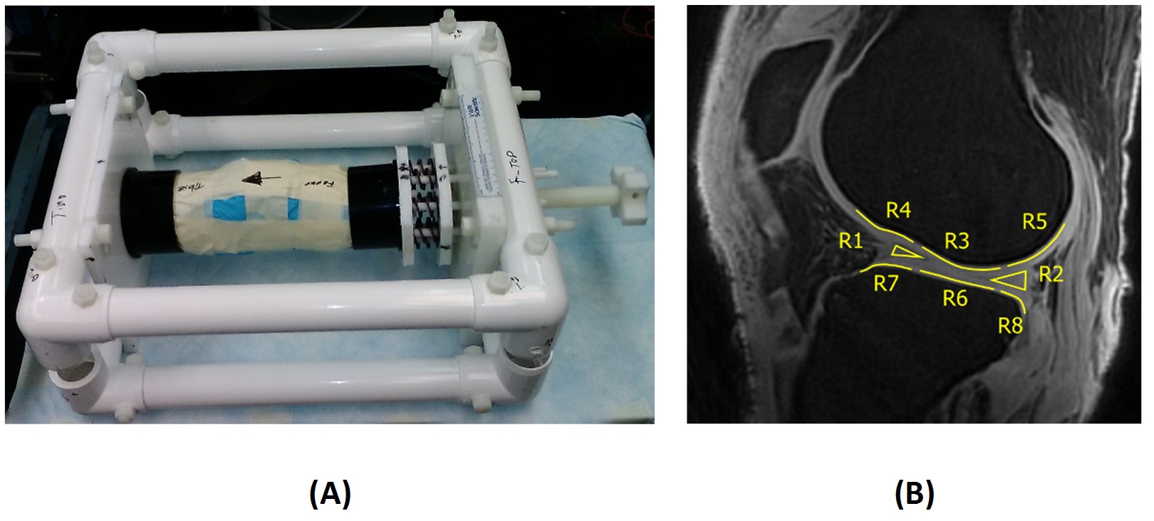
Sample preparation: Twelve human knee joints, from young (n=7, 42±12 years old) and old (n=5, 89±4 years old) donors were provided by a nonprofit donation company (United Tissue Network, AZ). The proximal and distal shafts were fit into an in-house designed loading device (Fig.1a). Eight sets of plastic springs (LL100125U40G, Lee Spring, NY) were used in this MRI compatible loading device (Fig.1). The compression load was adjustable manually using a 1-inch screw. The specimens were scanned at three steps of loading with 20 minutes rest in between; 1) 30, 2) 50 Kgf, and 3) unloaded (0 Kgf).
UTE sequences: Knee joints mounted in the loading device were placed parallel to B0 and scanned on a clinical 3T MR scanner (MR750, GE Healthcare Technologies, WI) using an eight-channel knee coil. For each loading stage, the following two imaging protocols were performed; A) 3D-UTE-MT-cones with three saturation pulse powers (q=500°,1000°,and 1500°) and five frequency offsets (Df=2, 5, 10, 20 and 50 kHz) (10) for MT modelling and B) 3D-UTE-cones with variable flip angles (FA=5, 10, 20 and 30, TR=20 ms) for T1 measurement as a prerequisite for MT modeling. T1 values were modified after correcting for B1 inhomogeneity. Other imaging parameters included: FOV=14cm, matrix=256×256, slice thickness=2mm, 40 slices. The total scan time was 65 mins.
Data analysis: Eight region of interests (ROIs) were defined (Fig.1b) on load 1 results (2ROIs for menisci, 3 ROIs for femoral AC and 3 ROIs for tibial AC) at lateral and medial joints. Defined ROIs were mapped on other loads results and modified by image processing experts to compensate the limited motions between loads.
Results
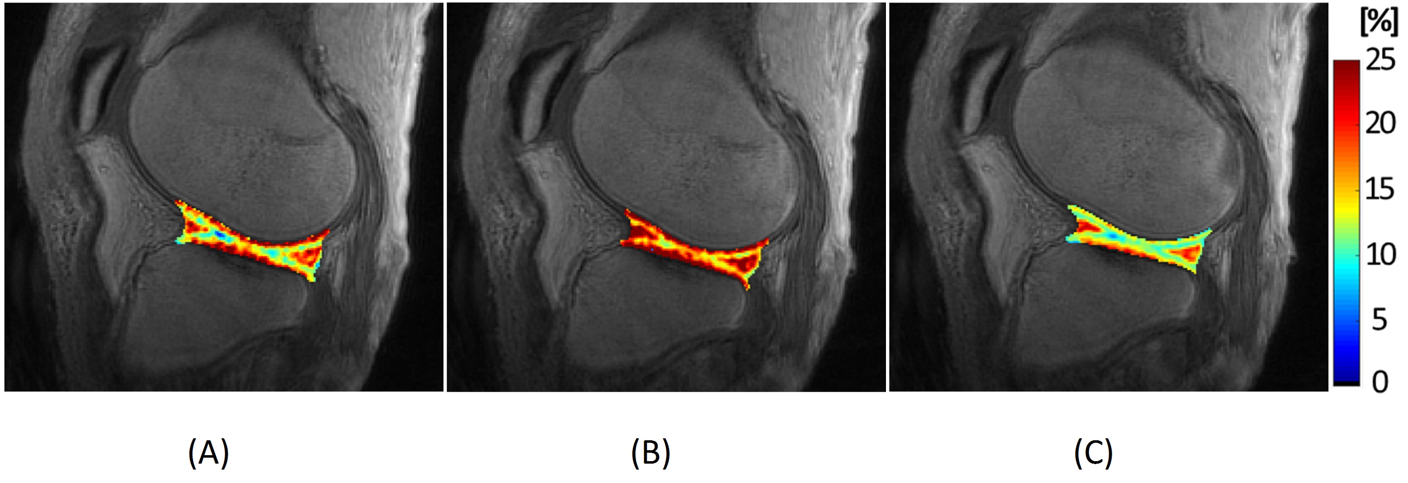
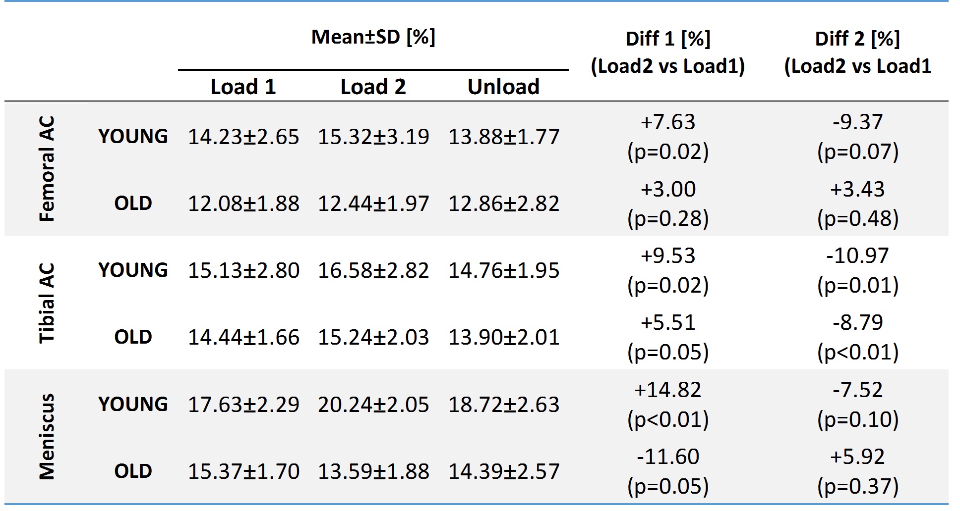
Figure 2 shows MMF maps at three loading stages on a representative knee joint. MMF demonstrated obvious increases with increasing the load to 50 Kgf and then decreased after unloading. Average MMF and its variations are presented in Figure.3 (Table) at the three loading steps. For young donors, average MMF increased significantly in cartilage and meniscus by increasing the load to 50 kgf. For elderly donors, MMF increase was not significant. Surprisingly, MMF decreased in meniscus, by increasing load to 50 Kgf. MMF reduction after unloading was significant only for tibial AC. Average MMF values were higher in young donors.Discussion
MMF variations with loading demonstrated different patterns in young and elderly donors. Tissue deformation under a certain load were likely higher in young donors compared with degenerated tissues in elderly donors. Mechanical load in elderly joints might be distributed differently from young joints such that menisci took less share in load distribution. Specifically, higher load might lead to menisci extrusion which is common in elderlies (11). MMF did not decrease significantly after unloading except for young tibial cartilage. Since this was a cadaveric study, cartilage and menisci may not have been able to restore their original shapes. MMF may help to distinguish the degenerated tissues based on two observations. First, MMF was higher for young donors, and second, larger and significant MMF variation by loading was observed in young knee joints.Conclusion
This study highlighted the feasibility of the UTE-MT modelling technique under loading to distinguish between healthy and degenerated joints. Significant variation of MMF with loading and unloading might be a characteristic of healthy tissue that indicates capability to distribute normal force. Localizing regions of nonsignificant MMF variations by loading might help to detect degenerated tissues.Acknowledgements
The authors acknowledge grant support from NIH (R01AR062581) and the VA Clinical Science and Rehabilitation R&D Awards (I01CX001388 and I01RX002604).References
1. Mansour JM. Biomechanics of Cartilage. Kinesiol. Mech. pathomechanics Hum. Mov. 2009:66–79. doi: 10.1002/art.23548.
2. Nishii T, Kuroda K, Matsuoka Y, Sahara T, Yoshikawa H. Change in knee cartilage T2 in response to mechanical loading. J. Magn. Reson. Imaging 2008;28:175–180. doi: 10.1002/jmri.21418.
3. Mayerhoefer ME, Welsch GH, Mamisch TC, Kainberger F, Weber M, Nemec S, Friedrich KM, Dirisamer A, Trattnig S. The in vivo effects of unloading and compression on T1-Gd (dGEMRIC) relaxation times in healthy articular knee cartilage at 3.0 Tesla. Eur. Radiol. 2010;20:443–449. doi: 10.1007/s00330-009-1559-3.
4. Souza RB, Kumar D, Calixto N, Singh J, Schooler J, Subburaj K, Li X, Link TM, Majumdar S. Response of knee cartilage T1rho and T2 relaxation times to in vivo mechanical loading in individuals with and without knee osteoarthritis. Osteoarthritis Cartilage [Internet] 2014;22:1367–76. doi: 10.1016/j.joca.2014.04.017.
5. Cotofana S, Eckstein F, Wirth W, Souza RB, Li X, Wyman B, Hellio-Le Graverand MP, Link T, Majumdar S. In vivo measures of cartilage deformation: Patterns in healthy and osteoarthritic female knees using 3T MR imaging. Eur. Radiol. 2011;21:1127–1135. doi: 10.1007/s00330-011-2057-y.
6. Shao H, Chang EY, Pauli C, Zanganeh S, Bae W, Chung CB, Tang G, Du J. UTE bi-component analysis of T2* relaxation in articular cartilage. Osteoarthr. Cartil. 2016;24:364–373. doi: 10.1016/j.joca.2015.08.017.
7. Qian Y, Williams AA, Chu CR, Boada FE. Multicomponent T2* mapping of knee cartilage: Technical feasibility ex vivo. Magn. Reson. Med. 2010;64:1427–1432. doi: 10.1002/mrm.22450.
8. Williams A, Qian Y, Chu CR. UTE-T2* mapping of human articular cartilage in vivo: A repeatability assessment. Osteoarthr. Cartil. [Internet] 2011;19:84–88. doi: 10.1016/j.joca.2010.10.018.
9. Ma Y-J, Shao H, Du J, Chang EY. Ultrashort echo time magnetization transfer (UTE-MT) imaging and modeling: magic angle independent biomarkers of tissue properties. NMR Biomed. 2016;29:1546–1552. doi: 10.1002/nbm.3609.
10. Ma Y-J, Chang EY, Carl M, Du J. Quantitative magnetization transfer ultrashort echo time imaging using a time-efficient 3D multispoke Cones sequence. Magn. Reson. Med. [Internet] 2017;00:1–9. doi: 10.1002/mrm.26716.
11. Svensson F, Felson DT, Zhang F, Guermazi A, Roemer FW, Niu J, Aliabadi P, Neogi T, Englund M. Meniscal body extrusion and cartilage coverage in middle-aged and elderly without radiographic knee osteoarthritis. Eur. Radiol. [Internet] 2018. doi: 10.1007/s00330-018-5741-3.
Figures