0393
Synthesizing rCBV maps from DCE-MRI of brain tumors using conditional adversarial networks1Imaging Physics, University of Texas MD Anderson Cancer Center, Houston, TX, United States, 2Diagnostic Radiology, University of Texas MD Anderson Cancer Center, Houston, TX, United States
Synopsis
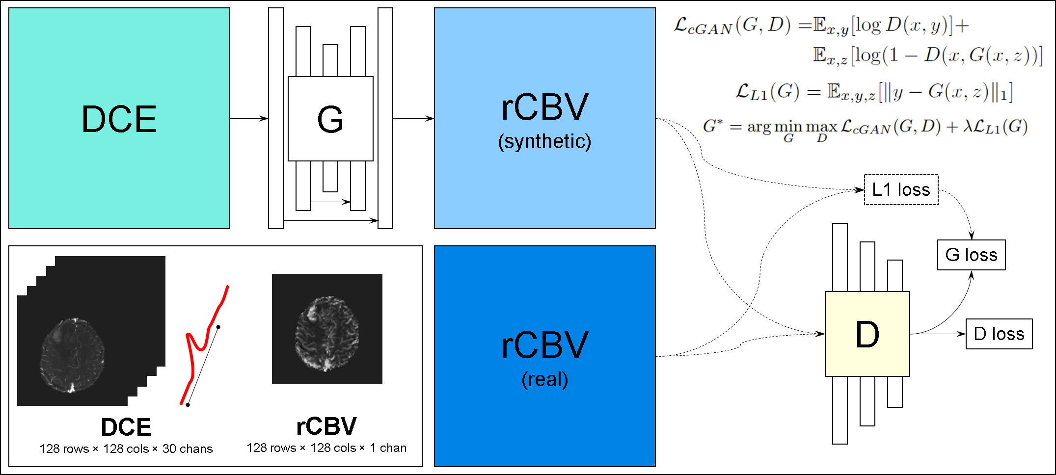
In this work we investigate conditional adversarial networks for synthesizing relative cerebral blood volume (rCBV) maps from dynamic contrast enhanced (DCE)-MRI. A network based on the pix2pix framework is trained to map DCE-MRI to rCBV maps using rCBV maps generated from dynamic susceptibility contrast (DSC)-MRI in the same patient cohort. The results demonstrate the feasibility of synthesizing realistic rCBV maps from DCE images, potentially improving MR perfusion imaging of the brain using a single contrast injection.
Introduction
Dynamic contrast enhanced (DCE)- and dynamic susceptibility contrast (DSC)-MRI are two perfusion imaging techniques that provide valuable information for diagnosis and evaluation of treatment response in patients with brain cancer[1]. In particular, DCE-MRI is best suited for evaluating vessel permeability whereas DSC-MRI provides robust relative cerebral blood volume (rCBV) imaging. Although the plasma volume can be obtained directly from pharmacokinetic modeling of the DCE-MRI data, the quality is generally inferior to the rCBV obtained from DSC-MRI due to the limitations in sensitivity and temporal resolution. Previous studies have demonstrated the two perfusion techniques are complimentary and using both improved diagnostic performance than with either one alone [2]. However, acquiring both DCE- and DSC-MRI in clinical practice requires two injections, often increasing the total dose of the gadolinium-based contrast agent. The purpose of this study was to investigate synthesizing rCBV maps (as computed from DSC-MRI) from DCE-MRI using conditional adversarial networks.Methods
DCE and DSC datasets from 113 patients with brain cancer undergoing treatment evaluation were analyzed. MRI was performed on a GE 3T MR750 scanner using an 8-channel brain coil. DCE-MRI was performed by using a 3D SPGR sequence with TR/TE/FA=3.6ms/1.3ms/30degrees, matrix=256x160, 20 slices, slice thickness=5mm, 60 phases, temporal resolution=5.4s. DSC-MRI was performed using a gradient-echo EPI sequence with TR/TE/FA=1500ms/25ms/90degrees, matrix=128x128, slice thickness and locations matched with DCE, 60 phases, temporal resolution=1.5s. A bolus injection (5mL/s) of full-dose of contrast agent (gadobutrol) was administered during each of the two perfusion scans with the order of DCE followed by the DSC, with an anatomical T1-weighted imaging in between.
The rCBV map was calculated from the DSC-MRI with leakage correction using nordicICE v2.3.14 (NordicNeuroLab, Berrgen, Norway). SPM12 (Wellcome Department of Cognitive Neurology, Institute of Neurology, London, UK) was used to register the DCE images and CBV maps. Edge slices that were truncated due to the registration process were removed from the dataset. In total, 1958 2D slices were available for analysis. We reserved 12 patients for testing (210 slices). We split the remaining data into 1574 slices for training and 174 slices for cross validation.
A modified version of the pix2pix framework [3] was implemented in TensorFlow v1.8.0 (Figure 1). We constructed a convolutional encoder-decoder generator based on the U-Net architecture [4]. The encoder consisted of 5 convolutional layers with reLU activations and the decoder consisted of 5 transpose convolutional layers with reLU activations (Figure 2). Batch normalization was used after all convolutional layers. Skip connections were used between the encoder and decoder. A tanh activation function was applied to the last layer of the decoder in the generator. The discriminator consisted of 5 convolutional layers with leaky reLU activations (α=0.2). Dropout was used after the transpose convolutional layers in the generator and after the convolutional layers in the discriminator. Gaussian noise (μ=0.0,σ=0.1) was applied to the input of the discriminator, and label smoothing was applied to the discriminator loss. We used the conditional GAN objective function with an L1 penalty on the pixels (L1+cGAN) as reported in the original pix2pix work [3]. Images from the first 30 time points of the DCE were used as 30 input channels into the network, and the rCBV maps were used as the outputs.
Model training was performed on an NVIDIA DGX-1 workstation. An Adam optimizer with a learning rate of 2*10-4 was used to train both the generator and discriminator. A batch size of 1 was used for training. For each training step, one training iteration was run on the discriminator followed by 3 training iterations on the generator.
Tumor-to-white matter rCBV ratios were computed by manually drawing ROIs in the enhancing lesion and in the contralateral normal appearing white matter areas. These ratios were measured and compared between the true and synthesized rCBV maps using correlation analysis.
Results
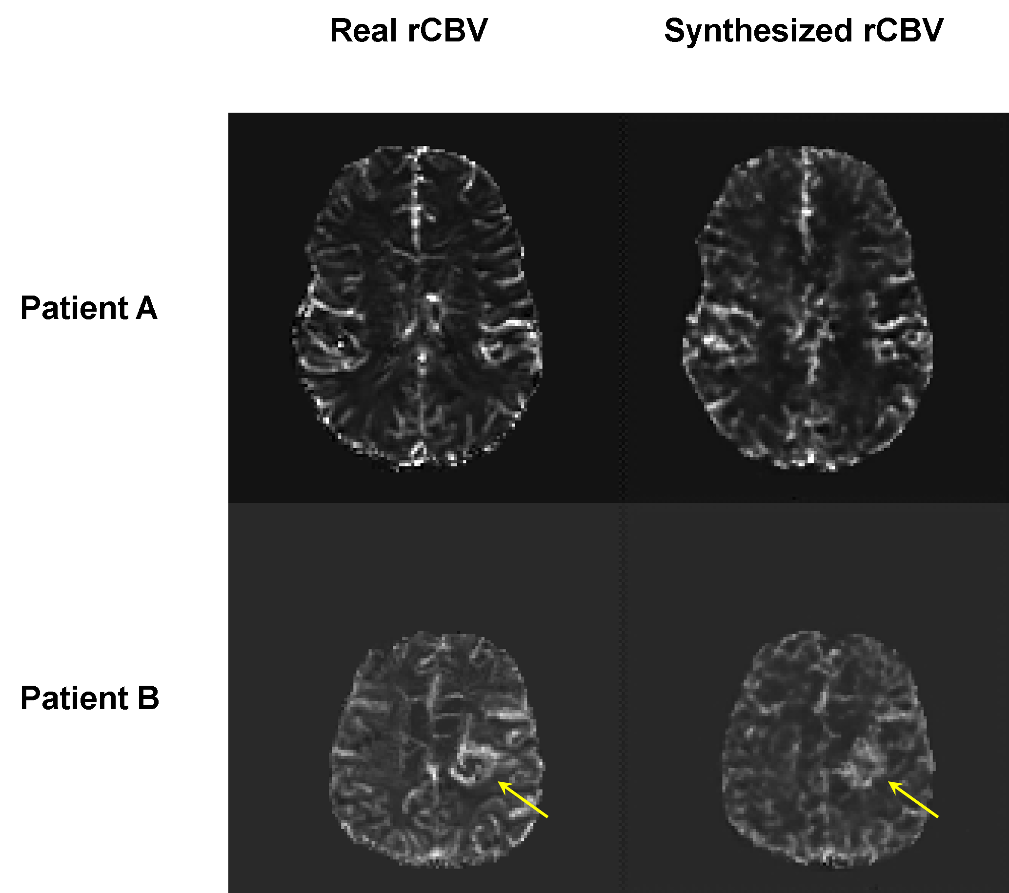
A comparison of the real and synthetic rCBV images are shown in Figure 3 along with the DCE input. Figure 4 shows that the tumor-to-white matter ratio obtained from the synthetic rCBV was significantly correlated with the real rCBV obtained from the DSC-MRI in the test patient group (R2=0.603, p=0.001).Discussion
Clinical DCE and DSC perfusion MRI offers complimentary information that are valuable for brain tumor evaluations. We have demonstrated the feasibility of synthesizing realistic rCBV maps from DCE images. This may enable MR perfusion imaging using a single contrast injection for assessing tumor response.Conclusion
Realistic rCBV maps can be synthesized from DCE images using conditional adversarial networks. The tumor-to-white matter rCBV ratio measured from the synthesized rCBV map is significantly correlated with those measured from the DSC-MRI.Acknowledgements
No acknowledgement found.References
[1] Shiroishi, M, Boxerman, J, Pope, W. Physiologic MRI for assessment of response to therapy and prognosis in glioblastoma, Neuro-Oncology; 18(4), pp. 467–478, 2015.
[2] Seeger A, Braun C, Skardelly M, et al. Comparison of three different MR perfusion techniques and MR spectroscopy for multiparametric assessment in distinguishing recurrent high-grade gliomas from stable disease. Acad Radiol;20(12): pp. 1557-65, 2013.
[3] Isola, P, Zhu, JY, Zhou, T, et al. Image-to-Image Translation with Conditional Adversarial Networks, arXiv:1611.07004v2.
[4] Ronneberger, O, Fischer, P, Brox, T. U-Net: Convolutional Networks for Biomedical Image Segmentation, arXiv:1505.04597.
Figures