0382
White matter fibrography of the extremely preterm brain: longitudinal connectome changes from childhood to adolescence.Ryan McNaughton1, Hernan Jara1, Mina Botros1, Baiyu Zhou1, Stephan W. Anderson2, Osamu Sakai2, Edward Sung2, Robert M. Joseph1, Karl Kuban2, and Michael T. O'Shea3
1Boston University, Boston, MA, United States, 2Boston University Medical Center, Boston, MA, United States, 3University of North Carolina at Chapel Hill Medical Center, Chapel Hill, NC, United States
Synopsis
Purpose: To study comparatively and longitudinally the connectome changes from childhood (age 10 years) to adolescence (age 15 years) using white matter fibrography (WMF). Methods: WMF was used to generate the connectomes of 9 extremely preterm born individuals using MRIs obtained at ages 10 and 15 years. Results: The most noticeable connectome change was a marked increase in the fiber density accompanied by fiber thinning. Conclusion: As anticipated, WMF connectomics of the extremely preterm brain demonstrate clearly observable WM architectural changes from 10 to 15 years of age from sparse fiber-thick to dense fiber-thin.
Purpose
Although survival rates for children born extremely preterm (EP) (gestational age <28 weeks) have greatly increased over the last 40 years, EP children remain at risk of developing a broad range of neurodevelopmental impairments including motor and sensory impairment, cognitive and learning disabilities, and psychiatric and behavioral disorders. The increased risk for development of neurological deficits in children born EP are probably linked to perturbations of critical maturational processes of the central nervous system (CNS) that occur during the first two trimesters of pregnancy and in the postnatal period (Fig. 1), which alter long term CNS architecture and impact later function. The purpose of this work was to study comparatively and longitudinally the connectome changes on a subcohort of the Extremely Low Gestational Age Newborn (ELGAN) study, from childhood (ELGAN-2: 10 years) to adolescence (ELGAN-3: 15 years) using white matter fibrography (WMF). WMF is a recently described application of Synthetic MRI.Materials & Methods
This study was approved by the Institutional Review Boards of the 12 participating institutions of the ELGAN study. Nine EP participants were scanned at 10 and 15 years of age with a 3T MRI protocol that included the dual echo turbo spin echo (DE-TSE) pulse sequence (TE1&2eff=12ms & 101ms, TR=10s). All studied brains were free from focal lesions. The directly-acquired images were used to create maps of the relaxation times (T2, and pseudo-T1), and of normalized proton density (qPD) using custom qMRI algorithms. Longitudinal relaxation rate (R1=1/T1) heavily-weighted images of the intracranium were generated with a synthetic MRI engine (all programs coded in Python 3.5, using the Canopy integrated development environment (Enthought, Austin, TX). The R1-weighted synthetic images, which show well-defined white matter structure, were processed with ImageJ (https://imagej.nih.gov/ij/): 3D-to-2D projected using the Volume Viewer plugin.Results
For all 9 participants, which did not have focal parenchymal lesions, the major connectome change from childhood to adolescence was a marked increase in fiber density accompanied by fiber thinning. This sparse-thick to dense-fine connectome child-to-adolescent developmental progression appears to occur homogeneously throughout the brain with no apparent lobar differences. In addition, the T2 values of white matter (WM) and gray matter (GM) decreased by approximately 10% from ages 10 to 15 years, in agreement with the scientific literature.Conclusion
Structural connectomics of the extremely preterm brain via WMF shows clear WM architectural changes from 10 to 15 years of age. These initial observations indicate that during this transition period from late childhood to adolescence, WM changes are predominantly in structural tissue characteristics – probably for diversification and specialization, from sparse-fiber thick to dense-fiber thin—and not necessarily via tissue quantity by volume. This work could be useful for understanding the normal and abnormal developmental pathways of the full-term and preterm human brain.Acknowledgements
This work was supported in part by the National Institute of Neurological Disorders and Stroke (5U01NS040069-05 and 2R01NS040069-09), National Institutes of Health Office of the Director (1UG3OD022348-01), and the National Institute of Child Health and Human Development (5P30HD018655-28).References
O'Shea TM, Allred EN, Dammann O, et al. The ELGAN study of the brain and related disorders in extremely low gestational age newborns. Early Human Development 2009;85(11):719-725.Figures
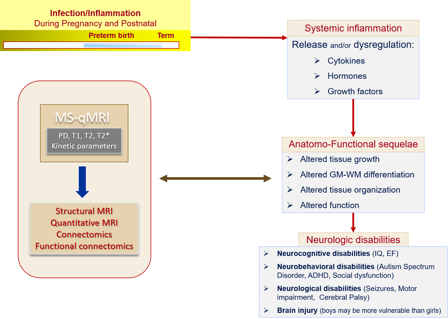
Figure 1. ELGAN Study hypothesis: Perinatal infection,
systemic inflammation lead to altered developmental pathways of the central
nervous system, which in turn can lead to neurologic disabilities.
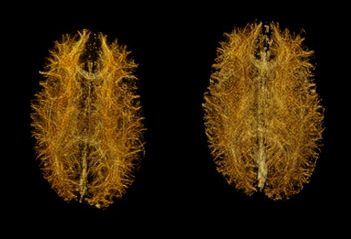
Figure 2. Connectome development from 9 to 15 years of a
female subject with impaired cognition (IQ = 55): exemplifies change from
sparse fiber-thick to dense fiber-thin.
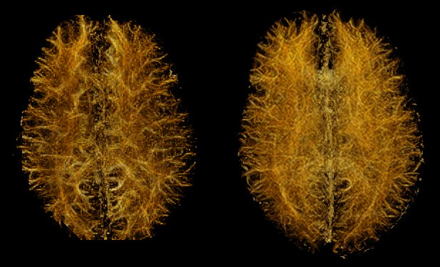
Figure 3. Connectome development from 9 to 15 years of a
male subject with normal cognition (IQ = 100): exemplifies change from sparse
fiber-thick to dense fiber-thin.