0376
Peri-tumoral radiomics on 3T MRI discriminative of D’Amico prostate cancer risk categories show association with epithelium, lumen and stromal densities on whole mount pathology1Case Western Reserve University, Cleveland, OH, United States, 2Self-employed, Lleida, Spain, 3University of Turku, Turku, Finland, 4Turku University Hospital, Turku, Finland, 5Icahn School of Medicine at Mount Sinai, New York, NY, United States
Synopsis
There is currently increasing interest in looking at role of radiomic features within the peri-tumoral region for disease characterization. In this work, we explore association of peri-tumoral radiomic features of prostate extracted from mpMRI with D’Amico risk. Additionally, we explore morphologic basis of these peri-tumoral features by analyzing the region on whole mount pathology. We observed greater epithelial content in high-risk compared to low, intermediate-risk lesions and vice versa with stroma. This heterogeneity within the peri-tumoral region may be captured by radiomic features that suggest peri-tumoral region of prostate may contain important information associated with risk of prostate cancer progression.
Introduction
Computer extracted texture features or radiomics quantify sub-visual image intensity relationships that capture underlying heterogeneity not discernible on routine imaging. In the context of prostate multi-parametric magnetic resonance imaging (mpMRI), radiomic features of the tumor have been shown1–3 to better discriminate PCa lesions from benign tissue and also distinguish aggressive from indolent disease compared to routine imaging. Very few studies have explored the morphologic basis of these radiomic features analyzing the corresponding pathology4. There is growing interest to explore the area immediately surrounding the lesion i.e. peri-tumoral region in which one may potentially find important information associated with the tumor5–8. Previous studies in the context of breast and lung cancer7,8 have shown that radiomic features of the peri-tumoral region may be predictive of disease prognosis and grade. However, this has not been explored in the context of PCa. In this work, we sought to investigate the role of radiomic features derived from peri-tumoral region of the prostate on mpMRI in distinguishing risk categories as defined by D’Amico criteria 9. Additionally, we explore ex vivo whole mount pathology of the prostate to understand the morphologic basis of peri-tumoral radiomic features of prostate cancer lesions on MRI.Methods
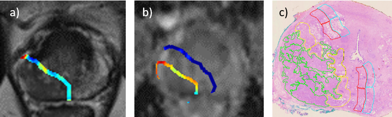
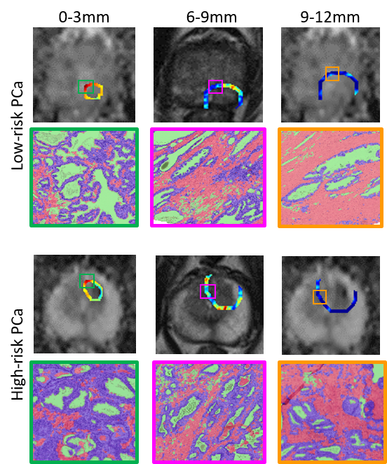
This retrospective, IRB approved and HIPAA compliant study consists of 18 patients who underwent 3T mpMRI prior to their first biopsy, diagnosed with PCa and underwent radical prostatectomy (RP)10. D’Amico criteria for these patients were determined using GS from 12-core ultra-sound biopsy, PSA and clinical stage prior to RP. 3 patients, one each belonging to low, intermediate and high-risk categories were selected whose biopsy GS was similar to that from RP for exploring the histo-morphometric basis while all the 18 patients were used for peri-tumoral radiomic analysis. Post RP, the prostate was stained with hematoxylin and eosin (H&E), sliced and digitized at 20x to obtain whole mount pathology (WMP)11. Correspondences between mpMRI and WMP were obtained based on anatomical landmarks and PCa regions of interest (ROIs) were annotated on mpMRI by an experienced radiologist using WMP as reference. An experienced pathologist delineated ROI’s on digitized WMP. Radiomics, including Haralick, Laws, Gabor and first order statistic features were derived from peri-tumoral region (0-3mm, 3-6mm, 6-9mm and 9-12mm) on T2W and apparent diffusion coefficient (ADC) maps. Peri-tumoral ROI’s were obtained on mpMRI and pathology by computing annular rings within the prostate extending beyond the tumor ROI (Figure 1). Previously presented tissue segmentation12,13 approaches were used to compute epithelium, lumen and stromal density within the peri-tumoral ROIs on WMP (Figure 2).Results
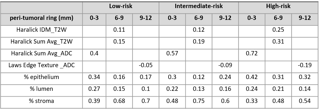
Haralick and Laws features from T2W and ADC were observed to be discriminative of low, intermediate and high risk categories. Haralick features were observed to be overexpressed in annular rings (0-3mm, 6-9mm) while Law’s feature under-expressed in annular ring (9-12mm) of high-risk lesion compared to low and intermediate-risk lesions. Density of stroma in the peri-tumoral ring just outside the tumor (0-3mm) was lower in high-risk PCa compared to low and intermediate risk and vice-versa with density of epithelium. The amount of lumen was fairly constant across all risk categories (Figure 3). Moving away from the tumor, the density of stroma increased for all risk categories, however, the rate of increase was greater in low and intermediate-risk compared to high-risk lesion.Discussion
Haralick features characterize underlying heterogeneity of mpMRI signal by quantifying spatial intensity relationships. Over-expression of Haralick features in 0-3mm, 6-9mm for high-risk indicates increased heterogeneity and was reflected in terms of higher epithelial density just surrounding the tumor. Our epithelial segmentation algorithm includes epithelial nuclei along with lymphocytes. Higher concentration of epithelium in high-risk may also be on account of increase in lymphocytes in surrounding tissue due to an immune response to contain PCa. Laws features characterizing edges in the horizontal and vertical directions observed to be underexpresed in high-risk indicating that edge orientations were along different directions. This again suggests heterogeneity similar to that captured by Haralick. We acknowledge that these are very preliminary findings on a small cohort and further studies on larger datasets are warranted to establish. However, differences observed in tissue pathology being reflected in peri-tumoral radiomics suggests that potentially discriminating information might exist in the peri-tumoral region on mpMRI.Conclusion
Higher heterogeneity in mpMRI signal intensities captures by radiomic features in the peri-tumoral region of prostate may be related to higher epithelial content on pathology. Differences in peri-tumoral pathology of PCa lesions may be captured by radiomic analysis, that can be potentially used in discriminating lesions of various risk categories as defined by D’Amico criteria.Acknowledgements
Research reported in this publication was supported by the National Cancer Institute of the National Institutes of Health under award numbers 1U24CA199374-01, R01CA202752-01A1R01CA208236-01A1R01 CA216579-01A1R01 CA220581-01A1National Center for Research Resources under award number1 C06 RR12463-01the DOD Prostate Cancer Idea Development Award; the DOD Lung Cancer Idea Development Award;the DOD Peer Reviewed Cancer Research Program W81XWH-16-1-0329the Ohio Third Frontier Technology Validation Fundthe Wallace H. Coulter Foundation Program in the Department of Biomedical Engineering and the Clinical and Translational Science Award Program (CTSA) at Case Western Reserve University. The content is solely the responsibility of the authors and does not necessarily represent the official views of the National Institutes of Health.References
1. Lemaître G, Martí R, Freixenet J, Vilanova JC, Walker PM, Meriaudeau F. Computer-Aided Detection and diagnosis for prostate cancer based on mono and multi-parametric MRI: a review. Comput Biol Med. 2015 May;60:8–31. doi:10.1016/j.compbiomed.2015.02.009 PMID: 25747341
2. Algohary A, Viswanath S, Shiradkar R, Ghose S, Pahwa S, Moses D, Jambor I, Shnier R, Böhm M, Haynes A-M, Brenner P, Delprado W, Thompson J, Pulbrock M, Purysko AS, Verma S, Ponsky L, Stricker P, Madabhushi A. Radiomic features on MRI enable risk categorization of prostate cancer patients on active surveillance: Preliminary findings: Radiomics Categorizes PCa Patients on AS. J Magn Reson Imaging. 2018 Feb 22; doi:10.1002/jmri.25983
3. Shiradkar R, Ghose S, Jambor I, Taimen P, Ettala O, Purysko AS, Madabhushi A. Radiomic features from pretreatment biparametric MRI predict prostate cancer biochemical recurrence: Preliminary findings. J Magn Reson Imaging JMRI. 2018 May 7; doi:10.1002/jmri.26178 PMID: 29734484
4. Penzias G, Singanamalli A, Elliott R, Gollamudi J, Shih N, Feldman M, Stricker PD, Delprado W, Tiwari S, Bohm M, others. Identifying the Histomorphometric Basis of MRI Radiomic Features in Distinguishing Gleason Grades of Prostate Cancer. Lab Invest. NATURE PUBLISHING GROUP 75 VARICK ST, 9TH FLR, NEW YORK, NY 10013-1917 USA; 2017. p. 400A–401A.
5. Shin HJ, Park JY, Shin KC, Kim HH, Cha JH, Chae EY, Choi WJ. Characterization of tumor and adjacent peritumoral stroma in patients with breast cancer using high-resolution diffusion-weighted imaging: Correlation with pathologic biomarkers. Eur J Radiol. 2016 May;85(5):1004–1011. doi:10.1016/j.ejrad.2016.02.017 PMID: 27130063
6. Roma AA, Magi-Galluzzi C, Kral MA, Jin TT, Klein EA, Zhou M. Peritumoral lymphatic invasion is associated with regional lymph node metastases in prostate adenocarcinoma. Mod Pathol Off J U S Can Acad Pathol Inc. 2006 Mar;19(3):392–398. doi:10.1038/modpathol.3800546 PMID: 16400321
7. Braman NM, Etesami M, Prasanna P, Dubchuk C, Gilmore H, Tiwari P, Pletcha D, Madabhushi A. Intratumoral and peritumoral radiomics for the pretreatment prediction of pathological complete response to neoadjuvant chemotherapy based on breast DCE-MRI. Breast Cancer Res BCR. 2017 May 18;19(1):57. doi:10.1186/s13058-017-0846-1 PMID: 28521821 PMCID: PMC5437672
8. Dou TH, Coroller TP, van Griethuysen JJM, Mak RH, Aerts HJWL. Peritumoral radiomics features predict distant metastasis in locally advanced NSCLC. PloS One. 2018;13(11):e0206108. doi:10.1371/journal.pone.0206108 PMID: 30388114
9. D’Amico AV, Whittington R, Malkowicz SB, Schultz D, Blank K, Broderick GA, Tomaszewski JE, Renshaw AA, Kaplan I, Beard CJ, Wein A. Biochemical outcome after radical prostatectomy, external beam radiation therapy, or interstitial radiation therapy for clinically localized prostate cancer. JAMA. 1998 Sep 16;280(11):969–974. PMID: 9749478
10. Jambor I, Kähkönen E, Taimen P, Merisaari H, Saunavaara J, Alanen K, Obsitnik B, Minn H, Lehotska V, Aronen HJ. Prebiopsy multiparametric 3T prostate MRI in patients with elevated PSA, normal digital rectal examination, and no previous biopsy. J Magn Reson Imaging JMRI. 2015 May;41(5):1394–1404. doi:10.1002/jmri.24682 PMID: 24956412
11. Jambor I, Borra R, Kemppainen J, Lepomäki V, Parkkola R, Dean K, Alanen K, Arponen E, Nurmi M, Aronen HJ, Minn H. Functional imaging of localized prostate cancer aggressiveness using 11C-acetate PET/CT and 1H-MR spectroscopy. J Nucl Med Off Publ Soc Nucl Med. 2010 Nov;51(11):1676–1683. doi:10.2967/jnumed.110.078667 PMID: 20956477
12. Whitney J, Corredor G, Janowczyk A, Ganesan S, Doyle S, Tomaszewski J, Feldman M, Gilmore H, Madabhushi A. Quantitative nuclear histomorphometry predicts oncotype DX risk categories for early stage ER+ breast cancer. BMC Cancer. 2018 May 30;18(1):610. doi:10.1186/s12885-018-4448-9 PMID: 29848291 PMCID: PMC5977541
13. Nguyen K, Sarkar A, Jain AK. Structure and context in prostatic gland segmentation and classification. Med Image Comput Comput-Assist Interv MICCAI Int Conf Med Image Comput Comput-Assist Interv. 2012;15(Pt 1):115–123. PMID: 23285542
Figures