0371
Microstrip Array Insert for Head Coils: Towards Layer fMRI at High Fields1Department of Radiology, Medical Physics, Medical Center ‐ University of Freiburg, Faculty of Medicine, University of Freiburg, Freiburg, Germany, 2German Cancer Consortium Partner Site Freiburg, German Cancer Research Center (DKFZ), Heidelberg, Germany
Synopsis
Rx coils for high field strengths are mostly based on loop coil design and cover relatively large FOVs, limiting the achievable voxel size due to the SNR and acquisition time constraints. Microstrip coils have limited sensitivity profile along the B1 direction, thus are useful for resolving cortical activity. In this study, we introduce flexible microstrip array insert coils (MSAi) that are compatible with existing volume coils. We demonstrate measurement of BOLD and highly confined activity wide spread in calcarine sulcus and motor cortex using MSAi at 3 T
Introduction
There is an increasing interest on laminar applications at ultra-high field strengths. Visual cortex and sensory motor systems of the human brain have been predominantly investigated [1,2]. Resolution depends not only on field strength but also image and temporal snr[3]. Use of dense Rx arrays 2Rx coils designed for imaging a specific region of brain [4], reduced FOV imaging methods [5]together with the SNR advantage of ultra-high fields enable sufficient SNR to resolve laminar activation. However, potential of high field systems (i.e. up to 3 T) have not been studied comprehensively. Existing Rx coils for high field strengths are mostly based on loop coil design and cover relatively large FOVs, limiting the achievable voxel size due to the SNR and acquisition time constraints. Microstrip coils have limited sensitivity profile along B1 direction [6], therefore useful for resolving cortical activity. In this study, we introduce flexible microstrip array insert coils (MSAi) that are compatible with existing coils such that their performance will not impeded by the volume coils they are inserted in. We demonstrate measurement of BOLD activity and grey matter activity around the drain vessels in calcarine sulcus and motor cortex using MSAi at 3 T.
Methods
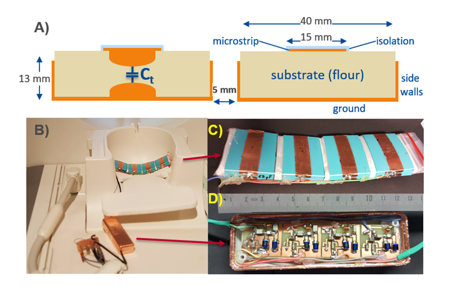
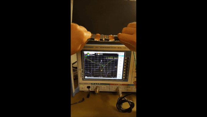
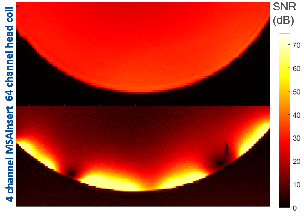
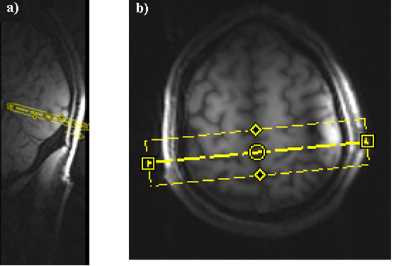
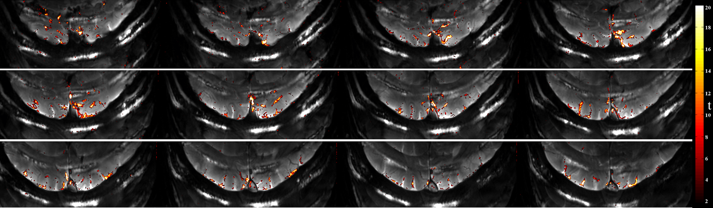
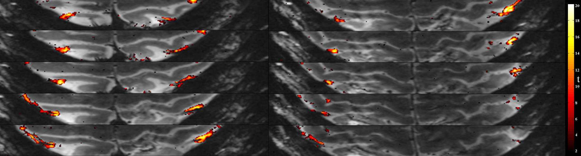
A four-element microstrip array insert (MSAi) of 5x18cm2 plane size was constructed using nylon formers and a continuous ground plane (Fig.1). Each microstrip block has dimensions of 1.27x4.2x5.3cm3. To keep the coil profile low and to prevent volunteer discomfort, remote tuning and matching was done after an l/4-long coaxial line section. Active detuning was achieved also by a remote inductance, as well as a serial matching capacitor for preamplifier decoupling. Each element was tuned to 123 MHz with an unloaded and loaded Q values of 59.2 and 20.2, respectively. An intrinsic decoupling of 14 dB between the neighboring strip elements resulted in a stable resonance even if the MSAi is bent 30° such that S11=-22.9+/-9.3 dB, fc=124.8+/-1.1 MHz, and S21=11.7+/-3.1 dB between the neighboring elements without preamplifier decoupling (Fig.2-Gif/video). A phantom experiment is conducted to compare image SNR Data was acquired on Siemens 3T MAGNETOM Prisma scanner with a commercial EPI sequence. The sequence parameters as follows: TE=36ms,TR=3s, FA=30o, bandwith 890 Hz/px, 0.8mm isotropic resolution with 38 slices. Field of view(FOV) is reduced to 144x54mm. Phase encoding direction is always chosen perpendicular to coil surface to avoid folding over, so anterior to posterio for occipital lobe and head to foot chosen for imaging the motor cortex. Slice prescriptions are shown in Fig.4. The visual paradigm was a flickering checkerboard with 8Hz reversal time displayed for 10seconds and 20s of resting period with a fixation cross. For motor task, subject was asked to perform finger tapping during displayed visual cues. A simple general linear model (GLM) is used to analyze functional data.Results & Discussion
2 to 3 fold SNR enhancement is achieved (Fig.3). GLM results showed confined activation patterns in central sulcus and calcarine sulcus (see inFigure5 & Figure6). Although there are many gray matter overlapping areas, some regions are very close to draining veins. Further studies needed to change sensitivity profile more on gray matter regions. In this study, We have demonstrated feasibility of sub-mm image acquisition in 3T by using commercially available sequences and only reducing FOV and without any sequence acceleration. Advantages of MSAi are; there is no need to move the patient-, could be insert inside the cushion and could easily be selected through the console and compatibly works with existing coils. MSAi can also be designed for ultra-high fields, which will improve the SNR in cortex even further and BOLD sensitivity will be more focused on capillaries instead of draining veins. Optimization of sensitivity profile might further improve necessary SNR. Integration of further MSAi elements also along the PE direction should be studied for better performance.Acknowledgements
No acknowledgement found.References
1. Huber, L. et al. High-Resolution CBV-fMRI Allows Mapping of Laminar Activity and Connectivity of Cortical Input and Output in Human M1. Neuron 96, 1253-1263.e7 (2017).
2.Koopmans, P. J., Barth, M. & Norris, D. G. Layer-specific BOLD activation in human V1. Hum. Brain Mapp. 31, 1297–1304 (2010).
3.Triantafyllou, C. et al. Comparison of physiological noise at 1.5 T, 3 T and 7 T and optimization of fMRI acquisition parameters. NeuroImage 26, 243–250 (2005).
4. Farivar, R., Grigorov, F., van der Kouwe, A. J., Wald, L. L. & Keil, B. Dense, shape-optimized posterior 32-channel coil for submillimeter functional imaging of visual cortex at 3T. Magn. Reson. Med. 76, 321–328 (2016).
5.Yuan, J., Zhao, T.-C., Tang, Y. & Panych, L. P. Reduced field-of-view single-shot fast spin echo imaging using two-dimensional spatially selective radiofrequency pulses. J. Magn. Reson. Imaging JMRI 32, 242–248 (2010).
6.Kumar, A. & Bottomley, P. A. Optimizing the Intrinsic Signal-to-Noise Ratio of MRI Strip Detectors. 166, 157–166 (2006).
Figures