0367
Dual-echo simultaneous multi-slice spiral acquisition for simultaneous CBF and BOLD fMRI at 7T1Maastricht University, Maastricht, Netherlands
Synopsis
Dual-echo acquisition with arterial spin labelling (ASL) allows for simultaneous measurements of CBF and BOLD signal changes which are helpful to understand the cerebral haemodynamics in health and disease. At ultra-high field (≥7T), however, this becomes impractical with established EPI based techniques since the readout duration precludes the use of appropriate echo times at commonly desired spatial resolutions. Single-shot spiral imaging presents a promising alternative: it allows the CBF signal to be acquired at very short TE while providing a flexible choice of TE for the second BOLD echo. In this work we show a dual-echo simultaneous multi-slice spiral out-in sequence for the acquisition of CBF and BOLD signals.
INTRODUCTION
Simultaneous acquisition of CBF and BOLD contrasts using arterial spin labeling (ASL) is used to investigate the cerebral haemodynamics in health and disease. At ultra-high fields however, due to the shorter transverse relaxation times (T2*), simultaneous acquisition becomes impractical with established EPI techniques as the readout duration precludes the use of suitable echo times for both CBF and BOLD, at least at commonly desired spatial resolutions. Single-shot spiral imaging presents a promising alternative, as it not only allows the CBF signal to be acquired with high SNR at very short (near zero) nominal TE and reduced sensitivity to off-resonance effects with spiral-out, but also provides a flexible choice of TE for the second BOLD echo. The benefits of spiral ASL have been shown in various implementations at 3T1,2 but are still lacking at 7T. The goal of this work was to implement a dual-echo simultaneous multi-slice (SMS) single-shot spiral out-in sequence for simultaneous acquisition of CBF and BOLD signals and bring it to in vivo application on healthy volunteers using a visuo-motor stimulation paradigm.METHODS
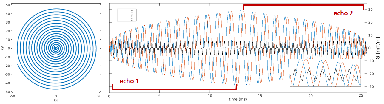
The CBF-BOLD spiral sequence was created in-house based on an SMS-EPI implementation with FAIR QUIPSS II spin labeling using a tr-FOCI inversion pulse3. Data on two subjects was acquired on a Siemens Magnetom 7T scanner (Siemens Healthineers, Erlangen, Germany) equipped with a 32-channel head coil (Nova Medical, Wilmington, MA, USA). The sequence parameters were as follows: FoV=200x200mm2, 24 slices with 2.0x2.0x2.0mm3 voxels, 50% slice gap, SMS-factor 2, ASL at TE1=2ms, BOLD at TE2=26ms, TI1/TI2/TR= 700/1800/2500 ms, flip angle=78o, variable density spiral trajectory with in-plane undersampling factor of 1.4 as described in4, 5120 ADC points per echo, 12.5ms duration, gradient amplitude 29mT/m, slew rate 132 mT/m/s. CAIPIRINHA blips were applied every 200us to sample two kz planes, analogous to a FoV/2 CAIPIRINHA shift5. For a smooth transition, the spiral-in (second echo) was slightly rotated w.r.t. to the the spiral-out but with otherwise the same parameters. The trajectory is shown in Figure 1. Blood equilibrium magnetization (M0) images were acquired with identical imaging parameters without inversion pulses and TR=20s for CBF quantification. Receive coil sensitivity and B0 off-resonance maps were obtained for image reconstruction. Images were reconstructed offline in MATLAB and Tensorflow using an SMS implementation of our Minimal Linear Networks approach to image reconstruction6 (https://github.com/giladddd/MLN.git). Two functional runs were obtained. Subjects were presented a radial flickering checkerboard for 20s while they simultaneously performed finger tapping followed with 40s rest periods, repeated in 10 blocks per run. Resting periods of 45 and 55s were present at the beginning and end of each run respectively. CBF (first echo) and BOLD (second echo) images were realigned to the M0 image using SPM12. Statistical analysis was carried out in FSL-FEAT separately for both echoes without spatial smoothing. Resulting z-score maps were averaged across the two runs for first and second echo separately. Two ROIs covering motor and visual cortices were defined based on z-scores thresholded at 1.5. Event-related averages were calculated from the mean time course signal. CBF was quantified using the PASL formula as described in7.RESULTS
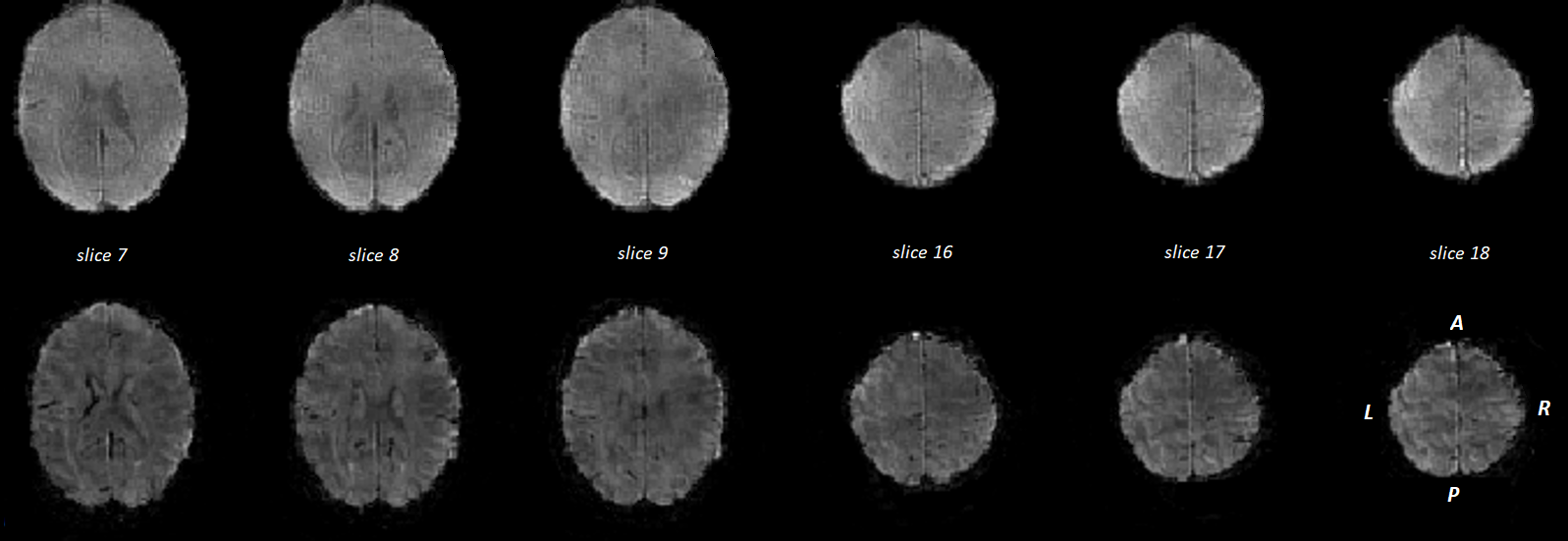
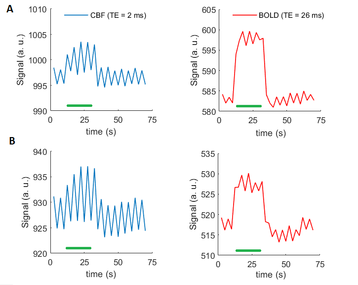
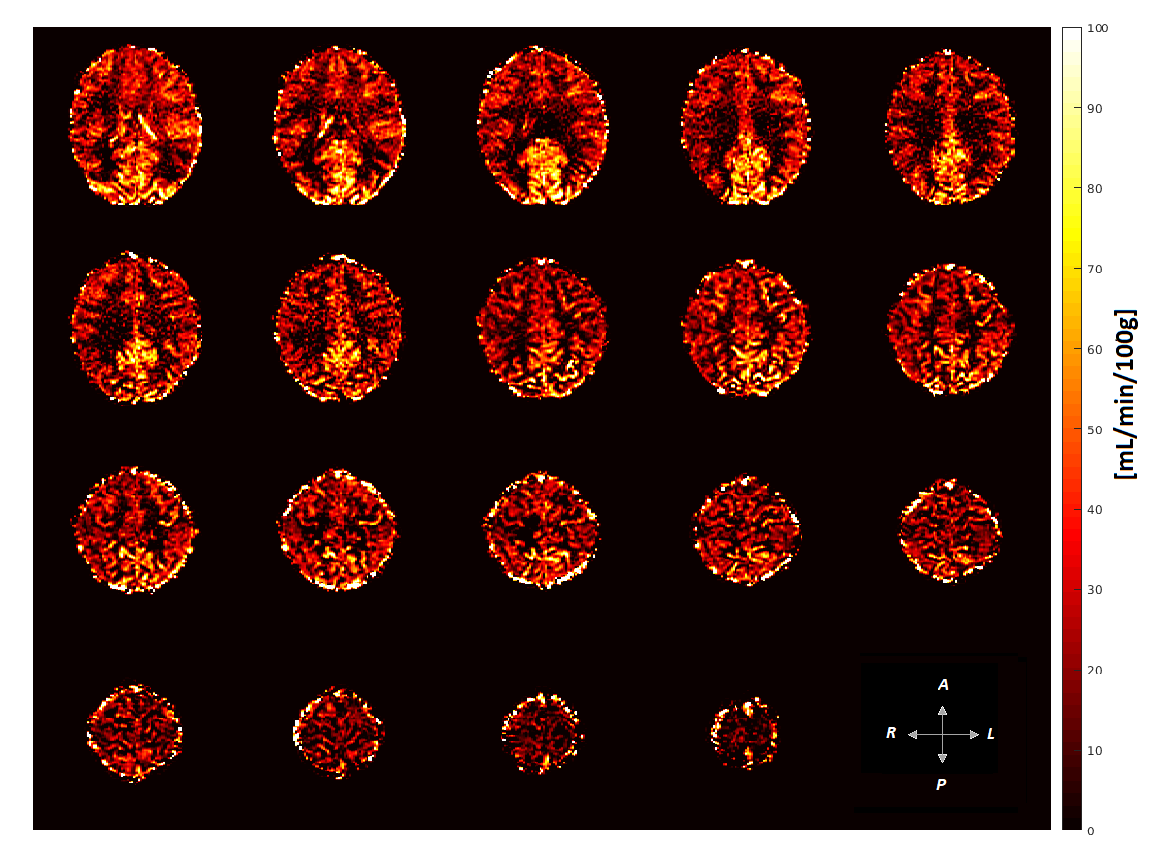
Reconstructed images from both echoes can be seen in Figure 2. Event-related averages from motor and visual cortices for CBF and BOLD are displayed in Figure 3. Higher baseline perfusion and CBF changes can be observed in the visual cortex compared to the motor cortex ROI. In contrast, the BOLD signal change is higher in the motor than visual cortex ROI. Quantitative CBF map from one subject is shown in Figure 4. High CBF values in the visual cortex are commonly observed in PASL approaches and have been previously reported8.DISCUSSION
To our knowledge, this work is the first demonstration of spiral ASL at 7T. The SMS technique in addition to the advantageous spiral sampling increases the achievable brain coverage within reasonable inversion times. It is important to note that the accelerated multi-echo spiral readout enables optimal TEs for both CBF and BOLD mapping irrespective of the resolution, in contrast to the most commonly employed EPI readouts where the achievable TEs are dictated by the desired matrix size and resolution. The presented technique will be particularly useful for high-resolution CBF and BOLD mapping in the subcortical brain regions, which have significantly shorter T2* than the cortex. The proposed approach may find application in both neuroscientific and clinical fMRI studies at 7T.Acknowledgements
This work was supported by the Netherlands Organization for Scientific Research (NWO 016.Vidi.178.052 to BAP). BAP is also partially and SK fully funded by the National Institute of Health (R01MH111444, PI Feinberg). Data were acquired at the facilities of Scannexus BV, Maastricht, NL.References
1. Chang YV, Vidorreta M, Wang Z, Detre JA. 3D-accelerated, stack-of-spirals acquisitions and reconstruction of arterial spin labeling MRI. Magn Reson Med. 2017;78(4):1405-1419. doi: 10.1002/mrm.26549.
2. Nielsen JF, Hernandez-Garcia L. Functional perfusion imaging using pseudocontinuous arterial spin labeling with low-flip-angle segmented 3D spiral readouts. Magn Reson Med. 2013;69(2):382-90. doi: 10.1002/mrm.24261.
3. Ivanov D, Poser BA, Huber L, Pfeuffer J, Uludağ K.Optimization of simultaneous multislice EPI for concurrent functional perfusion and BOLD signal measurements at 7T. Magn Reson Med. 2017;78(1):121-129. doi: 10.1002/mrm.26351.
4. Kim DK, Adalsteinsson E, Spielman DM. Simple analytic variable density spiral design. Magn Reson Med. 2003;50(1):214–219.
5. Zahneisen B, Poser BA, Ernst TE, Stenger VA. Three-dimensional Fourier-encoding of simultaneously excited slices: generalized acquisitions and Reconstruction framework. Magn Reson Med. 2014; 71(6):2071-81.
6. Liberman G, Poser BA. Minimal Linear Networks for MR Image Reconstruction. Proceedings of the ISMRM Machine Learning Workshop Part 2, Washington; October 2018.
7. Alsop DC1, Detre JA, Golay X, Günther M, Hendrikse J, Hernandez-Garcia L, Lu H, MacIntosh BJ, Parkes LM, Smits M, van Osch MJ, Wang DJ, Wong EC, Zaharchuk G. Recommended implementation of arterial spin-labeled perfusion MRI for clinical applications: A consensus of the ISMRM perfusion study group and the European consortium for ASL in dementia. Magn Reson Med. 2015;73(1):102-16. doi:10.1002/mrm.25197.
8. Ivanov D, Gardumi A, Haast RAM, Pfeuffer J, Poser BA, Uludağ K. Comparison of 3T and 7T ASL techniques for concurrent functional perfusion and BOLD studies. Neuroimage. 2017;156:363-376. doi: 10.1016/j.neuroimage.2017.05.038
Figures