0360
Attention modulation of layer-specific signals in human visual cortexChengwen Liu1,2, Chencan Qian1, Zihao Zhang1,3, Kaibao Sun1, Jing An4, Danny J.J. Wang5,6, and Peng Zhang1,2
1State Key Lab of Brain and Cognitive Science,Institute of Biophysics,Chinese Academy of Sciences, Beijing, China, 2University of Chinese Academy of Sciences, Beijing, China, 3The Innovation Center of Excellence on Brain Science, Chinese Academy of Sciences, Beijing, China, 4Siemens Shenzhen Magnetic Resonance Ltd, Shenzhen, China, 5Department of Neurology, University of California Los Angeles, Los Angeles, CA, United States, 6Stevens Neuroimaging and Informatics Institute, University of Southern California, Los Angeles, CA, United States
Synopsis
Attention mechanisms at different cortical
layers of the human visual cortex remain poorly understood. Here we investigated
the attention modulation of layer-specific activities in the human visual
cortex, using submillimeter-resolution BOLD fMRI at 7 Tesla. Results showed that
compared to the middle-layer activities, attention increased signals in the superficial
and deep layers of V1, and in the superficial layers of V2 and V3. Contrast
modulation was strongest in the middle layer of V1, consistent with the feedforward
input from the LGN. These findings suggest that top-down spatial attention mainly
modulates output signals in the superficial layers of human visual cortex.
Introduction
Attention are important for selectively processing relevant information in clutered visual scences. Human early visual cortex consists of six layers of neurons, with distinct roles in input, output and intracortical connections. The laminar circuits of attention in the human visual cortex is largely unknown. One possibility is that attention mainly improves output neural signals in the superficial layer that project to higher cortical areas, or the activites in the deep-layer neurons that project to the thalamus1. Another possibility is that attention may primarily modulate signals in the input layers that project locally within the cortical column2. To test these hypothesis, we used high-resolution BOLD fMRI at ultra-high field (7T) to dissociate feedforward and feedback signals from different cortical layers in the human visual cortex, and investigated the effect of attention on the layer-specific visual activities.Methods
Figure 1 shows the stimuli and procedure for the attention task. Subjects paid attention either to the left or to the right checkerboard patterns to detect occational spatial frequency change (Exp1, n=10) or small contrast increment (Exp2, n=7) of the attended stimulus.The checkerboard patterns were presented at five contrast levels (2.5%, 6%, 14%, 35% and 83%) in Exp 1 and at two contrast levels (2.5%, 50%) in Exp 2. Task difficulties were matched across different contrast conditions.MRI data were collected with a 7T research system (Siemens Healthcare,Erlangen,Germany) using a 1TX/32RX head coil(Nova Medical, Cambridge, MA, USA).Subjects used bitebars to restrict head motion. Functional data were collected with a T2*-weighted 2d GE-EPI sequence in Exp 1 (0.75 mm isotropic voxels with 48 slices, TE =23ms, TR=4s, FOV=120*75mm, iPAT=3) and a T2-weighted 3d passband balanced-SSFP sequence in Exp 2 (0.8mm isotropic voxels with 10 slices, 20% oversampling on both sides of z direction, TE=2.53ms, TR=5.06ms, volume acquisition time = 6s, FOV=102*102mm, iPAT=2, FA = 20 deg). T1, PD and T2*-weighted anatomical volumes were acquired at 0.7 mm isotropic resolution with MPRAGE sequences.MRI data were analyzed using AFNI/SUMA and FreeSurfer software.The GM/pial surface was manually edited to remove sinus and other artifacts to ensure correct segmentation. Gray matter was sub-divided into three equi-volume layers with two additional surfaces, together with the WM and pial surfaces3. For each voxel, we calculated the percentage of volume in the WM, CSF and the three GM layers. These layer-weights were subsequently used in a spatial regression approach to unmix layer activities. Pial veins were masked out based on PD/T2*-weighted images and signal change over 5% in the functional data. Gray matter voxels under the vein were also excluded.Results
Figure 2A shows that attention increased BOLD signals from all three cortical layers. In figure 2B, the layer responses were normalized by the response in the middle layer, the superficial layer showed greater activity in the attended than in the unattended condition. This can be clearly observed in all three visual areas, for both GE-EPI and bSSFP sequences. V1 deep layer also showed increased response in the attended condition (for GE-EPI). The layer reponse profile of bSSFP is flatten than of GE-EPI, suggesting less sensitivity to the pial surface veins. In figure 2C, we computed an attention modulation index (AMI, attended-unattended / attended+unattended) for each cortical layer.This AMI was also significantly larger in the superficial layers compared to the middle layers of V1,V2 and V3.Figure 3A shows the normalized contrast response functions (CRF) in different layers of V1-V3. In figure 3B, a parameters C50 denotes the contrast at which the response reaches the half maximum level. It shows that C50 was significantly larger in the middle than in the superficial layer of V1, suggesting more linear contrast response in the middle layer of V1, which is consistent with a feedforward input from the LGN.Discussion and Conclusion
Both T2*-weighted GE-EPI and T2-weighted b-SSFP sequences showed consistent results that top-down spatial attention mainly modulated activities in the superficial layers of human visual cortex V1, V2 and V3. A deep-layer signal increase was also observed in V1 from GE-EPI results. These findings were consistent with the recent monkey neurophysiology study that feedback signals mainly target superficial and deep layers in early visual cortex4. To conclude, our results suggest that attention might serve to increase the output signals in the early visual cortex to prioritize the processing of relevant information.Acknowledgements
This research was supported by grants from Bureau of International Cooperation, Chinese Academy of Sciences (153311KYSB20160030),the Project of State Key Laboratory of Ophthalmology, Optometry and Visual Science,Wenzhou Medical University,Wenzhou 325027,and The General Program of National Natural Science Foundation of China(31871107).References
- Self, Matthew W., et al. "Benchmarking laminar fMRI: neuronal spiking and synaptic activity during top-down and bottom-up processing in the different layers of cortex." Neuroimage(2017).
- Nandy, Anirvan S., Jonathan J. Nassi, and John H. Reynolds. "Laminar organization of attentional modulation in macaque visual area V4." Neuron 93.1 (2017).
- Kok, Peter, et al. "Selective activation of the deep layers of the human primary visual cortex by top-down feedback." Current Biology 26.3 (2016).
- Van Kerkoerle, Timo, Matthew W. Self, and Pieter R. Roelfsema. "Layer-specificity in the effects of attention and working memory on activity in primary visual cortex." Nature communications 8 (2017).
Figures
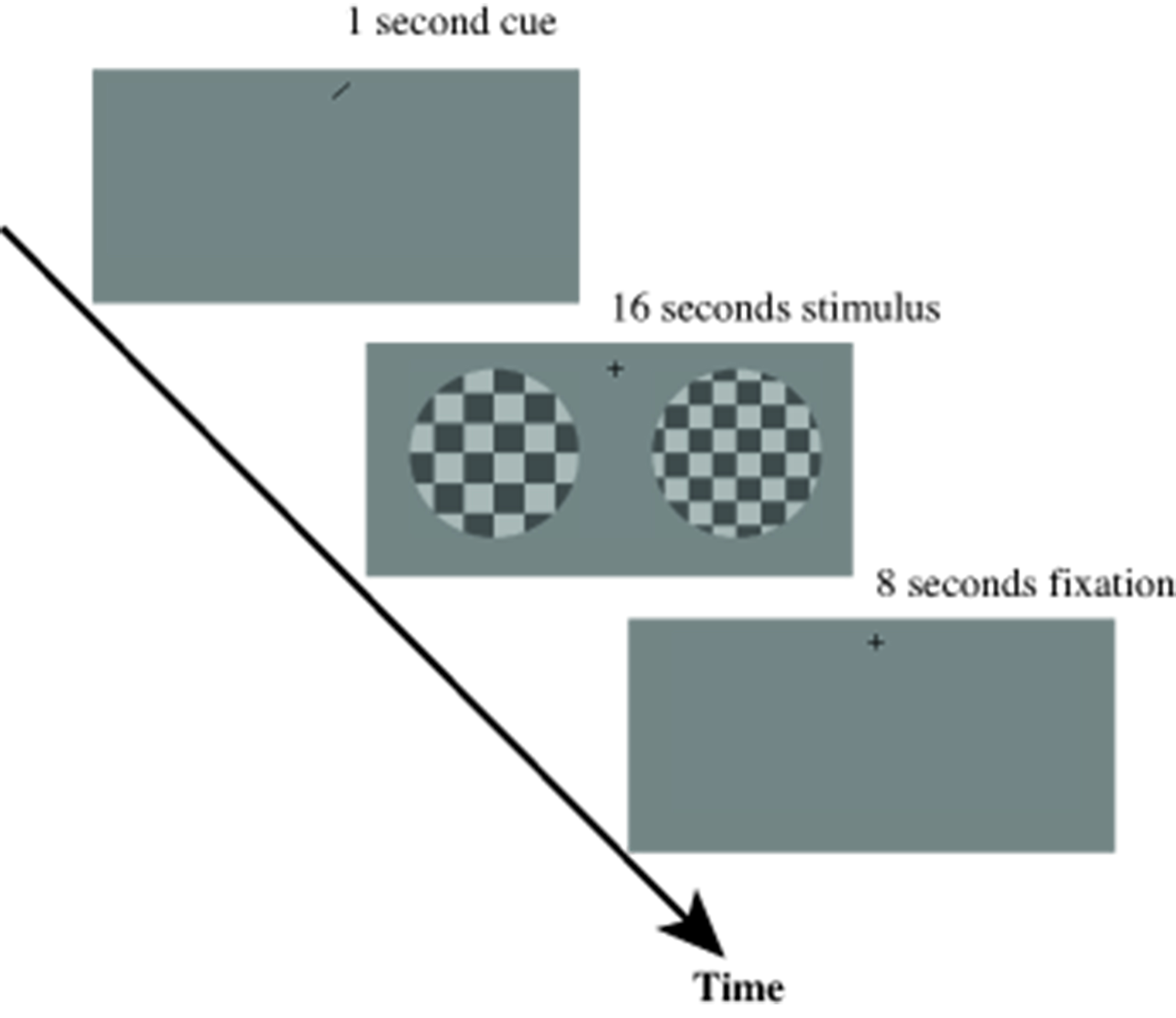
Figure1. During scanning, subjects detect spatial frequency change on
one side of the checkerboard stimuli while keeping fixation.
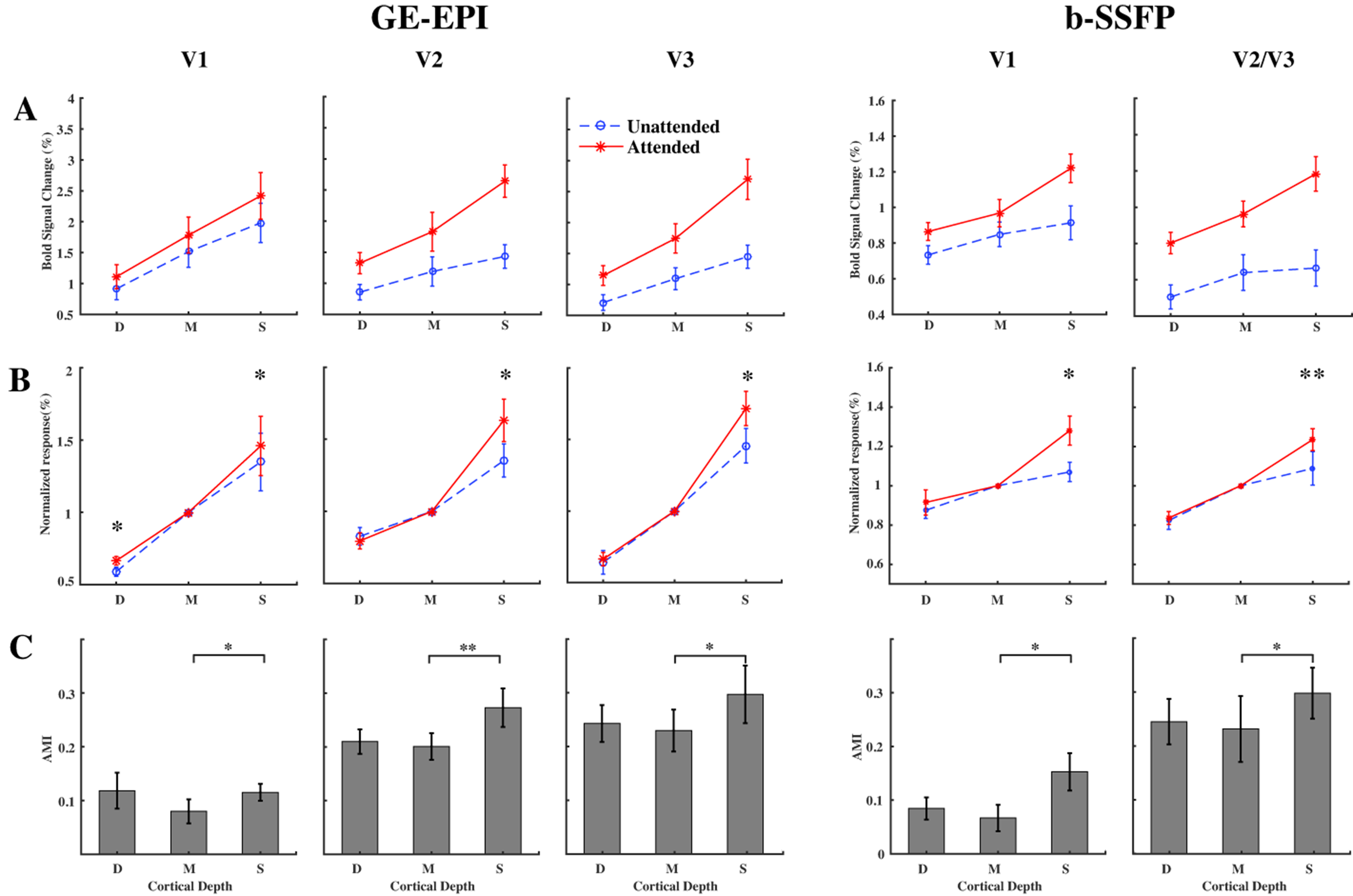
Figure2. (A) BOLD response from
difference depth of V1-V3 in attended and unattended conditions (B)Same as (A),
except that layer response profile were normalized to the middle layer reponse.
(C)Attention modulation index(AMI) for different depth of visual areas.
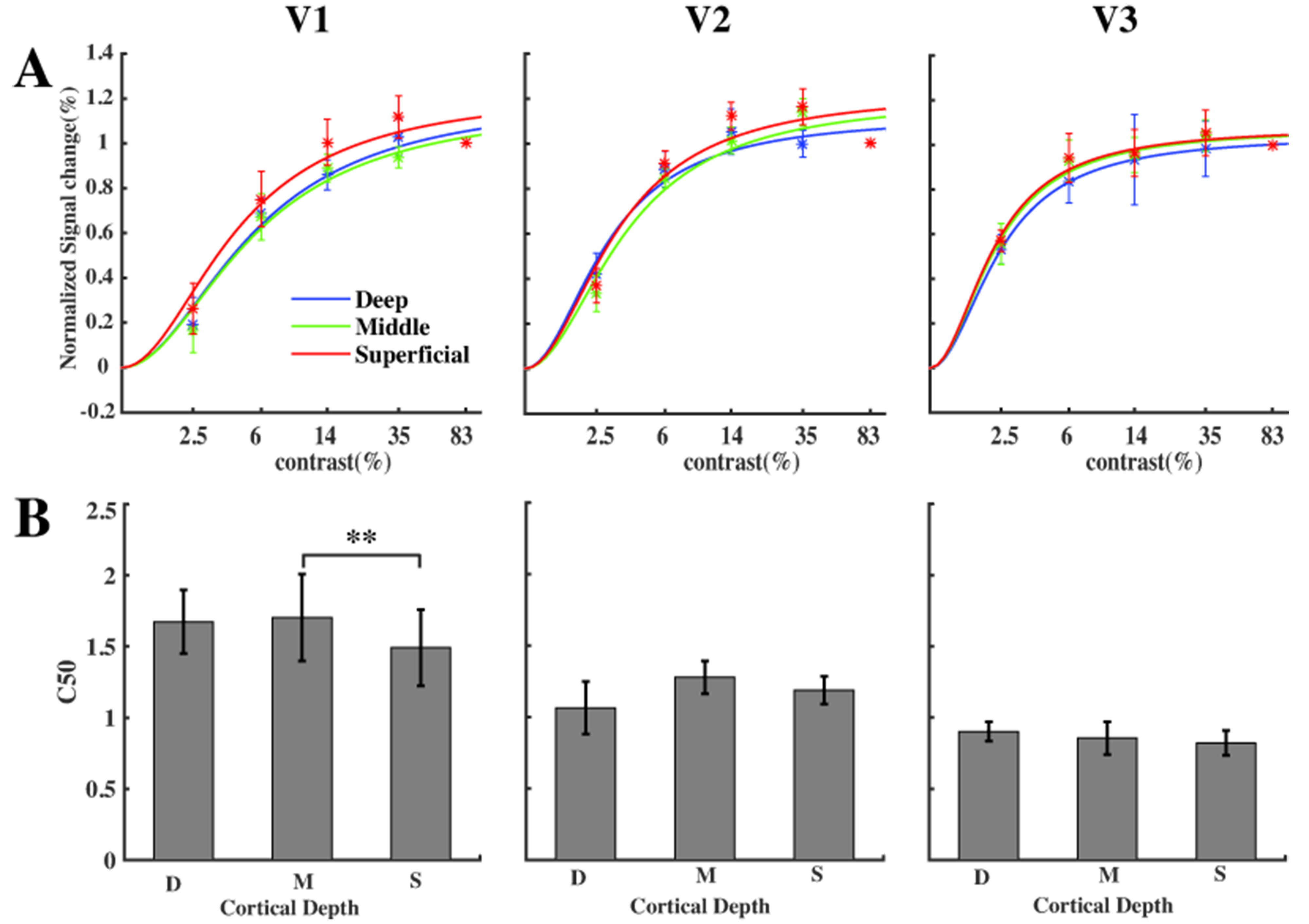
Figure3.
(A)Normalized contrast reponse function(CRF) become less linear from V1 to V3. (B)CRF
fitting parameter C50 (contrast at half maximum response) was larger (more
linear) in the middle layer of V1