0357
The Predominant Role of the Vagus Nerve in Gastric Electrical Stimulation Evoked Gut-Brain Axis1Biomedical Engineering, Purdue University, West Lafayette, IN, United States, 2Electrical and Computer Engineering, Purdue University, West Lafayette, IN, United States, 3Psychological Science, Purdue University, West Lafayette, IN, United States
Synopsis
The gut-brain axis serves the bidirectional communication between the enteric nervous system and the central nervous system and links the gastrointestinal function with cognitive and emotional centers of the brain. Three different pathways have been identified to be responsible for the gut-brain axis, including the chemical modulation, the spinal nerve, and the vagus nerve. However, the functional role of each pathway is unclear and still under investigation. Using functional magnetic resonance imaging (fMRI), we report that gastric electrical stimulation (GES) can modulate the gut-brain axis and evokes fMRI activity in multiple brain regions including both cognitive and sensory systems. Most of the GES evoked brain regions are attributable to the vagal function, suggesting a central role of the vagus nerve in the gut-brain axis, especially in modulating the cognitive and emotional centers.
Purpose
The gut-brain axis serves the bidirectional communication between the enteric nervous system and the central nervous system. It links the peripheral gastrointestinal function with cognitive and emotional centers of the brain and relates to gut-brain diseases such as irritable bowel syndrome. Three different pathways have been identified to be responsible for the gut-brain axis, including the chemical modulation, the spinal nerve, and the vagus nerve. However, the functional role of each pathway is unclear and still under investigation. In this study, we focused on the vagal function in the gut-brain axis and addressed the vagal contribution to brain activity during gastric stimulation. Functional magnetic resonance imaging (fMRI) was applied to map the whole-brain activity, and the gastric electrical stimulation (GES) was used to modulate the gut-brain axis.Method
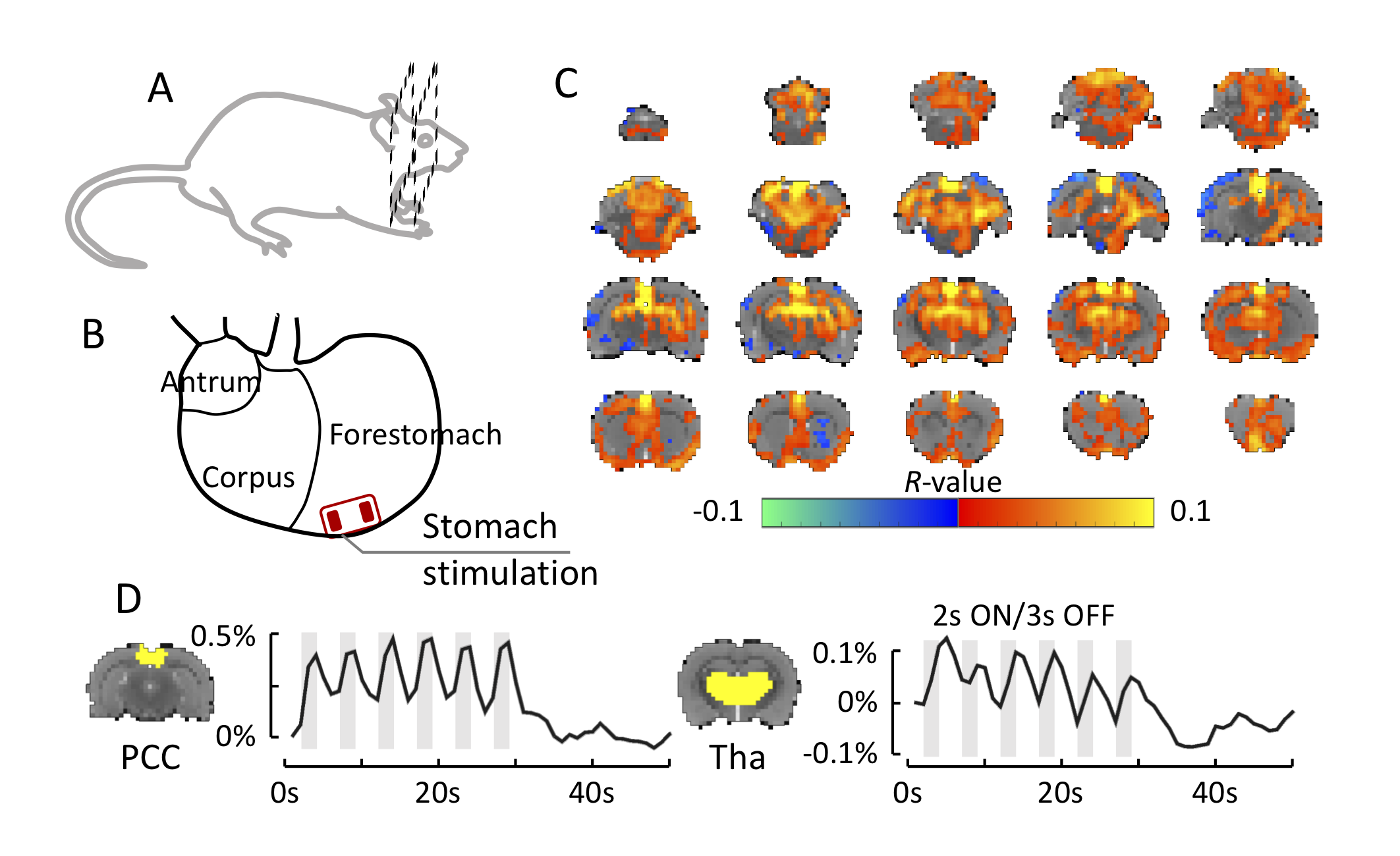
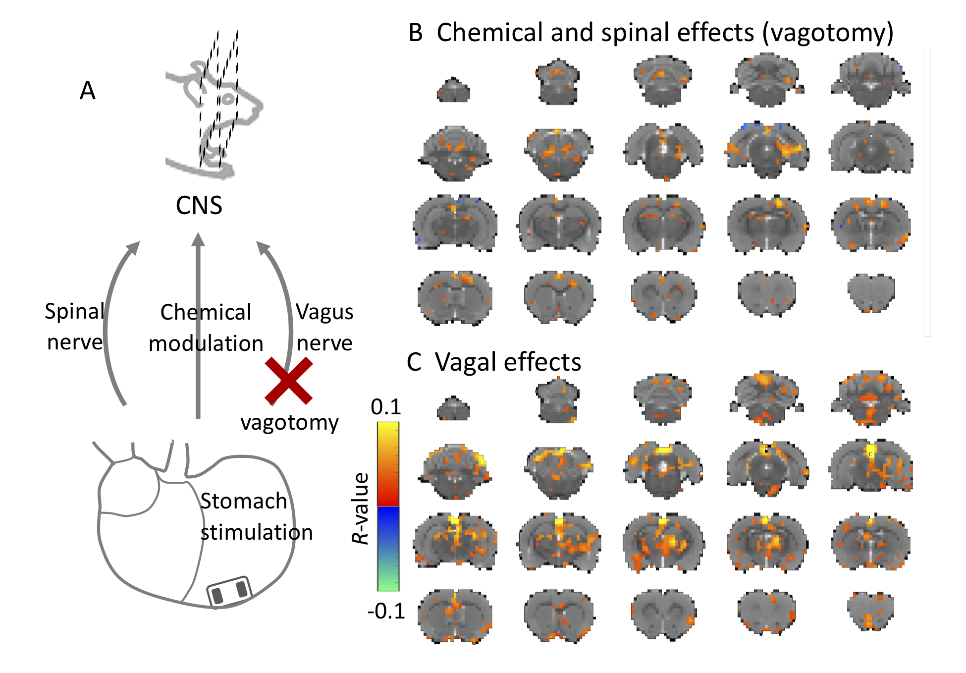
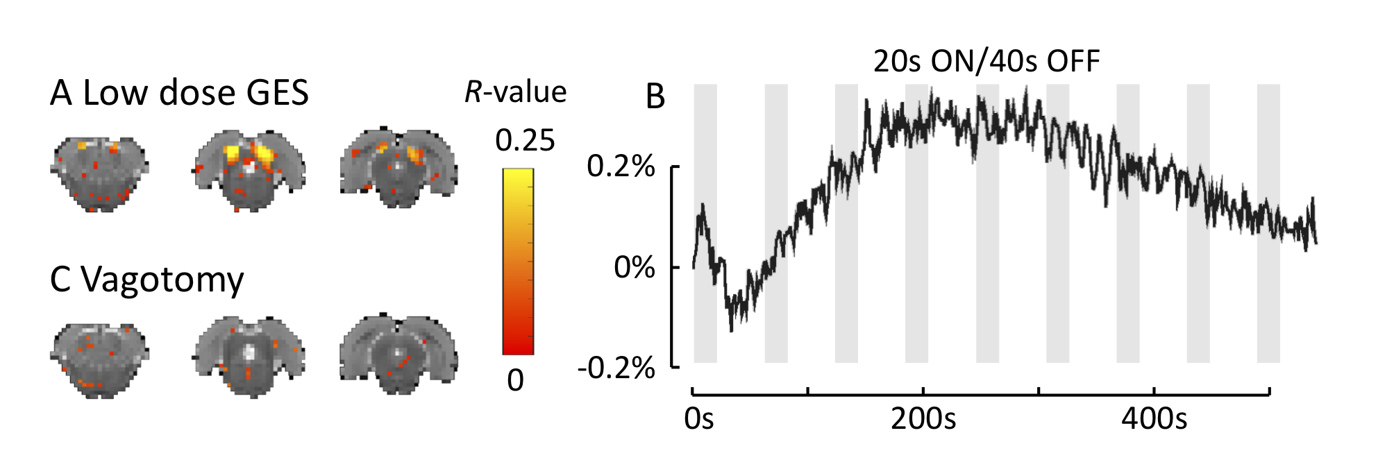
Ten Sprague Dawley rats (male, 250-400 g) were scanned with fMRI during GES. Each animal was chronically implanted with an MRI-compatible patch electrode on the stomach wall (Fig. 1B). After recovering from surgery, the animal was scanned with 2-D single-shot gradient-echo echo-planar imaging (EPI, 1s repetition time, 16.5ms echo time, 60° flip angle, 0.5×0.5×1 mm3 voxel size) in a 7-tesla small-animal MRI system (BioSpec 70/30, Bruker). During imaging sessions, the animal was anesthetized with continuous dexmedetomidine (SC-infusion, 0.015 mg/Kg/h) and isoflurane (0.1-0.5% mixed in O2). Each animal went through multiple imaging sessions with high-dose or low-dose GES protocols. The high-dose GES (6mA, 0.3ms, 20Hz) was adjusted from the clinical dosage. It was delivered in a 30s-ON-30s-OFF block-design paradigm. For each ON period, the 2s-ON-3s-OFF pulse train was repeated for six times (Fig. 1D). The low-dose GES (0.6mA, 0.2ms, 5Hz) was designed by optimizing the gastric motility. It was applied in a 20s-ON-40s-OFF block design paradigm with continuous pulse train filling the 20s ON period (Fig. 3C). Both high-dose and low-dose stimulation were repeated one more time on the same animal after surgically damaging the cervical vagus nerve (vagotomy) on both sides. In data analysis, fMRI data were handled with standard preprocessing steps. After that, a response model was derived by convolving GES with a hemodynamic response function. The response model was correlated with blood-oxygen-level-dependent (BOLD) time series from each voxel. In addition, BOLD time series were cross-correlated with the time series from a different imaging session under the same stimulation protocol. The correlation coefficient was presented in the activation map, and the threshold was determined by t-test (p<0.05).Results
Ten rats were scanned with fMRI during high-dose GES applied to the forestomach (Fig. 1B). By correlating the BOLD activity and the GES-derived response model, we found multiple regions activated by GES. These regions included the posterior cingulate cortex, thalamus, somatosensory cortex, prefrontal cortex, hippocampus, and some midbrain and brainstem nucleus (Fig. 1C). The same stimulation was applied again on four rats after performing vagotomy. Without the vagus nerve, only the somatosensory cortex and a part of the subiculum were activated by GES (Fig. 2B). The comparison before and after vagotomy revealed that the vagus nerve contributed to most GES evoked brain regions, including the posterior cingulate cortex, thalamus, amygdala, and some brainstem nucleus (Fig. 2C). We also tested the low-dose GES on nine rats with the vagus nerve attached and three rats with vagotomy. The correlation between the BOLD activity and the stimulation did not generate strong brain responses. However, the inferior colliculus showed reproducible activity in multiple repetitions of the same GES paradigm (Fig. 3A), showing a slow drift over minutes (Fig. 3B). This slow-drift response was also diminished by vagotomy (Fig. 3C).Conclusion
GES modulates the gut-brain axis and evokes BOLD activity in multiple brain regions including both cognitive and sensory systems. Most of the GES evoked brain regions are attributable to the vagal function, suggesting a central role of the vagus nerve in the gut-brain axis, especially in modulating the cognitive and emotional centers.Acknowledgements
This study was funded by National Institutes of Health’s SPARC - Stimulating Peripheral Activity to Relieve Conditions - program (OT2OD023847).References
No reference found.Figures