0355
Can preoperative Diffusion-weighted MR predict the Microvascular invasion of hepatocellular carcinoma? A deep learning evaluation1School of Medical Information Engineering, Guangzhou University of Chinese Medicine, Guangzhou, China
Synopsis
Microvascular invasion (MVI) of hepatocellualr carcinoma(HCC) is regarded as the most important factor associated with the success of curative resection and the outcome after liver transplation. As the MVI prediction can only be ultimately determined by the histopathological features of the tumor cells, numerous works have attempted to predict the MVI of HCC based on noninvasively preoperative images. Diffusion-weighted MR has also shown to be effective for MVI prediction based on signal intensities of lesions and the apparent diffusion coefficient (ADC). In this work, the emerging deep learning technique is used for MVI prediction of HCC based on DWI.
Introduction
Preoperative prediction of microvascular invasion (MVI) of hepatocellular carcinoma (HCC) is remarkably significant and helpful for prognosis, treatment strategy and patient management1. The prediction of MVI cannot be diagnosed before surgery or biopsy and can only be ultimately determined by the histopathological features of the tumor cells. Many works have been predicting MVI of HCCs using radiological or radimoics features extracted from preoperative images2,3. Diffusion-weighted MR has also shown to be effective for MVI prediction based on signal intensities of lesions and the apparent diffusion coefficient (ADC)4. As deep learning technique can learn increasing high-level features of images that can optimally represent the characteristics of the data, we anticipate that deep feature derived from Diffusion weighted MR (DWI) might be better than ADC for MVI prediction. The purpose of this study is to investigate whether DWI can be useful for MVI prediction of HCC using a deep learning evaluation.Method
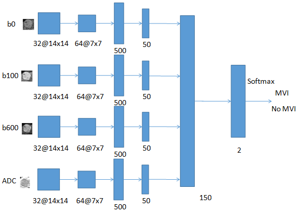
This study was approved by the local institutional review board and the patients’ informed consent was waived due to its retrospective properties. In this retrospective study, consecutive 54 subjects with fifty-four HCCs from July 2012 to May 2017 with resected HCC were retrieved (50 male, 4 female, aged 50.26±12.32 years within an age range of 27 to 76 years). The histology information of MVI in HCC was retrieved from the archived clinical histology report, where tumor size, differentiation, the presence or absence of microvascular invasion, the surgical resection margin, and the presence or absence of fibrosis or cirrhosis of the HCCs were described. Of the 54 lesions, twenty-six were pathologically determined as the absence of MVI, while twenty-eight were pathologically determined as the presence of MVI. All subjects with DWI examinations were acquired using a 3.0 Tesla MR scanner (Signa Excite HD 3.0T, GE Healthcare, Milwaukee, WI, USA). DWI examinations were performed with a single-shot echo-planar imaging and a breath-hold routine after CE-MR examinations. DWI parameters were: three b values of 0,100,600 sec/mm2; TR/TE 1800/35ms; flip angle, 90; a matrix of 128×128;slice thickness: 8 mm; interslice gap, 1 mm. First, ADC images were computed by mono-exponentially fitting the three b-value points. Then, multiple 2D axial patches (28×28) of HCCs from b0, b100, b600 and ADC images were extracted to increase the dataset for training the convolutional neural network. Furthermore, deep features were separately extracted from b0, b100, b600 and ADC based on the CNN for MVI prediction. Finally, fusion of deep features derived from three b-value images and ADC was conducted for MVI prediction. Figure 1 showed the framework of the proposed deep fusion model with DWI for MVI prediction. Values of prediction performance in differentiating the presence and absence of MVI were denoted as mean±standard deviation as a result of four-folded cross-validation with 10 repetitions on the data set.Results
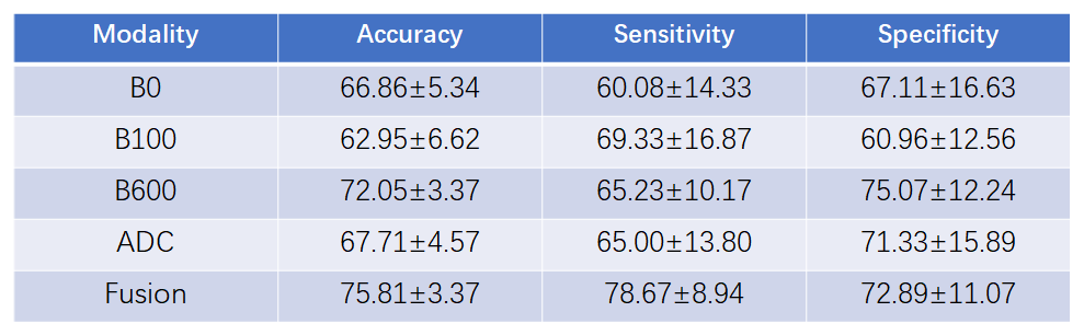
Table 1 showed the characterization performance of deep features using different image sets for MVI prediction. Deep feature in higher b values (b600) yielded better performance for MVI prediction than that of the lower b values (b0 and b100). Comparatively, deep feature in the ADC did not obtain promising results for MVI prediction, which was much lower than that of the higher b value (b600). Furthermore, fusion of deep features from the b0, b100, b600 and ADC images yielded best results for MVI prediction.Discussion
Our study suggests that deep feature derived from higher b value yields better performance for MVI prediction, implying that DWI imaging with higher b value might be better for MVI prediction. In the present study, deep feature from higher b value image obtained better performance than that from the ADC image for MVI prediction, inferring that image features from higher b value image can be more representative than that from the ADC image. Furthermore, our study also suggests that fusion of deep features derived from original b0, b100, b600 and ADC images results in improved performance compared with that of the single image, demonstrating that multiple b value images and ADC image can be taken full advantage to yield better performance for MVI characterization.Conclusion
Our study suggests that fusion of deep features derived from DWI image with respect to the three b-value images and ADC image yields better performance for MVI prediction. It can be believed that the proposed fusion model may be broadly used for MVI prediction with DWI images in clinical practice.Acknowledgements
This research is sponsored by the grants from National Natural Science Foundation of China (81771920).References
[1] Redriguez-Peralvarez M, Luong T, Andreana L, et al. A Systematic Review of Microvascular Invasion in Hepatocellular Carcinoma: Diagnostic and Prognostic Variability. Ann Surg Oncol 2013;20:325-339.
[2] Unal E, Ldilman L, AkataD, Ozmen M, Karcaaltincaba M. Microvascular invasion in hepatocellular carcinoma. Diagn Interv Radiol 2016;22: 125-132.
[3] Renzulli M, Brocchi S, Cucchetti A, et al. Can Current Preoperative Imaging Be Used to Detect Microvascular Invasion of Hepatocellular Carcinoma? Radiology 2016; 279(2):432-442.
[4] Suh YJ, Kim MJ, Choi JY, Park MS, Kim KW. Preoperative prediction of the Microvascular invasion of hepatocellular carcinoma with Diffusion-weighted imaging. Liver Transp 2012; 18:1171-1178.
Figures