0352
Fast High-Resolution Diffusion Tensor Imaging of the Cervical Spinal Cord Using Point-Spread-Function Encoded EPI (PSF-EPI)1Department of Biomedical Engineering, Center for Biomedical Imaging Research, Beijing, China, 2Philips Healthcare, Beijing, China, 3Beijing Chao Yang Hospital, Beijing, China, 4Beijing ChaoYang Hospital, Beijing, China
Synopsis
Diffusion tensor imaging (DTI) of spinal cord holds great promise to aid diagnosis of spine-related diseases. However, it is greatly limited in clinics by the physiological motion induced artifacts and susceptibility inhomogeneity induced distortions. To reduce distortions in DTI, various multi-shot EPI (MS-EPI) methods have been proposed. However, the acquisition time is prolonged due to multiple shots. In this study, we achieved cervical spine DTI using only 6 shots based on a distortion- and blurring- free MS-EPI technique, Point-Spread-Function Encoded EPI (PSF-EPI). The acquisition time is within 5 minutes. The efficacy of PSF-EPI is demonstrated on healthy volunteers and patients.
Introduction
Diffusion tensor imaging (DTI) of spinal cord plays a vital role in a variety of clinical applications, such as pre- and post- spinal surgery evaluation 1. However, spinal cord imaging has been significantly limited by physiological motions and magnetic susceptibility inhomogeneity surrounding the spine 2. This can induce severe distortions, impeding diagnostic interpretation. Furthermore, given that the average diameter of spinal cord is approximately 1 cm, high resolution DTI is a necessity 2. This further pushes the need of high fidelity spinal cord DTI with practical sampling efficiency.
Single-shot echo planar imaging (SS-EPI) is the mostly implemented technique for DWI due to fast speed and insensitivity to bulk motion. However, SS-EPI suffers from T2* blurring and severe geometric distortion. To remedy these difficulties, various multi-shot EPI (MS-EPI) methods have been proposed 3,4. Interleaved EPI (iEPI) is a MS-EPI technique which has shown reduced distortion and decreased blurring artifacts in diffusion weighted imaging of the spinal cord 5. However, the acquisition time is prolonged due to multiple shots.
In this study, we adopted a distortion- and blurring- free MS-EPI technique, Point-Spread-Function Encoded EPI (PSF-EPI) with Tilted-CAIPI 6 and achieved cervical spine DTI using only 6 shots. The acquisition time is below 5 minutes. The efficacy of PSF-EPI is demonstrated on both healthy volunteers and patients by comparing it with iEPI results.
Material and Methods
The scans were performed on a Philips 3T Achieva TX scanner (Philips Healthcare, Best, The Netherlands) using a 16-channel head and neck phase array coil. Cardiac triggering was used to reduce ghost artifacts induced by bulk physiological motions, such as swallowing, CSF pulsation and respiratory motion 2. To correct inter-shot phase variation, we acquired an additional navigator to support high acceleration rates along the anterior-posterior phase encoding direction 7. Additionally, two saturation bands were placed on the two sides of the spinal cord to suppress unwanted signals from oral cavity and surrounding lipids.
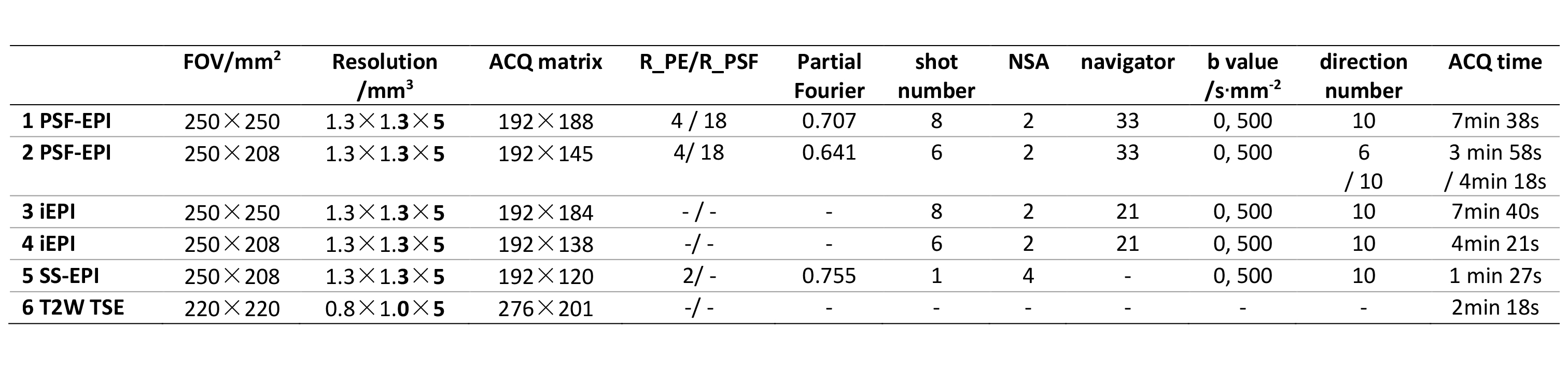
In the PSF-EPI sequence, an additional spin-warp phase encoding (PE) is exerted on the phase encoding direction in 2D EPI 8. This generates a three dimensional k-space consisting of $$$kx$$$ (readout), $$$ky$$$ (EPI-PE) and $$$ks$$$ (PSF-PE) dimensions. PSF-EPI can achieve distortion- and blurring- free imaging along $$$ks-kx$$$. By using the signal correlation along $$$ky$$$, parallel imaging is allowed to achieve high acceleration, especially combined with a Tilted-CAIPI reconstruction strategy 6,9. Here the acceleration rates along EPI-PE and PSF-PE are denoted by R_PE and R_PSF, respectively. T2W anatomical images, SS-EPI DWI, PSF-EPI DWI and iEPI DWI were acquired for all participants. Additionally, a calibration scan for reconstruction was acquired. It took about 30 seconds. The specific acquisition parameters for each sequence are summarized in Table 1 (PSF-EPI prescan time excluded).
Three healthy volunteers and five patients received cervical spine surgery were recruited for the experiment. This study was approved by the Institutional Review Board and undertaken with each participant informed consent.
Results
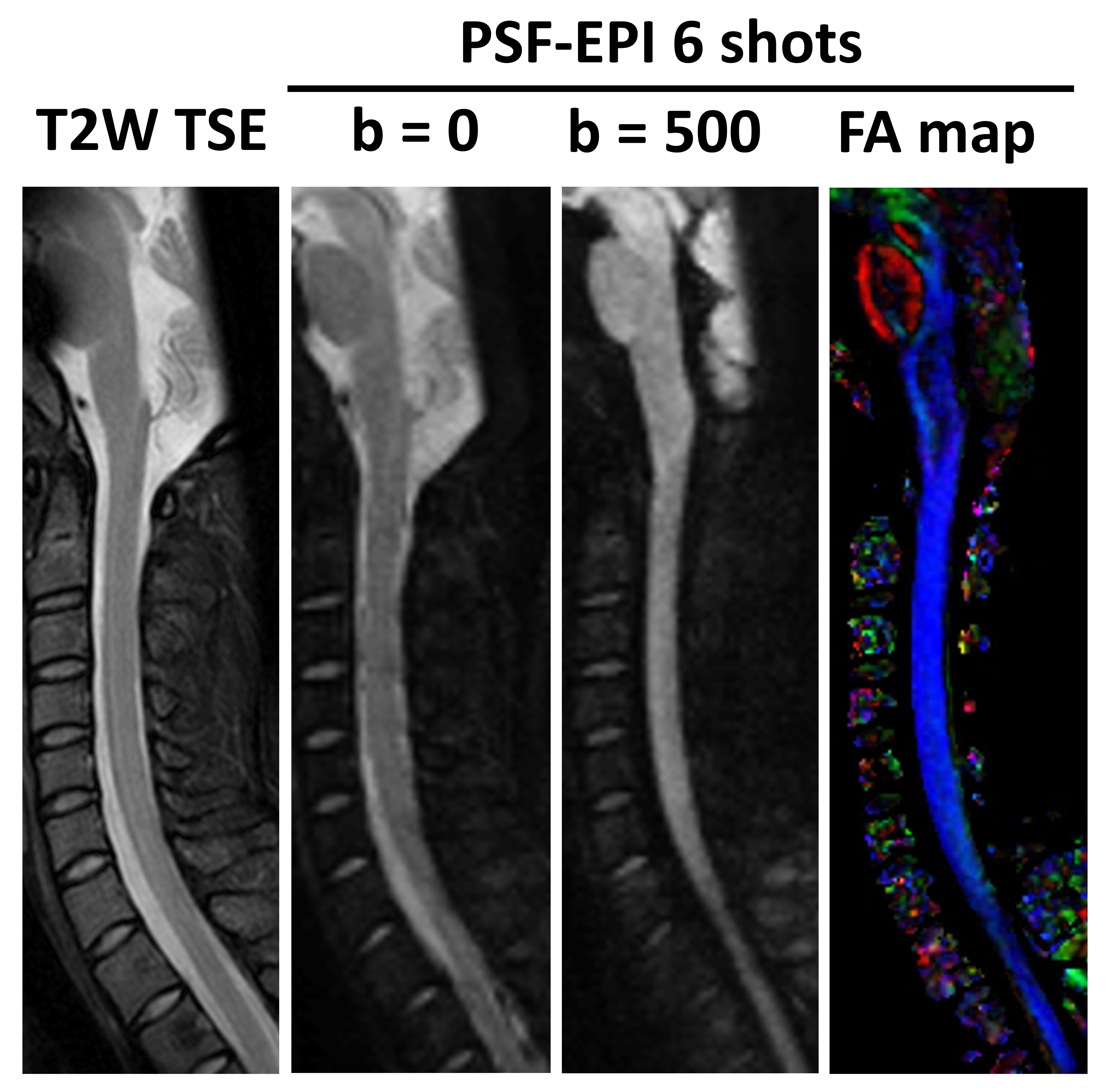
Fig. 1 demonstrates the 6-shot DWI results of a healthy volunteer. The SS-EPI result shows severe distortions around intervertebral disks, which are effectively reduced by iEPI. However, the spinal cord in iEPI images shifts along the AP direction below C7. In addition, the iEPI images show ghost artifacts proximal to the brainstem, possibly due to susceptibility inhomogeneity at complicated tissue interface. In the contrary, the 6-shot PSF-EPI result maintains high consistency with the structural reference.
Fig. 2 shows the 6-shot PSF-EPI DWI results from another healthy volunteer. The total acquisition time is around 4min 30s, which is practical for clinical applications. Due to metal implants in the post-operative patients, 8-shot DWI shows better metal artifacts control than 6-shot DWI.
Fig. 3 shows the 8-shot DWI results of a patient with an anterior cervical plate system in the spine. Very severe distortions around metallic fixations in C4-C6 are observed in the iEPI images. In comparison, the PSF-EPI images show improved fidelity, which is more consistent with the T2W TSE image.
Discussion and Conclusion
In this study, we demonstrated the feasibility of high-resolution DTI using only 6 shots for intact cervical spine cords or 8 shots for spinal cords with metal implants around based on the PSF-EPI sequence. With clinically practical acquisition time, the PSF-EPI results show effective distortion correction, improved anatomical fidelity and reasonable SNR levels for both healthy volunteers and patients.
Compared with the previously proposed PSF-mapping technique for distortion correction in spinal cord DTI 10, the current PSF-EPI technique does not require complex image registration and takes similar scan time.
More patients will be recruited to evaluate the performance of this technique in future studies.
Acknowledgements
We acknowledge the assistance of Xiaodong Ma.References
1. Maier, S. E. (2007). Examination of spinal cord tissue architecture with magnetic resonance diffusion tensor imaging. Neurotherapeutics, 4(3), 453-459.
2. Andre, J. B. , & Bammer, R. (2010). Advanced diffusion-weighted magnetic resonance imaging techniques of the human spinal cord. Topics in Magnetic Resonance Imaging, 21(6), 367-78.
3. Wheeler‐Kingshott, Claudia A.M, Parker, G. J. M. , Symms, M. R. , Hickman, S. J. , Tofts, P. S. , & Miller, D. H. , et al. (2002). Adc mapping of the human optic nerve: increased resolution, coverage, and reliability with csf‐suppressed zoom‐epi. Magnetic Resonance in Medicine, 47(1), 24–31.
4. Holdsworth, S. J., Skare, S., Newbould, R. D., & Bammer, R. (2010). Robust grappa-accelerated diffusion-weighted readout-segmented (rs)-epi. Magnetic Resonance in Medicine, 62(6), 1629-1640.
5. Bammer, R. , Augustin, M. , Prokesch, R. W. , Stollberger, R. , & Fazekas, F. . (2002). Diffusion‐weighted imaging of the spinal cord: interleaved echo‐planar imaging is superior to fast spin‐echo. Journal of Magnetic Resonance Imaging, 15(4), 364-73.
6. Dong, Z., Wang, F., Reese, T. G., Manhard, M. K., Bilgic, B., Wald, L. L., ... & Setsompop, K. (2018). Tilted‐CAIPI for highly accelerated distortion‐free EPI with point spread function (PSF) encoding. Magnetic resonance in medicine.
7. Dai, E. , Zhang, Z. , Ma, X. , Dong, Z. , Li, X. , & Xiong, Y. , et al. (2018). The effects of navigator distortion and noise level on interleaved epi dwi reconstruction: a comparison between image- and k-space-based method. Magnetic Resonance in Medicine.
8. Zaitsev M, Hennig J, & Speck O. (2004). Point spread function mapping with parallel imaging techniques and high acceleration factors: fast, robust, and flexible method for echo‐planar imaging distortion correction. Magnetic Resonance in Medicine, 52(5): 1156-1166.
9. In, M. H., Posnansky, O., & Speck, O. (2017). High-resolution distortion-free diffusion imaging using hybrid spin-warp and echo-planar psf-encoding approach. Neuroimage, 148, 20.
10. Xu, J., Shimony, J. S., Klawiter, E. C., Snyder, A. Z., Trinkaus, K., Naismith, R. T., ... & Song, S. K. (2013). Fast diffusion tensor imaging and tractography of the whole cervical spinal cord using point spread function corrected echo planar imaging. Magnetic Resonance in Medicine, 69(1), 144-149.
Figures