0345
Distortion-free diffusion-weighted imaging of the eyeballs and the optic nerves using PSF-EPI1Center for Biomedical Imaging Research, Department of Biomedical Engineering, School of Medicine, Tsinghua University, Beijing, China, 2Philips Healthcare, Beijing, China
Synopsis
Distortion-free diffusion-weighted imaging can provide high fidelity information for the diagnosis of ophthalmological diseases. Traditional echo-planar imaging technique suffers from geometric distortion in the skull base. This study applies a point spread function encoded technique in eyeball imaging. This technique can generate distortion-free DWI of the eyeballs and the optic nerves with high efficiency. Its resistance to geometric distortion and ability to generate accurate ADCs were validated on phantom and healthy subjects. This technique has the potential in providing accurate information for diagnosis and characterizing diseases that are difficult to distinguish by traditional imaging methods.
Introduction
DWI probes the diffusion of water, and has the capability to characterize orbital tumors and inflammatory lesions 1-3, especially extraocular tumors and optic neuritis, which cannot be detected by ophthalmoscopy 4,5. Single-shot echo planar imaging (SS-EPI) based DWI is used extensively in the clinic because of its high efficiency and resistance to motion artifacts. However, SS-EPI suffers from geometric distortion near the skull base 6. Interleaved EPI (iEPI) is a multi-shot EPI variant that can reduce distortion, but it can’t completely suppress the distortion 7.
This study applies a distortion-free DWI technique, point spread function (PSF) encoded echo planar imaging (PSF-EPI), on eyeball imaging. By applying two phase-encoding gradients, phase-encoding (PE) and PSF-encoding gradients, and integration along the PE direction, this technique shows immunity to field inhomogeneity at air-tissue interfaces like the skull base 8-15. It also provides sufficient acquisition efficiency by undersampling along both phase-encoding directions and employing tilted-CAIPI reconstruction 9,14. The geometric integrity of PSF-EPI is validated on phantom and healthy subject.
Methods
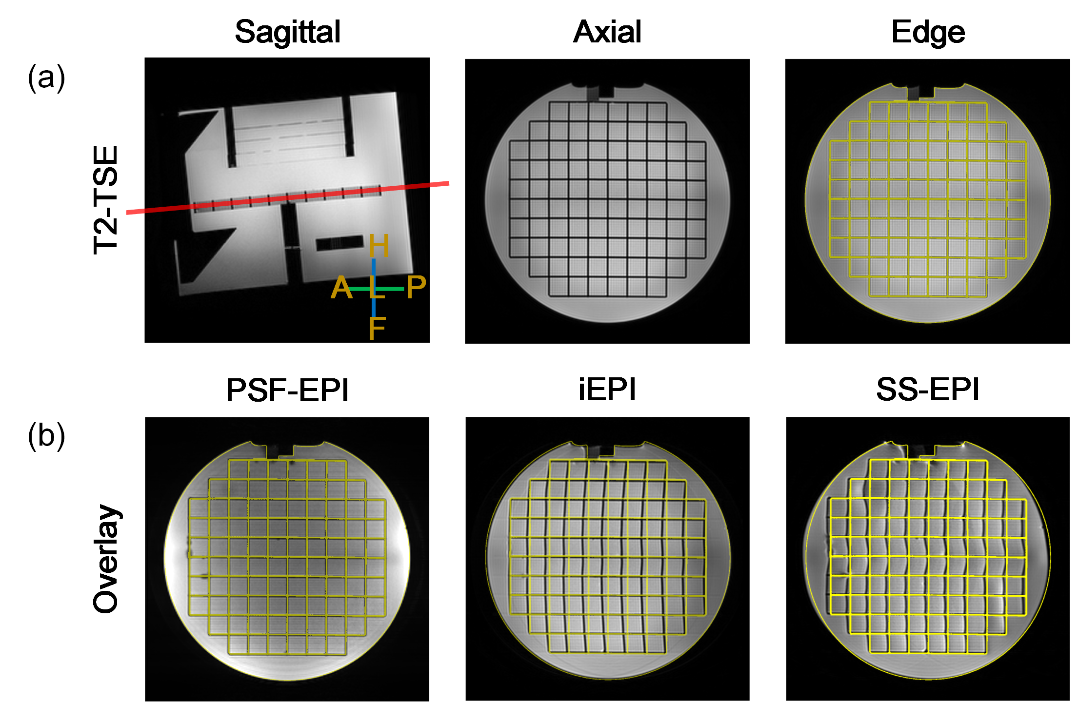
Non-diffusion-weighted images of an ACR phantom were acquired to evaluate PSF-EPI in terms of geometric distortion. Because there are often angles between the optic nerves and the axial plane, the slices were tilted in order to imitate in-vivo condition and validate the robustness of PSF-EPI.
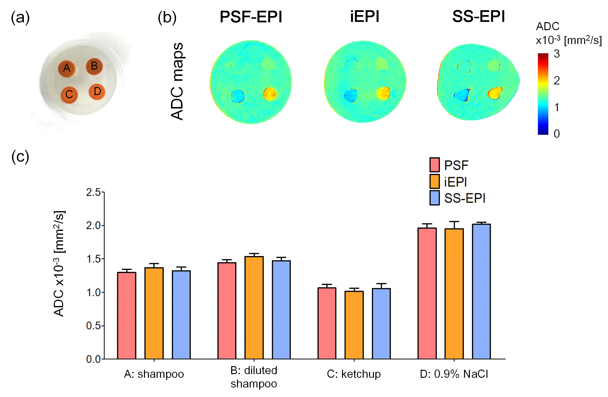
For ADC quantification, a cylindrical phantom with different compartments was used. It contained four polyethylene tubes filled with shampoo, diluted shampoo, ketchup and 0.9% NaCl solution 15. The ADC values were analyzed by placing regions of interest (ROI) on the ADC maps.
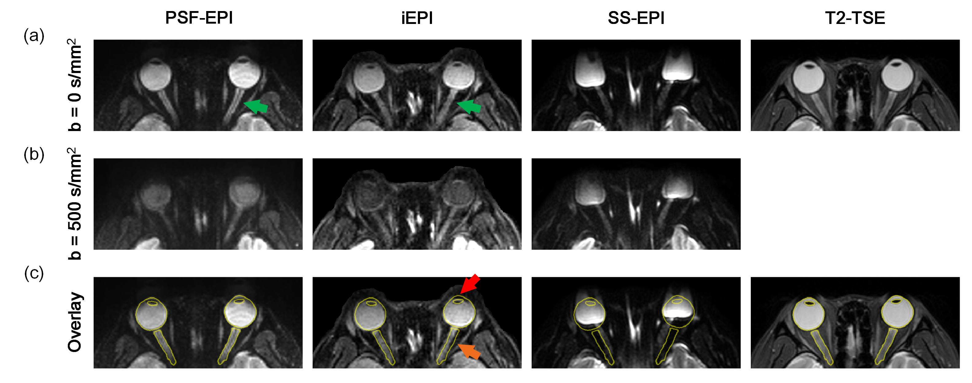
In the acquisition of in-vivo images, one healthy subject was asked to conduct an eye fixation task to reduce eye motion. Slice orientation was determined according to the location of the eyeballs and the optic nerves. ADC values of vitreous humor were calculated via ROI analysis.
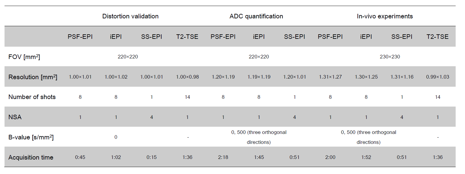
For comparison, SS-EPI and iEPI were also applied in both phantom and in-vivo experiments. PSF-EPI technique used in this study was combined with tilted-CAIPI reconstruction for acquisition acceleration and self-navigation 14. Therefore, the distortion reduction ability of PSF-EPI and iEPI can be compared, using the similar acquisition time and the same numbers of shots. T2 weighted turbo spin echo (T2-TSE) images were acquired as distortion-free reference. All of the acquisition parameters are listed in Table 1.
All images in this study were acquired on a 3.0T Philips Achieva TX scanner (Philips Healthcare, Best, The Netherlands), using a 32-channel head coil. This study was approved by the Institutional Review Board and written informed consent was obtained from all the participants.
Results and Discussion
The results of the phantom experiment are summarized in Figure 1. Images of SS-EPI and iEPI showed different levels of distortion, while PSF-EPI preserved high structural consistency with T2-TSE in spite of the slice orientation. PSF-EPI had better performance in distortion reduction than iEPI and SS-EPI did.
Figure 2 summarizes the results of ADC quantification experiments. The tubes in SS-EPI and iEPI were found distorted. ADCs of iEPI showed different levels of bias in four different compartments. The results of PSF-EPI accorded well with those of SS-EPI and the published results 16. PSF-EPI generated more correct ADCs than iEPI did.
The images of in-vivo experiment are summarized in Figure 3. SS-EPI suffered from severe distortion and signal loss. iEPI provided images with incompletely suppressed distortion. The eyeballs and the optic nerves in PSF-EPI matched well with the reference edge. Besides, the contrast of the optic nerves and the fluid in the optic sheath was better in PSF-EPI. PSF-EPI outperformed iEPI and SS-EPI in reducing distortion of the eyeballs and the optic nerves. It could also maintain their structural integrity, using the similar acquisition time to that of iEPI.
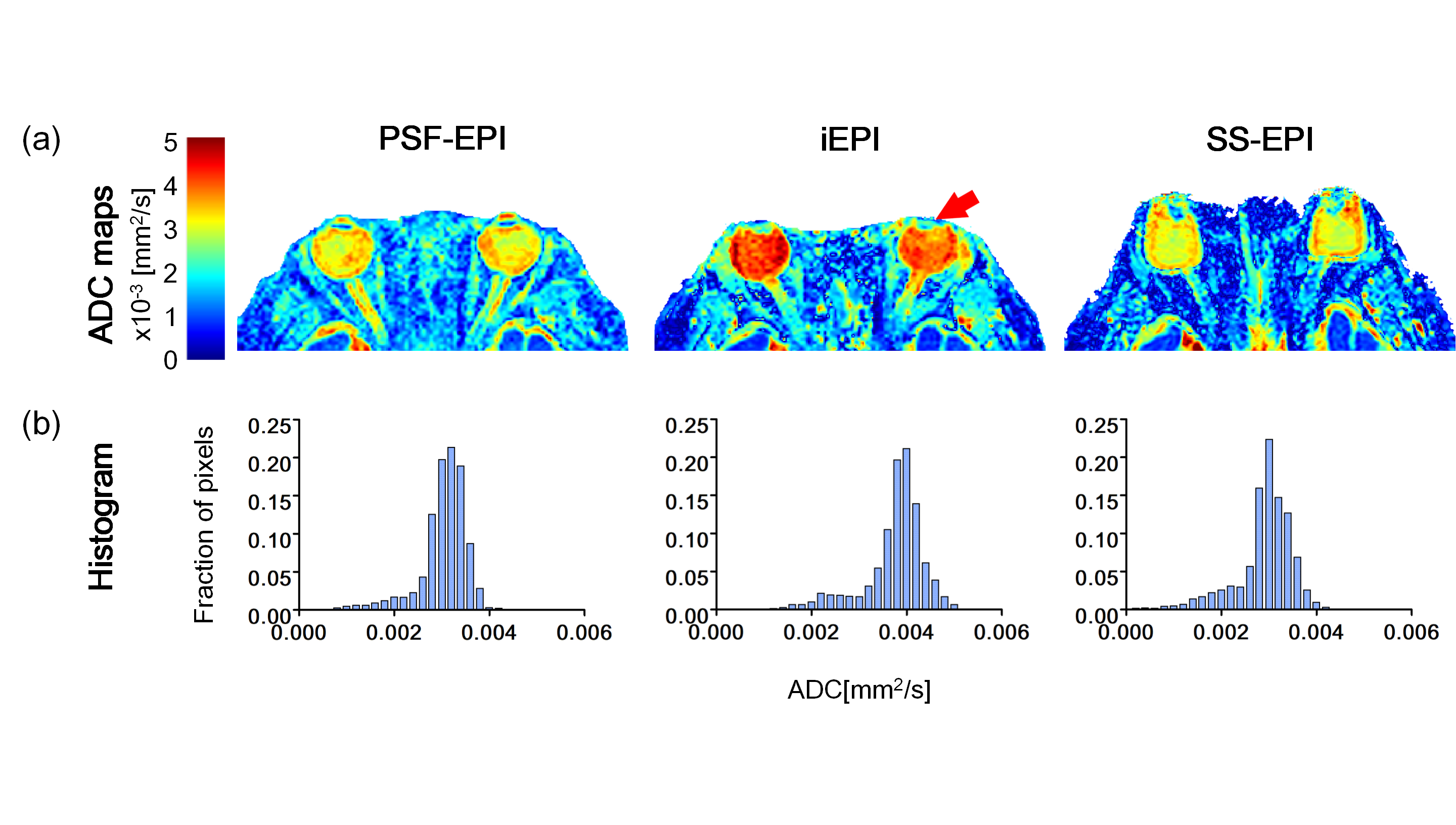
Figure 4 shows the ADC maps of in-vivo experiment. The ADC maps exposed different levels of signal loss of iEPI and SS-EPI. iEPI also introduced bias of ADC values. PSF-EPI not only maintained structural integrity but also generated ADC values in agreement with the results in previous work 17-19. Therefore, PSF-EPI could provide accurate ADC values on both phantom and healthy subject.
Conclusion
This study demonstrates the ability of PSF-EPI to achieve distortion-free DWI of the eyeballs and even the optic nerves with high efficiency. This technique is able to provide accurate, both structural and diffusion-weighted information of intraocular and extraocular masses. It has the potential to provide more accurate guidance for diagnosis of ophthalmological disease. Furthermore, it also has great potential to help characterize diseases that are difficult to distinguish by traditional diffusion-weighted EPI and structural imaging.Acknowledgements
No acknowledgement found.References
1. Kapur R, Sepahdari AR, Mafee MF, et al. MR Imaging of Orbital Inflammatory Syndrome, Orbital Cellulitis, and Orbital Lymphoid Lesions: The Role of Diffusion-Weighted Imaging. 2009;30(1):64-70.
2. Sepahdari AR, Aakalu VK, Setabutr P, Shiehmorteza M, Naheedy JH, Mafee MF. Indeterminate Orbital Masses: Restricted Diffusion at MR Imaging with Echo-planar Diffusion-weighted Imaging Predicts Malignancy. 2010;256(2):554-564.
3. Sepahdari AR, Kapur R, Aakalu VK, Villablanca JP, Mafee MF. Diffusion-Weighted Imaging of Malignant Ocular Masses: Initial Results and Directions for Further Study. 2012;33(2):314-319.
4. Khurana A, Eisenhut CA, Wan W, et al. Comparison of the diagnostic value of MR imaging and ophthalmoscopy for the staging of retinoblastoma. 2013;23(5):1271-1280.
5. Fatima Z, Motosugi U, Muhi A, Hori M, Ishigame K, Araki T. Diffusion-Weighted Imaging in Optic Neuritis. Canadian Association of Radiologists Journal 2013;64(1):51-55.
6. Xu X, Wang Y, Hu H, et al. Readout-segmented echo-planar diffusion-weighted imaging in the assessment of orbital tumors: comparison with conventional single-shot echo-planar imaging in image quality and diagnostic performance. 2017;58(12):1457-1467.
7. Wang Y, Ma X, Zhang Z, et al. A comparison of readout segmented EPI and interleaved EPI in high-resolution diffusion weighted imaging. Magnetic Resonance Imaging 2018;47:39-47.
8. In M-H, Posnansky O, Speck O. PSF mapping-based correction of eddy-current-induced distortions in diffusion-weighted echo-planar imaging. 2016;75(5):2055-2063.
9. In M-H, Posnansky O, Speck O. High-resolution distortion-free diffusion imaging using hybrid spin-warp and echo-planar PSF-encoding approach. NeuroImage 2017;148:20-30.
10. Oh S-H, Chung J-Y, In M-H, et al. Distortion correction in EPI at ultra-high-field MRI using PSF mapping with optimal combination of shift detection dimension. 2012;68(4):1239-1246.
11. Robson MD, Gore JC, Constable RT. Measurement of the point spread function in MRI using constant time imaging. 1997;38(5):733-740.
12. Zaitsev M, Hennig J, Speck O. Point spread function mapping with parallel imaging techniques and high acceleration factors: Fast, robust, and flexible method for echo-planar imaging distortion correction. 2004;52(5):1156-1166.
13. Zeng H, Constable RT. Image distortion correction in EPI: Comparison of field mapping with point spread function mapping. 2002;48(1):137-146.
14. Dong Z, Wang F, Reese TG, et al. Tilted-CAIPI for highly accelerated distortion-free EPI with point spread function (PSF) encoding. Magnetic Resonance in Medicine 2018;0(0).
15. In M-H, Posnansky O, Beall EB, Lowe MJ, Speck O. Distortion Correction in EPI Using an Extended PSF Method with a Reversed Phase Gradient Approach. PLOS ONE 2015;10(2):e0116320.
16. Kıvrak AS, Paksoy Y, Erol C, Koplay M, Özbek S, Kara F. Comparison of apparent diffusion coefficient values among different MRI platforms: a multicenter phantom study. Diagnostic and interventional radiology 2013;19 6:433-437.
17. Erb-Eigner K, Willerding G, Taupitz M, Hamm B, Asbach P. Diffusion-Weighted Imaging of Ocular Melanoma. 2013;48(10):702-707.
18. Meral İ, Bilgili Y. Diffusion Changes in the Vitreous Humor of the Eye during Aging. 2011;32(8):1563-1566.
19. Paul K, Huelnhagen T, Oberacker E, et al. Multiband diffusion-weighted MRI of the eye and orbit free of geometric distortions using a RARE-EPI hybrid. 2018;31(3):e3872.
Figures