0341
The Relationship between Glutamate and BOLD signal changes During Face-Name Paired-Associates Encoding and Retrieval Memory Task in Healthy Adults concerning age, performance level and genetic risk-A combined 1H-MRS and fMRI study1Department of Diagnostic Radiology, The University of Hong Kong, Hong Kong, Hong Kong, 2State Key Laboratory of Brain and Cognitive Sciences, The University of Hong Kong, Hong Kong, Hong Kong, 3Department of Educational Psychology, Chinese University of Hong Kong, Hong Kong, Hong Kong, 4Department of Social Work and Administration, The University of Hong Kong, Hong Kong, Hong Kong, 5Philips Healthcare, Hong Kong, Hong Kong, 6Alzheimer's Disease Research Network, The University of Hong Kong, Hong Kong, Hong Kong
Synopsis
Glutamate is hypothesized to be the neurotransmitter in mediating BOLD fMRI. In this study, fMRI technique was combined with Magnetic Resonance Spectroscopy(MRS) to investigate the relationship between glutamate and the BOLD signal changes during face-name memory task. Three different task-based fMRI face name memory experiments were performed respectively focusing on age difference, performance level and genetic risk. [Glx]abs in left hippocampus of elderly(Experiment1), high-performance(Experiment2) and low genetic risk(Experiment3) showed high correlation with BOLD signal changes in activated regions. On the whole, glutamate appears to be excitatory and lead to compensatory excitations.
Introduction
Glutamate as an excitatory neurotransmitter participates in various processes for memory. The receptors of glutamate are distributed on pre- and post-synaptic positions to help with the neuronal communication and signal processing. In our former study, ageing as a primary risk factor for Alzheimer’s disease (AD) has positive influence on the rising of glutamate concentration.1 However, there was a controversy related to the phenomenon: beneficial for compensation mechanism or toxic as the excessive release? To clarify this point, fMRI technique was combined with Magnetic Resonance Spectroscopy(MRS) to discover the relationship between neurotransmitter glutamate and BOLD signal changes during face name memory task2. Not only focusing on age-related difference, two related conditions(performance level and genetic risk) were also included in the data analysis. The aim of this study is to discover the role of glutamate in neuronal activations during memory task, namely, young versus elderly, high-performance versus low-performance, high genetic risk versus low genetic risk.Method
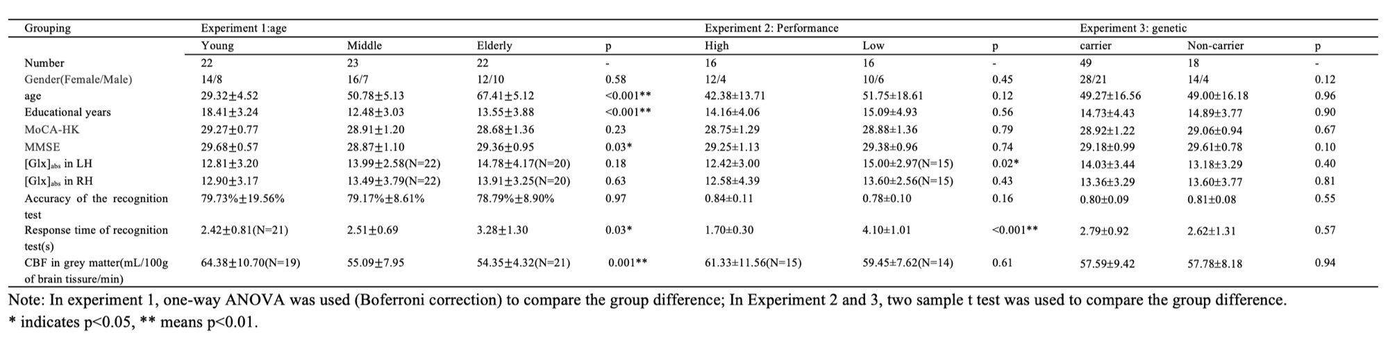
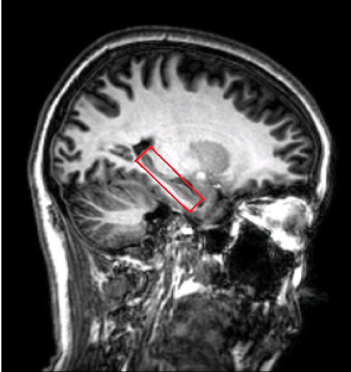
In total 67 healthy, cognitively normal subjects (age: from 20 to 84, meanSD: 49.2±16.3 years old, gender:43F/25M) were studied. All subjects underwent an MRI examination with a Phlips-3T MR scanner using a standard 8 channel head coil. Structural images were acquired with 3D fast field echo sequence (3D-T1-FFE sagittal, TR=7ms, TE=3.2ms, Flip angle=8, voxel size=111, FOV=256). Single Voxel Spectroscopy(SVS) was performed on bilateral hippocampus with the following parameters (TR/TE=2,000/39 ms, number of signals averaged=128, phase cycles=16, spectral width=2,000 Hz with spectral resolution of 1.95 Hz per point, and free induction decay=1024). Absolute concentration of Glx ([Glx]abs) (summation of glutamate and glutamine) was measured and quantified using internal water as reference by QUEST in jMRUI (4.0) with cerebrospinal fluid, grey matter and white matter water content corrected. Voxels were placed on the high-resolution structural images acquired before MRS. The position of voxels with size of 2.5×1.5×1 cm3 was shown in Figure1.
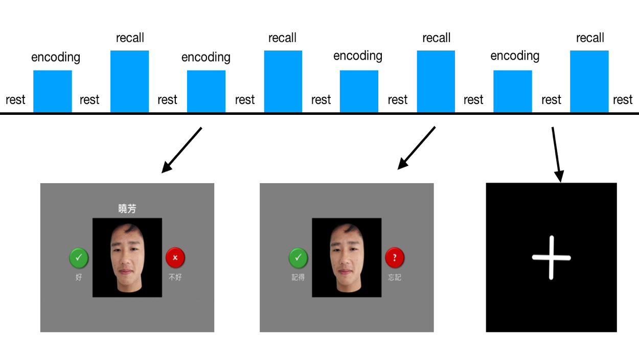
Three different task-based fMRI face name memory experiments were performed. Experiment1: three different age groups with young subjects aged between 20 to 40, middle-age between 40 to 60 and elderly aged over 60; Experiment2: low performance versus high performance(according to behavioral test, low: below 25th percentile, high: top 25th percentile); Experiment3: low genetic risk versus high genetic risk(non-ApoE4 carriers vs. ApoE4 carriers).(Table1) fMRI images were collected by using a gradient-echo echo-planar sequence (parameters: TR=2000ms, TE=30ms, flip angle=90, voxel size=334) sensitive to blood-oxygen-level-dependent(BOLD) contrast. During the fMRI scanning session, face-name paired-associates encoding and retrieval task was presented. This block-designed task performed at the end of the paradigm, and was adapted from a previously published paper2. (Figure 2) The processing and statistical calculations were performed using Statistical Parametric Mapping (SPM12) based on MATLAB.
Experiment 1: based on the two sample t results between different age groups, the peak t value of the activated regions which showed significant difference(False Discovery Rate correction, p<0.05, cluster size>30 voxels) was correlated with the [Glx]abs in bilateral hippocampus.(Pearson correlation method)
Experiment 2: based on the two sample t results between performance difference, the peak t value of the activated regions (Alphasim correction, p<0.01, cluster size>16 voxels) was correlated with the [Glx]abs in bilateral hippocampus.
Experiment 3: based on the activated regions obtained from experiment 1 and 2, the peak t value of the activated regions was correlated with the [Glx]abs in bilateral hippocampus.
Results
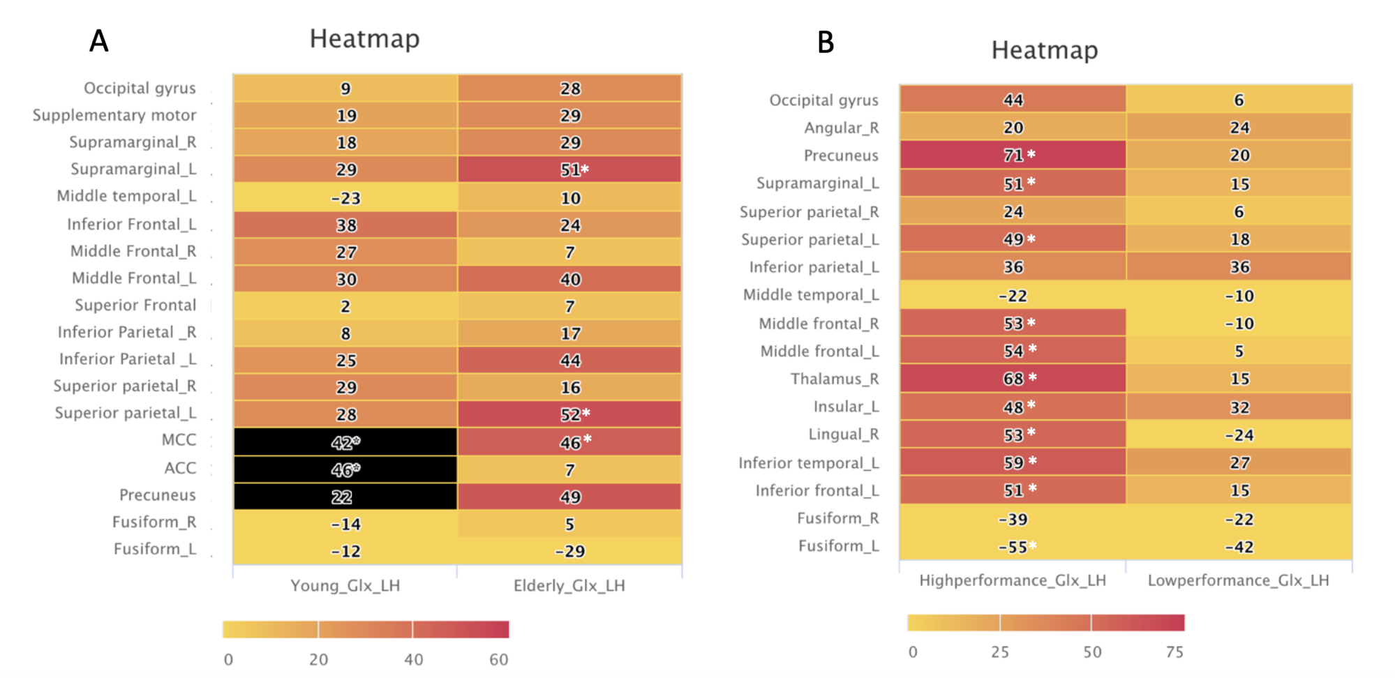
Experiment1: Age-related differences. With increased [Glx]abs in LH, the BOLD signal changes with elderly subjects in precuneus, middle cingulate cortex, left superior parietal and left supramarginal had more significant correlation with [Glx]abs than young-age group during encoding-fixation. (Fig.3A)
Experiment2: Performance-related differences. With lower [Glx]abs in the LH, high-performance group showed high correlation with BOLD signal changes in performance-related activated regions like precuneus, supramarginal and frontal gyrus(Fig. 3B). No correlation between [Glx]abs with BOLD signal changes in the low performance group.
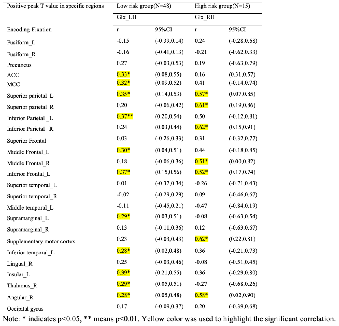
Experiment3: Genetic-risk difference. With slightly higher [Glx]abs, low-risk group showed significant positive correlation with in LH, but we found higher correlation coefficients with [Glx]abs in RH in the high risk group(slightly higher [Glx]abs level than LH).(Regions were shown in Table2)
Discussion and conclusion:
Many former works had already demonstrated that compensatory mechanisms existed in elderly subjects and ApoE4 carriers.3 In current study, we found compensatory activations occurred in the elderly group which were highly correlated with glutamate. Similarly, ApoE4 carriers might compensate for their risks, i.e. with activations being correlated with glutamate in the RH. However, low performance group could not compensate since no significant correlation between BOLD signal changes with [Glx]abs was found. To conclude, glutamate as an excitatory neurotransmitter facilitates neuronal activations in a compensatory manner.Acknowledgements
This work was supported by the Hong Kong Research Grant Council (HKU17108315 to Henry Mak).References
1. H Zhang, PW Chiu , SWH Wong, et al. The Relationship between Glutamate and BOLD signal changes During Face-Name Paired-Associates Encoding and Retrieval Task in Healthy Adults - A combined 1H-MRS and fMRI study. ISMRM. 2018
2. Putcha D, O'Keefe K, LaViolette P, et al. Reliability of functional magnetic resonance imaging associative encoding memory paradigms in non-demented elderly adults. Hum Brain Mapp. 2011;32:2027-2044
3. Bondi MW, Houston WS, Eyler LT, et al. Fmri evidence of compensatory mechanisms in older adults at genetic risk for alzheimer disease. Neurology. 2005;64:501-508
Figures