0339
Reduced functional connectivity of resting state networks in the healthy brain is associated with R2*changes consistent with myelin breakdown1Invicro, London, United Kingdom, 2High Field MR Centre, Medical University of Vienna, Vienna, Austria, 3AGE Research Unit, Imperial College London, London, United Kingdom
Synopsis
Functional connectivity of select resting state networks has been shown to diminish with age. Reported observations of reductions in visual and salience network strength appear to be supported by findings of vulnerability of the associated parietal, occipital, and frontal lobes to structural changes in the healthy brain.
We present data suggesting that measures of R2* in parietal and frontal lobar regions are correlated to visual and salience network connectivity in healthy individuals. These observations may indicate that early breakdown in myelin, associated with R2* shortening in white matter, may be responsible for age related decline in these resting state networks.
Purpose
An age related decline in functional connectivity has been reported previously in visual and salience resting state networks(RSNs) using rs-fMRI[1][2]. Furthermore, associated parietal, occipital, and frontal lobar regions of the brain have been shown to be particularly vulnerable to structural changes[3]. Myelin plays a crucial part in transmission of neuronal signalling, which is impacted by healthy aging as well as in a number of brain disorders. Recently, reductions in R2* in the absence of changes to magnetic susceptibility estimates (using QSM), were attributed to the breakdown of intact myelin[4]. In this work, we compare R2* and QSM estimates in select brain regions with RSN connectivity to probe the relationship between network function and white matter damage in the healthy brain.Methods
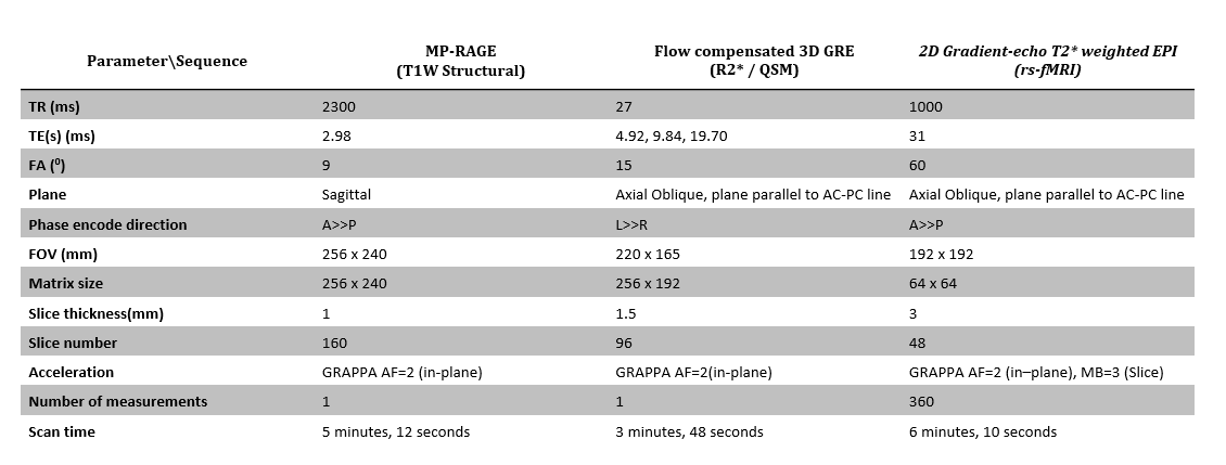
Data were acquired at 3T (Siemens, TrioTim) as part of a multi-parametric MRI brain protocol (MIND MAPS) using a 32-channel head coil in nine healthy volunteers (male and female, ages 46-75yrs). Imaging data for T1W structural MRI (sMRI), rs-fMRI, and R2*/QSM were acquired using the pulse sequences detailed in Figure 1. The DICOM data was converted into a Brain Imaging Data Structure[5] compliant format prior to further processing.
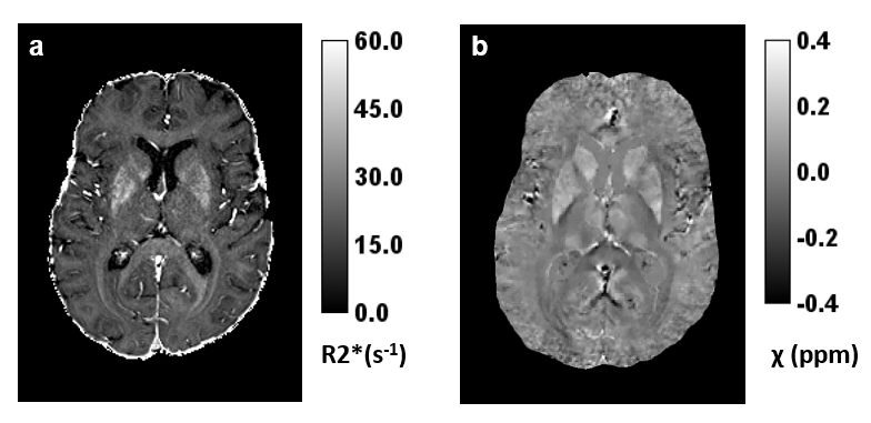
To generate R2* and Quantitative Susceptibility maps, phase data collected for each head coil element was combined using a Matlab implementation of the ASPIRE[6] method. MEDI toolbox software (Cornell MRI Research Lab) was then used to perform phase unwrapping, removal of background field contributions and calculations of R2* and magnetic susceptibility (Figure 2).
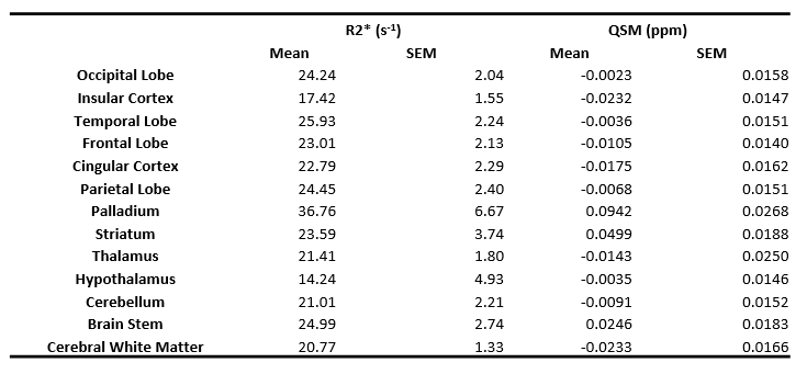
Anatomical atlas[7] labels were transformed into the subject sMRI space using parameters from a nonlinear registration of the sMRI data to the atlas (DARTEL, SPM12). The labels were then transformed a second time from subject sMRI space onto the R2*/QSM subject images using parameters from a rigid-body registration(SPM8) of sMRI data to R2* mapping data. Mean regional estimates of R2* and magnetic susceptibility were calculated using these labels in brain regions listed in the table in Figure 3.
Pre-processing of the rs-fMRI data and registration to a T1-weighted image was performed using fMRIprep [8]. Pre-processing included brain tissue segmentation and spatial normalisation for the T1-weighted image and slice timing correction for the functional images. Then, a group independent component analysis (ICA) was carried out (MELODIC ICA, FSL), using a multi-session temporal concatenation and 50 output components. Fourteen of the networks were identified as non-noise and individualised versions of these were produced for each subject using a dual-regression analysis[9]. Mean regression coefficients (β) were then extracted for each map, and each individual, providing measures of mean network strength/connectivity.
Pearson correlations were then used to investigate relationships between RSN ICA β values and mean regional estimates of R2* and Magnetic Susceptibility.
Results
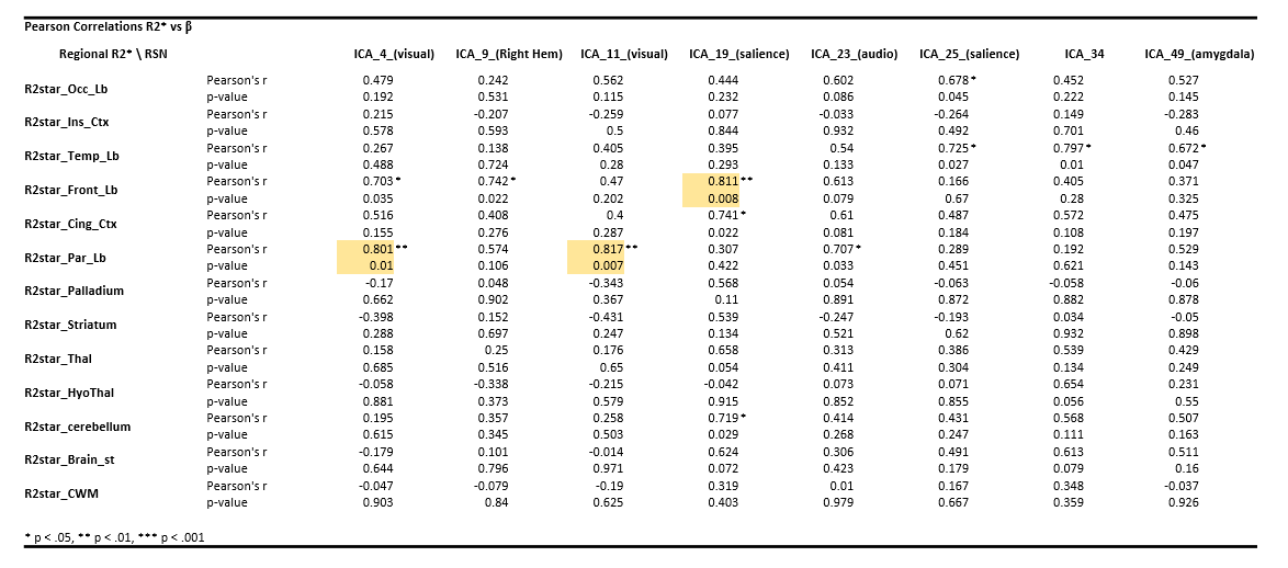
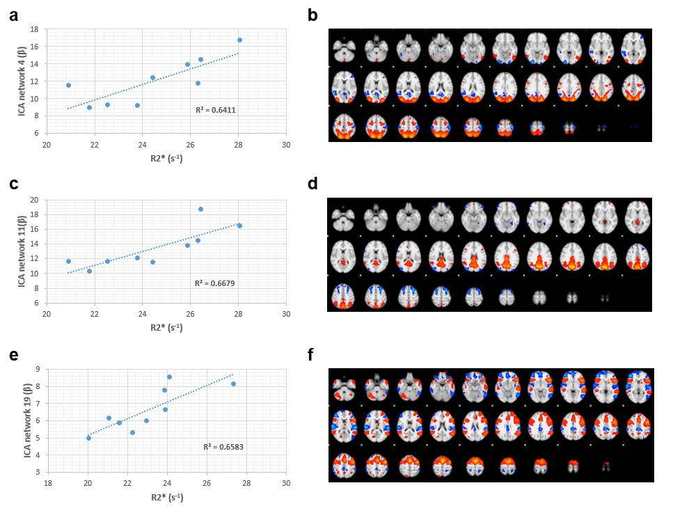
Regional estimates of R2* and Magnetic Susceptibility are presented in the table in Figure 3. Correlations between regression coefficient β and regional R2* estimates were observed for eight RSNs (Figure 4). Three correlations achieved p values of 0.01 or less with a positive increase in R2* with β (plotted in Figure 5). These included two RSNs labelled by MELODIC ICA as visual networks (network 4(p =0.01 ,r=0.80) and network 11(p=0.007,r=0.82)) that correlated with R2* in the Parietal Lobe (Figure 4, a-d). Additionally, increasing connectivity in ICA network 19, labelled as salience, correlated with increasing R2* in the Frontal Lobe(p=0.008,r=0.81) (Figure 5e,f). There were no significant correlations observed between QSM estimates and β for any RSNs tested in the ICA analysis.Discussion and Conclusions
Observations of R2* shortening accompanied by a lack changes in QSM measurements have been shown to reflect early breakdown of myelin prior to later stages of removal of myelin debris and iron accumulation in the development of white matter lesions in Multiple Sclerosis [4]. In this work, a relationship of reduced R2* in the parietal and frontal lobes was observed with reduced β in visual and salience RSNs respectively. The lack of correlations with QSM estimates suggest weakening of RSN connectivity is driven by early white matter breakdown in the frontal and parietal lobes – both known to be vulnerable to aging effects and neurodegenerative diseases. Further work will involve inclusion of more subjects and MRI data (ASL, DTI, and sMRI volumetrics) from the MIND MAPS project to lend support to these initial findings.Acknowledgements
The MIND
MAPS consortium.
References
[1] V. La Corte et al., “Cognitive Decline and Reorganization of Functional Connectivity in Healthy Aging: The Pivotal Role of the Salience Network in the Prediction of Age and Cognitive Performances,” Front. Aging Neurosci., vol. 8, p. 204, Aug. 2016.
[2] R. F. Betzel, L. Byrge, Y. He, J. Goñi, X.-N. Zuo, and O. Sporns, “Changes in structural and functional connectivity among resting-state networks across the human lifespan,” Neuroimage, vol. 102, pp. 345–357, 2014.
[3] S. M. Resnick et al., “One-year Age Changes in MRI Brain Volumes in Older Adults,” Cereb. Cortex, vol. 10, no. 5, pp. 464–472, May 2000.
[4] Y. Zhang et al., “Quantitative Susceptibility Mapping and R2* Measured Changes during White Matter Lesion Development in Multiple Sclerosis: Myelin Breakdown, Myelin Debris Degradation and Removal, and Iron Accumulation,” AJNR. Am. J. Neuroradiol., vol. 37, no. 9, pp. 1629–1635, Sep. 2016.
[5] K. J. Gorgolewski et al., “The brain imaging data structure, a format for organizing and describing outputs of neuroimaging experiments,” Sci. Data, vol. 3, p. 160044, Jun. 2016.
[6] K. Eckstein et al., “Computationally Efficient Combination of Multi-channel Phase Data From Multi-echo Acquisitions (ASPIRE),” Magn. Reson. Med., vol. 79, no. 6, pp. 2996–3006, Oct. 2017.
[7] A. C. Tziortzi et al., “Imaging dopamine receptors in humans with [11C]-(+)-PHNO: Dissection of D3 signal and anatomy,” Neuroimage, vol. 54, no. 1, pp. 264–277, 2011.
[8] O. Esteban et al., “FMRIPrep: a robust preprocessing pipeline for functional MRI,” bioRxiv, Jan. 2018.
[9] L. D. Nickerson, S. M. Smith, D. Öngür, and C. F. Beckmann, “Using Dual Regression to Investigate Network Shape and Amplitude in Functional Connectivity Analyses ,” Frontiers in Neuroscience , vol. 11. p. 115, 2017.
Figures