0336
Hippocampal NAA/mIns ratio and spatial working memory across the lifespan1Danish Research Center for Magnetic Resonance, Copenhagen University Hospital Hvidovre, Copenhagen, Denmark, 2Department of Clinical Medicine, Faculty of Health and Medical Sciences, University of Copenhagen, Copenhagen, Denmark, 3Department of Neurology, Copenhagen University Hospital Bispebjerg, Copenhagen, Denmark, 4Center for Magnetic Resonance, Department of Electrical Engineering, Technical University of Denmark, Lyngby, Denmark
Synopsis
The NAA/mIns ratio has been proposed as a marker of unhealthy ageing. We found that the NAA/mIns ratio is decreased in the hippocampus for middle-aged and old individuals compared to young individuals. For old individuals, declining NAA/mIns ratios are correlated with poorer performance on a spatial working memory task but this relationship is not found in middle-aged and young individuals. Our results suggest that the NAA/mIns ratio decrease in the hippocampus is observable before the negative functional effects.
Introduction
The hippocampus is a key age-sensitive brain region, affecting cognitive functions such as spatial working memory1. The N-acetyl aspartate (NAA) /myo-inositol (mIns) ratio has been proposed as a possible biomarker for unhealthy ageing but this has never been investigated in the hippocampus2. The ratio reflects the relationship in the brain between neuronal (NAA) and glial (mIns) integrity and lower levels are suggestive of negative biological developments such as neuroinflammation3. We hypothesised that the NAA/mIns ratio will be lower in the hippocampus with higher age. We further expected this ratio to correlate negatively with spatial working memory.
Methods
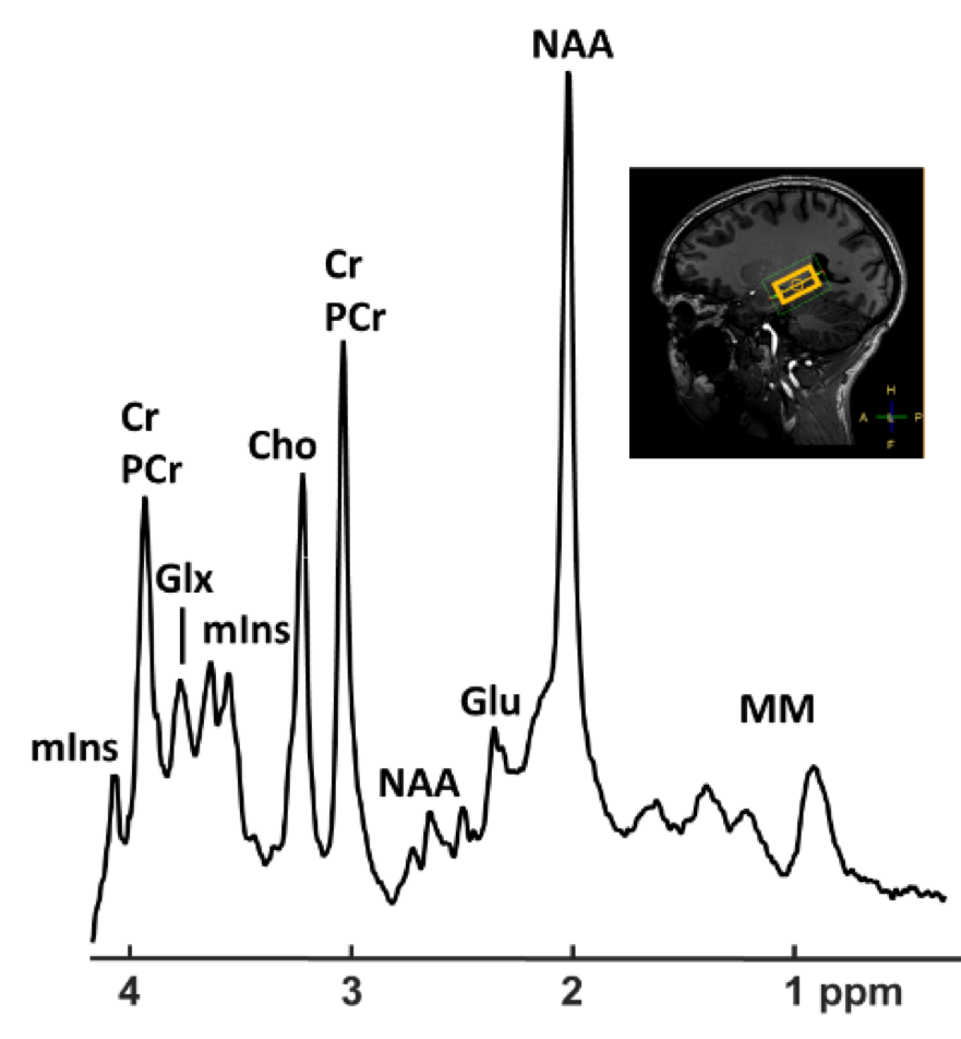
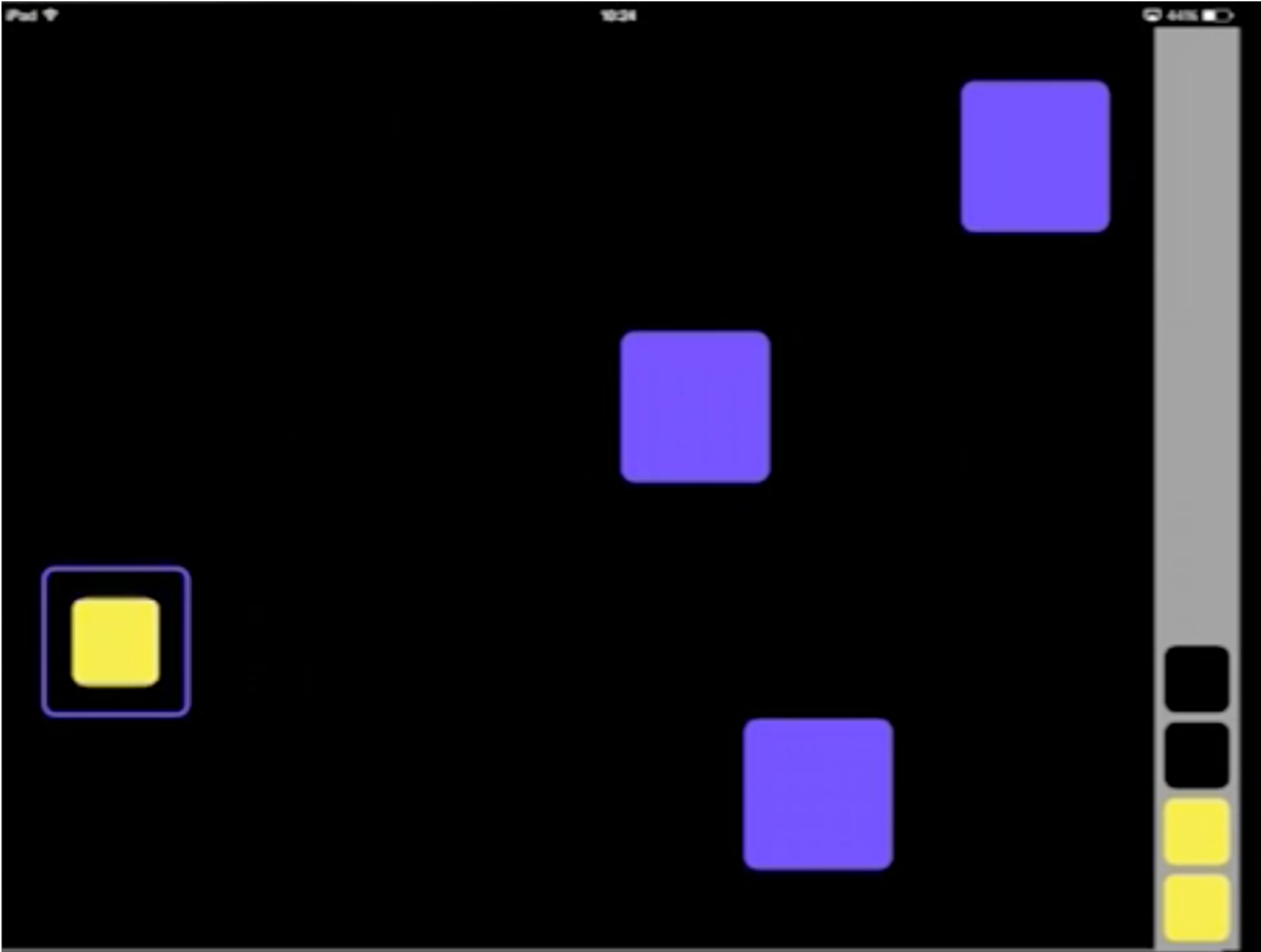
For this ongoing study, 60 healthy volunteers are being recruited in three age groups: young (18-26 years), middle-aged (39-50 years) and old (69-80 years). NAA and mIns have been measured using proton magnetic resonance spectroscopy (1H-MRS). A 7 tesla whole-body MR scanner (Philips, Best, The Netherlands) was used in combination with a dual transmit coil and a 32-channel receive head coil (Nova Medical, Wilmington, MA, USA). To assess metabolite levels in the left hippocampus, a sLASER sequence was used (30x15x15 mm3, TR/TE=3700/32 ms, 64 averages) (Fig.1). Prior to MRS, an anatomical image was acquired for placement and segmentation of the MRS voxel (MPRAGE, 380 slices, slice thickness = 0.5 mm, TR=8 ms, TE=3.2 ms, FOV=256x256x190 mm, voxel size=1x1x0.5mm). Segmentation was done with the SPM4 CAT tool (The Structural Brain Mapping Group, University of Jena, Jena, Germany). 1H-MRS was analyzed with LCModel5 using a custom basis set with 20 metabolites and macromolecules. Metabolite concentrations were corrected for partial volume effects6. All participants underwent cognitive testing using CANTAB, including a spatial working memory (SWM) task (Cambridge Cognition, 2018) (Fig.2). Statistics were performed in Matlab (MATLAB and Statistics Toolbox Release 2018a, The MathWorks, Inc., Natick, MA, USA) where significance was assessed with one-way ANOVA followed by Tukey-Kramer posthoc tests (p<0.05) and correlation was measured using the Pearson correlation coefficient. Experiments were performed according to the local ethical protocols. Data acquisition is expected to be finished ultimo November 2018.Results
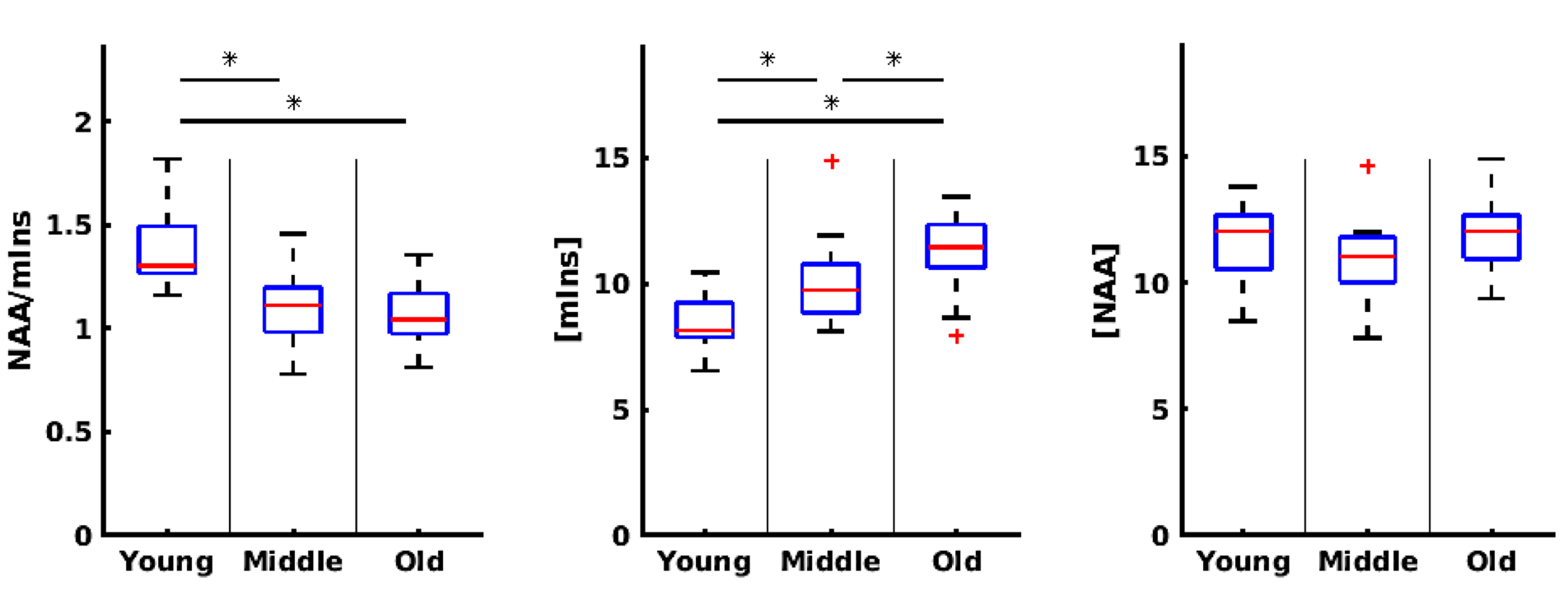
The preliminary analyses were based on data from 19 young (10 females), 20 middle-aged (10 females) and 19 old (9 females) participants. All measured spectra were of sufficient quality to be included and all CRLBs were below 16. the NAA/mIns ratio significantly differed between groups (F(2,55)=19.77, p<3*10-7) and the posthoc test showed that NAA/mIns ratio was lower in both the old (m=1.07, SD=0.04) and the middle-aged (m=1.11, SD=0.04) individuals as compared to the young individuals (m=1.38, SD=0.04) (Fig. 3). The concentration of mIns was also significantly different across groups (F(2,55)=18.65, p=6*10-7) with higher concentrations in old individuals (m=11.2, SD=0.32) as compared to middle-aged (m=9.98, SD=0.31) and young individuals (m=8.43, SD=0.32); mIns concentrations were also significantly increased in middle-aged individuals as compared to young individuals. No significant between group difference was observed for NAA (F(2,55)=2.38, p=0.1).
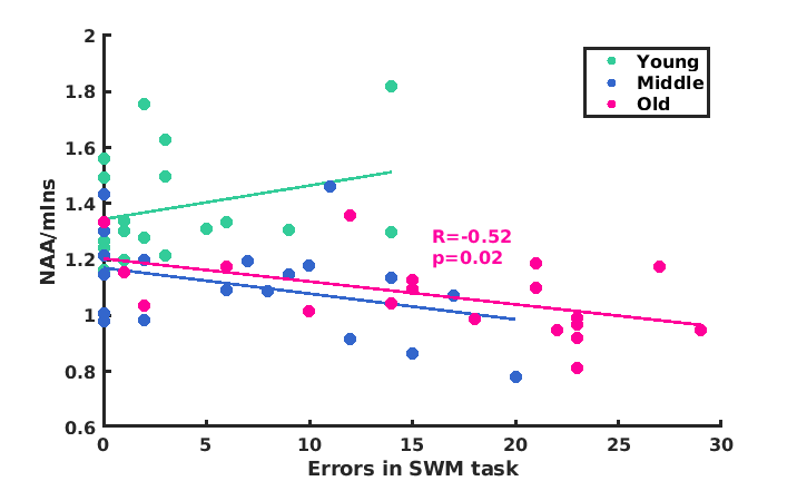
The number of errors made in SWM was used as the outcome score. For old individuals, a negative correlation was observed between the NAA/mIns ratio and the SWM error score (p=0.021, R=-0.52) (Fig. 4). No correlations between the NAA/mIns ratio and the SWM error score were observed for the other groups (p>0.05).
Discussion
We found that a decrease in the NAA/mIns ratio was detectable between both the young and middle-aged individuals and the young and old individuals. The process causing this shift may therefore already start before the age of 40. Notably, only for the old individuals did the NAA/mIns ratio correlate with SWM performance. The functional meaning of the lower hippocampal NAA/mIns ratio is, thus, observed only at higher ages. Based on these findings, it could be very interesting for future studies to investigate if the hippocampal NAA/mIns ratio measured in middle-age could be a biomarker of hippocampus related cognitive decline later in life.Conclusion
Overall, the NAA/mIns ratio in the hippocampus was decreased in middle-aged and old individuals as compared to young individuals. Only in old individuals, did a lower level of NAA/mIns correlate with worse performance on a spatial working memory task.Acknowledgements
The work was supported by The Danish Agency for Science, Technology and Innovation grant no. 0601-01370B, and The John and Birthe Meyer Foundation.References
1. Valenzuela, M. J. et al.Posterior compensatory network in cognitively intact elders with hippocampal atrophy. Hippocampus25,581–593 (2015).
2. Waragai, M., Moriya, M. & Nojo, T. Decreased N-Acetyl Aspartate/Myo-Inositol Ratio in the Posterior Cingulate Cortex Shown by Magnetic Resonance Spectroscopy May Be One of the Risk Markers of Preclinical Alzheimer’s Disease: A 7-Year Follow-Up Study. J. Alzheimer’s Dis.60,1411–1427 (2017).
3. Chang, L., Munsaka, S. M., Kraft-Terry, S. & Ernst, T. Magnetic resonance spectroscopy to assess neuroinflammation and neuropathic pain. J. Neuroimmune Pharmacol.8,576–593 (2013).
4. Ashburner, J. SPM: A history. Neuroimage62,791–800 (2012).
5. Provencher, S. W. Automatic quantitation of localized in vivo 1H spectra with LCModel. NMR Biomed.14,260–64 (2001).
6. Gasparovic, C. et al.Neurometabolite Concentrations in Gray and White Matter in Mild Traumatic Brain Injury: An 1H–Magnetic Resonance Spectroscopy Study. J. Neurotrauma26,1635–1643 (2009).
Figures