0332
Age-Related Microstructural Alterations in Human Corpus Callosum Measured by High-Gradient Diffusion MRI1Radiology, Massachusetts General Hospital, Charlestown, MA, United States, 2Harvard Medical School, Boston, MA, United States, 3Neurology, Massachusetts General Hospital, Charlestown, MA, United States, 4Harvard-MIT Division of Health Sciences and Technology, Massachusetts Institute of Technology, Cambridge, MA, United States
Synopsis
Cerebral white matter exhibits degenerative changes during normal aging. Noninvasive approaches to measure these microstructural alterations would be invaluable for understanding the substrate and regional variability of age-related white matter degenerations. Recent advances in diffusion MRI have leveraged high gradient strengths to increase sensitivity toward axonal size and density in living human brains. Here, we examined the relationship between age and microstructural properties measured using high-gradient diffusion MRI. We observed an increase in apparent axon diameter and decrease in density with advancing age in the corpus callosum, with changes most pronounced in the genu and relatively absent in the splenium.
Introduction
Alterations in fiber composition within the corpus callosum interfere with the efficiency of interhemispheric transfer in older adults and likely contribute to cognitive aging.1,2 On histology, an increase in the number of large myelinated callosal fibers has been observed with increasing age,3 with less myelinated fibers in the genu found to be particularly susceptible to the deleterious effects of aging4,5. These trends have been corroborated on numerous neuroimaging studies6-13. DTI offers useful insight into the microstructural properties of white matter but is not specific to axonal and myelin integrity. Noninvasive approaches to estimate axon diameter and density in the living human brain would be invaluable for understanding the microstructural substrate of age-related white matter changes.
In recent years, a number of advanced diffusion MRI techniques for inferring axon diameter and packing density have become more readily translated to studying white matter structure in the living human brain, largely through the availability of higher gradient strengths on human MRI scanners.14,15 The goal of this study is to explore age-related differences in apparent axon diameter and density estimated using high-gradient diffusion MRI in the corpus callosum.
Methods
Participants A total of 36 healthy, cognitively normal adults (aged 22-72, 23F) participated in this study.
Data Acquisition Imaging data were acquired on the 3T Connectome scanner equipped with 300 mT/m maximum gradient strength14,16,17 using a custom-made 64-channel phased array head coil18 for signal reception. Sagittal 2-mm isotropic resolution diffusion-weighted spin-echo EPI images were acquired with whole brain coverage. The following parameters were used: TR/TE = 4000/77ms, δ=8ms, Δ=19/49ms, 8 diffusion gradient strengths linearly spaced from 30-290mT/m per Δ, 32-64 diffusion directions, parallel imaging (R=2) and simultaneous multislice (MB=2). Five b=0 images with reversed phase encoding direction were acquired for distortion correction.
Data Analysis Diffusion data were corrected for gradient nonlinearity17, motion, susceptibility and eddy current distortions using the TOPUP and EDDY tools in FSL19-21. A previously validated method22 was employed for the voxel-wise fitting for axon diameter, restricted and hindered volume fraction, and hindered diffusivity using Markov-Chain Monte-Carlo (MCMC) sampling. Corpus callosum masks were created from FreeSurfer labels and manually edited to ensure exclusion of voxels outside the corpus callosum (e.g., fornix and CSF). The corpus callosum was further divided into five sub-sections, which were derived from evenly spaced partitions along the primary eigenaxis using FreeSurfer’s automatic labeling23. Correlation analyses were performed between age and the ROI-averaged axonal metrics.
Results
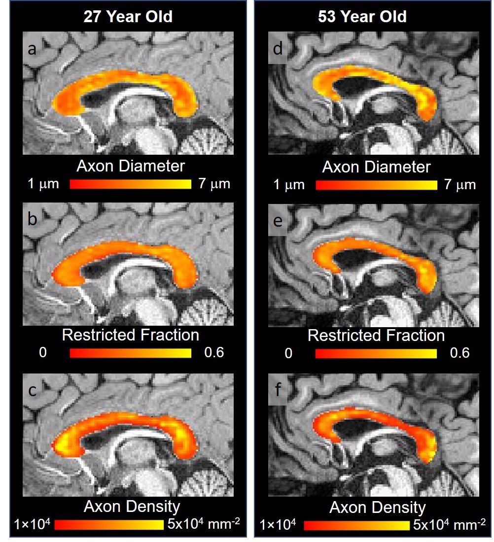
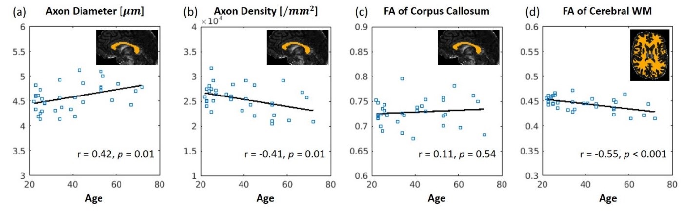
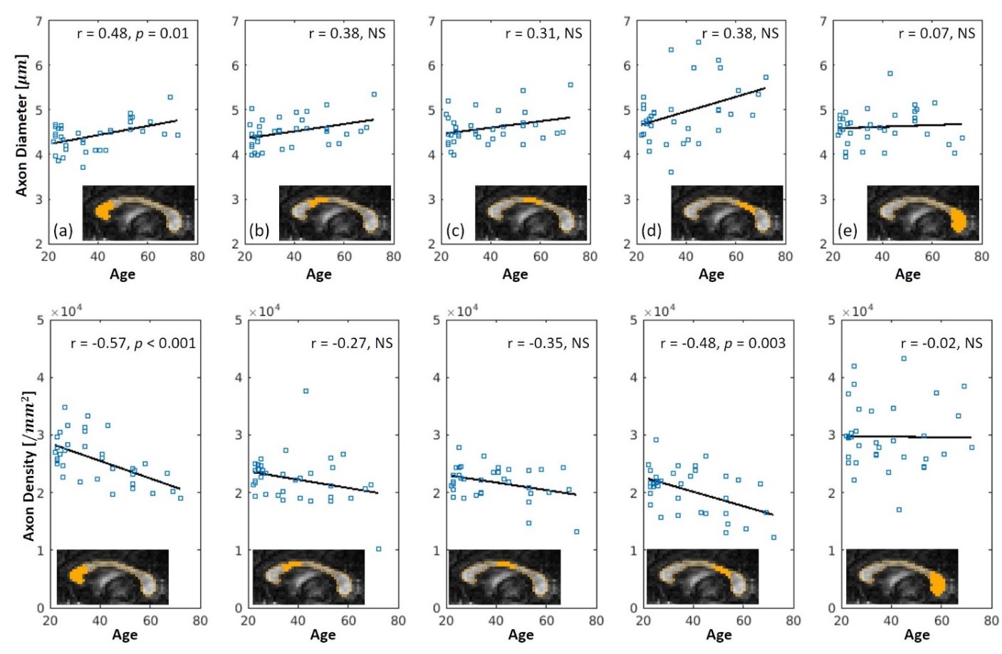
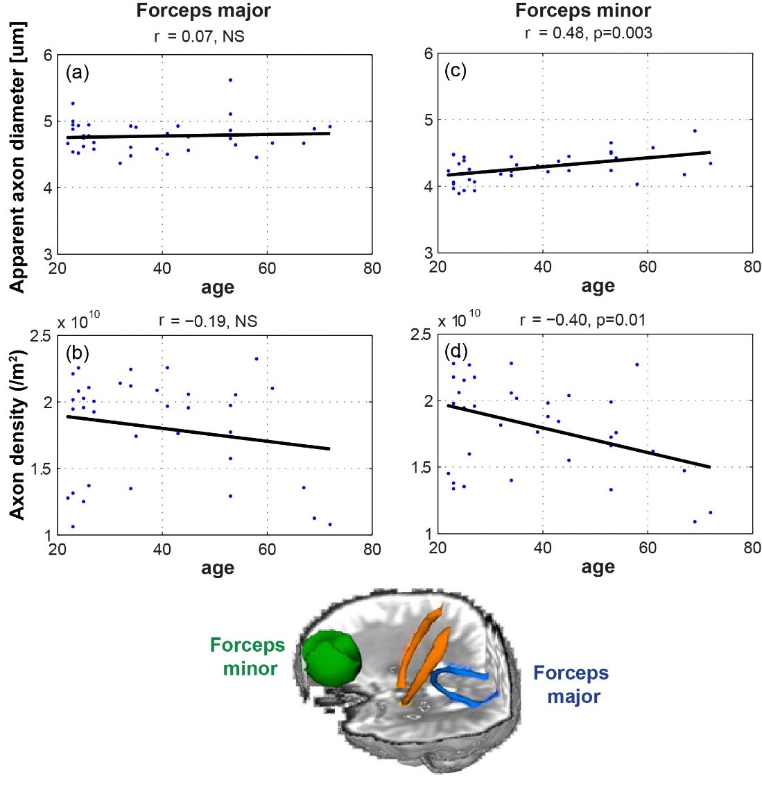
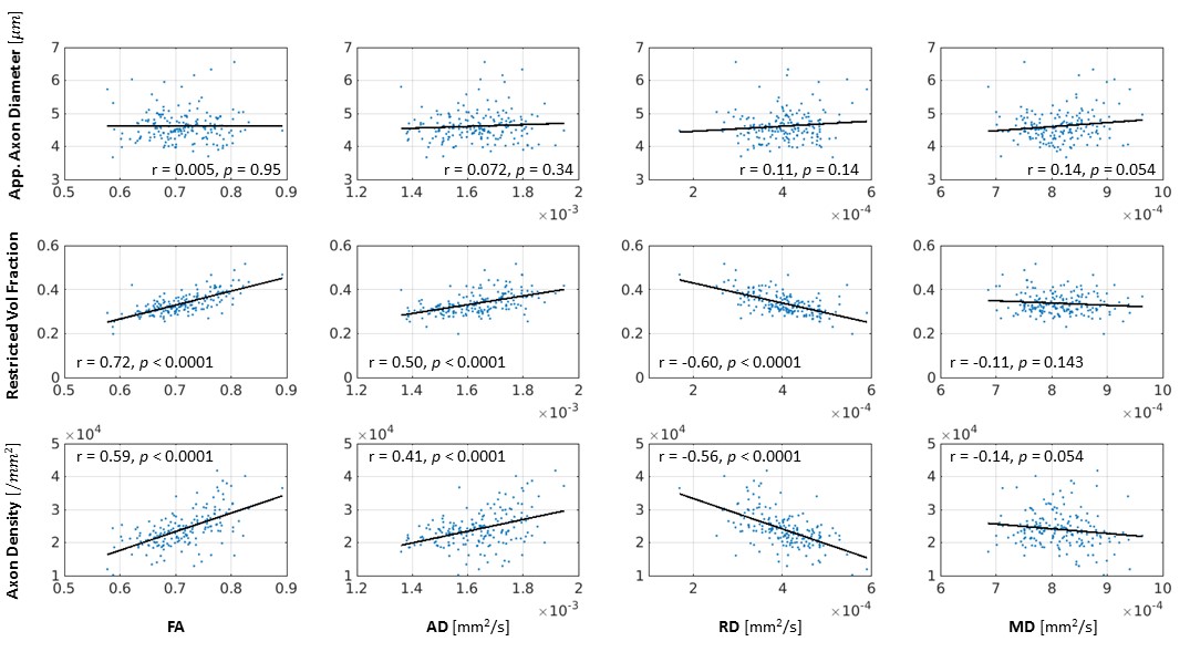
Figure 1 shows representative maps of apparent axon diameter, restricted volume fraction and axon density in younger and older adult participants. Apparent axon diameter and axon density were significantly correlated with age in the whole corpus callosum (Figure 2a,b). While the previously reported correlations between FA and age were replicated in the cerebral white matter (Figure 2d), no significant correlation was observed for the corpus callosum (Figure 2c). Similar analyses in sub-regions of the corpus callosum showed the strongest age-related effects in the axonal metrics in the genu of the corpus callosum (Figure 3). Increased apparent axon diameter and decreased axon density were found in the forceps minor but not in the forceps major (Figure 4). The relationship between the axonal microstructural metrics and more widely studied DTI metrics related to aging are shown in Figure 5.Discussion
We observed regionally selective, age-related microstructural axonal differences in the corpus callosum and adjacent white matter tracts estimated from high-gradient diffusion MRI. A global increase in apparent axon diameter and decrease in axon density was seen throughout the corpus callosum with increasing age, with the effect being most pronounced in the genu of the corpus callosum. The findings were mirrored by similar trends in the adjacent forceps minor and forceps major.
Our results support the hypothesis that select fiber bundles are preferentially affected by aging, and that these trends follow a regional distribution that reflects the selective vulnerability of certain anterior fiber bundles to age-related degeneration. More importantly, the axonal imaging metrics provide unique and complimentary regional markers of microstructural changes relative to DTI. This approach offers a more specific microstructural interpretation of the axonal changes underpinning the previously noted age-related differences in FA within anterior versus posterior fiber bundles, suggesting that the underlying substrate of age-related degeneration may relate to fiber size and packing density.
Acknowledgements
This work was funded by a National Institutes of Health Blueprint for Neuroscience Research Grant U01MH093765, as well as National Institutes of Health funding from the National Center for Research Resources P41EB015896, National Institute of Biomedical Imaging and Bioengineering R01EB006847 and R00EB015445, National Institute of Neurological Disorders and Stroke K23NS096056, and K23NS078044, and Instrumentation Grants S10-RR023401, S10-RR023043, and S10-RR019307. Funding support was also received from the National Multiple Sclerosis Society, the American Heart Association Postdoctoral Fellowship Award (17POST33670452), a Radiological Society of North America Research Resident Grant (RR1427), the Conrad N. Hilton Foundation (17330) and the Massachusetts General Hospital Executive Committee on Research Fund for Medical Discovery Fellowship Award and Claflin Distinguished Scholar Award.References
1. Janowsky, J.S., Kaye, J.A. & Carper, R.A. Atrophy of the corpus callosum in Alzheimer's disease versus healthy aging. J Am Geriatr Soc 44, 798-803 (1996).
2. Jeeves, M.A. & Moes, P. Interhemispheric transfer time differences related to aging and gender. Neuropsychologia 34, 627-636 (1996).
3. Aboitiz, F., Rodriguez, E., Olivares, R. & Zaidel, E. Age-related changes in fibre composition of the human corpus callosum: sex differences. Neuroreport 7, 1761-1764 (1996).
4. Aboitiz, F., Scheibel, A.B. & Zaidel, E. Morphometry of the Sylvian fissure and the corpus callosum, with emphasis on sex differences. Brain 115 ( Pt 5), 1521-1541 (1992).
5. Kemper, T.L. Neuroanatomical and neuropathological changes during aging and dementia. in Clinical neurology of aging, 2nd ed. 3-67 (Oxford University Press, New York, NY, US, 1994).
6. Salat, D.H., et al. Age-related changes in prefrontal white matter measured by diffusion tensor imaging. Ann N Y Acad Sci 1064, 37-49 (2005).
7. Davis, S.W., et al. Assessing the effects of age on long white matter tracts using diffusion tensor tractography. Neuroimage 46, 530-541 (2009).
8. Pfefferbaum, A., Adalsteinsson, E. & Sullivan, E.V. Frontal circuitry degradation marks healthy adult aging: Evidence from diffusion tensor imaging. Neuroimage 26, 891-899 (2005).
9. Pfefferbaum, A., et al. Age-related decline in brain white matter anisotropy measured with spatially corrected echo-planar diffusion tensor imaging. Magn Reson Med 44, 259-268 (2000).
10. Lebel, C., Caverhill-Godkewitsch, S. & Beaulieu, C. Age-related regional variations of the corpus callosum identified by diffusion tensor tractography. Neuroimage 52, 20-31 (2010).
11. Sullivan, E.V. & Pfefferbaum, A. Diffusion tensor imaging and aging. Neurosci Biobehav Rev 30, 749-761 (2006).
12. Sullivan, E.V., Adalsteinsson, E. & Pfefferbaum, A. Selective age-related degradation of anterior callosal fiber bundles quantified in vivo with fiber tracking. Cereb Cortex 16, 1030-1039 (2006).
13. Bennett, I.J. & Madden, D.J. Disconnected aging: cerebral white matter integrity and age-related differences in cognition. Neuroscience 276, 187-205 (2014).
14. McNab, J.A., et al. The Human Connectome Project and beyond: initial applications of 300 mT/m gradients. NeuroImage 80, 234-245 (2013).
15. Huang, S.Y., et al. The impact of gradient strength on in vivo diffusion MRI estimates of axon diameter. Neuroimage 106, 464-472 (2015).
16. Setsompop, K., et al. Pushing the limits of in vivo diffusion MRI for the Human Connectome Project. Neuroimage 80, 220-233 (2013).
17. Fan, Q., et al. MGH-USC Human Connectome Project datasets with ultra-high b-value diffusion MRI. NeuroImage 124, 1108-1114 (2016).
18. Keil, B., et al. A 64-channel 3T array coil for accelerated brain MRI. Magn Reson Med 70, 248-258 (2013).
19. Andersson, J.L., Skare, S. & Ashburner, J. How to correct susceptibility distortions in spin-echo echo-planar images: application to diffusion tensor imaging. Neuroimage
20, 870-888 (2003). 20. Andersson, J.L.R. & Sotiropoulos, S.N. An integrated approach to correction for off-resonance effects and subject movement in diffusion MR imaging. NeuroImage 125, 1063-1078 (2016).
21. Andersson, J.L.R., Graham, M.S., Zsoldos, E. & Sotiropoulos, S.N. Incorporating outlier detection and replacement into a non-parametric framework for movement and distortion correction of diffusion MR images. Neuroimage 141, 556-572 (2016).
22. Fan, Q., et al. Validation of diffusion MRI estimates of compartment size and volume fraction in a biomimetic brain phantom using a human MRI scanner with 300mT/m maximum gradient strength. Neuroimage (2018).
23. Fischl, B., et al. Whole brain segmentation: automated labeling of neuroanatomical structures in the human brain. Neuron 33, 341-355 (2002).
Figures