0330
Age-related microstructural and physiological changes in normal brain assessed via anomalous diffusion derived γ, DTI, DKI and NODDI metrics1SAIMLAL, Sapienza University of Rome, Rome, Italy, 2Institute for Complex Systems, CNR, Rome, Italy, 3Department of Computer Science & Centre for Medical Image Computing, University College London, London, United Kingdom, 4Laboratory for Structural, Physiologic and Functional Imaging, Perelman School of Medicine University of Pennsylvania, Philadelphia, PA, United States, 5ImpAct Team, Neuroscience Research Center, Lyon, France, 6Neuroimaging Laboratory, Santa Lucia Foundation, Rome, Italy
Synopsis
In this study we used γ-metrics, derived from anomalous diffusion signal representation, as well as DTI, NODDI, DKI derived parameters to assess physiological (i.e. the iron content) and microstructural (myelin damage, axonal disintegration, neuron cell loss) modifications in cerebral WM and scGM of middle- and older-aged subjects. We found that γ-metrics are remarkably sensitive and provide complementary information compared to DTI-metrics, MK and NODDI to detect modifications in frontal WM, where substantial changes are expected with aging. Also, the combined use of these techniques may unravel different patterns of modifications of the ageing brain.
Introduction
Nowadays, the expanding longevity coupled with declining cerebral nervous system functions, suggests the need for continued development of new imaging contrast mechanisms to assist in the differential diagnosis of age-related declines. Recently, it has been developed a new imaging contrast metrics derived from anomalous diffusion signal representation and obtained from diffusion-weighted (DW) data collected by varying diffusion gradient strengths1-4. It has been highlighted that the new metrics, named γ-metrics, depend on the local inhomogeneities due to magnetic susceptibility differences between tissues and diffusion compartments, thus providing information about myelin orientation and iron content within cerebral regions5. The major structural modifications occurring in brain aging are myelinated fibers damage in nerve fibers and iron accumulation in gray matter nuclei6,7. In this work, we investigated the potential of γ-metrics in relation to other conventional diffusion metrics such as DTI, DKI and NODDI in detecting age-related structural changes within white matter (WM) and subcortical gray matter (scGM).Materials and Methods
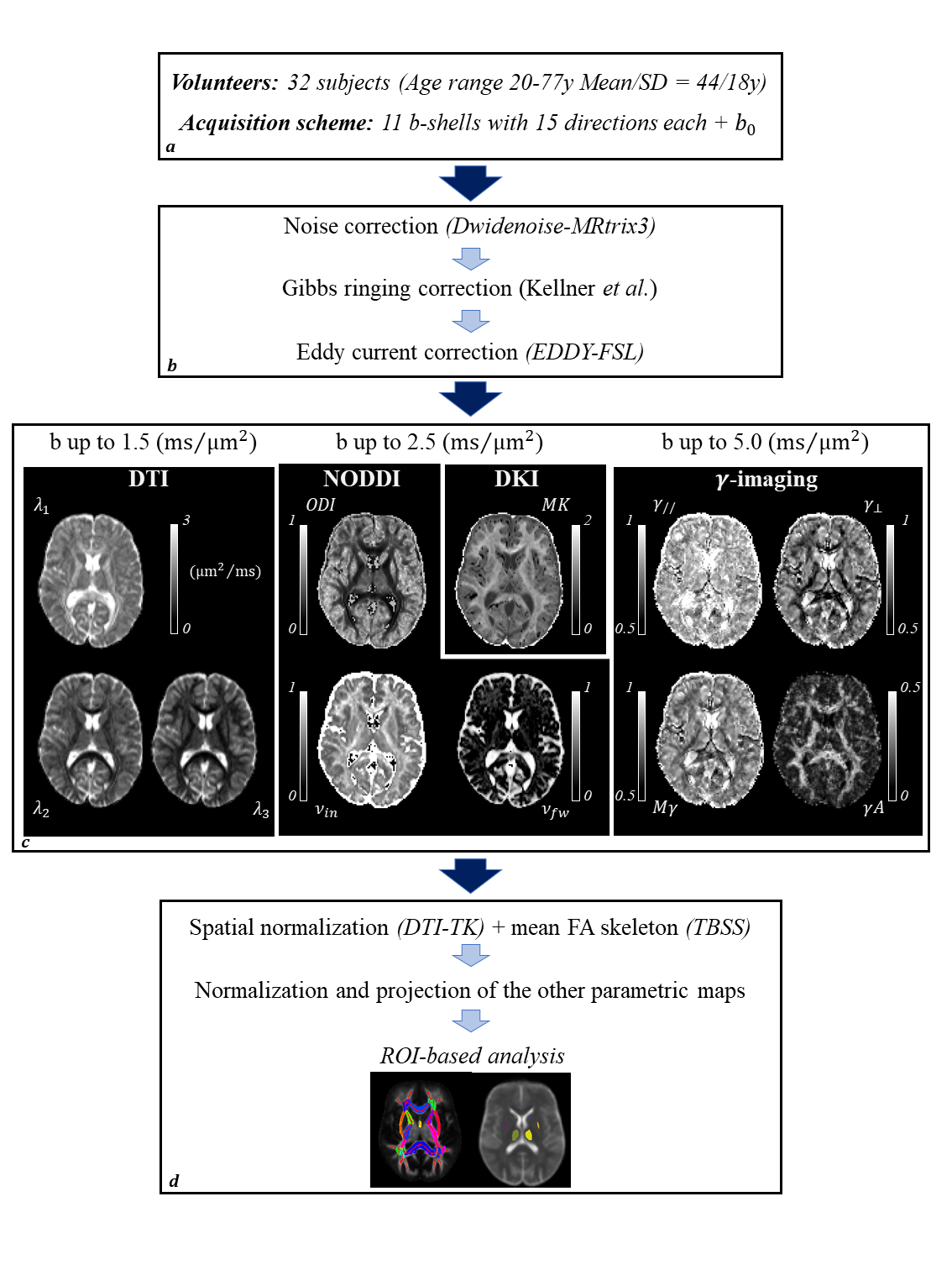
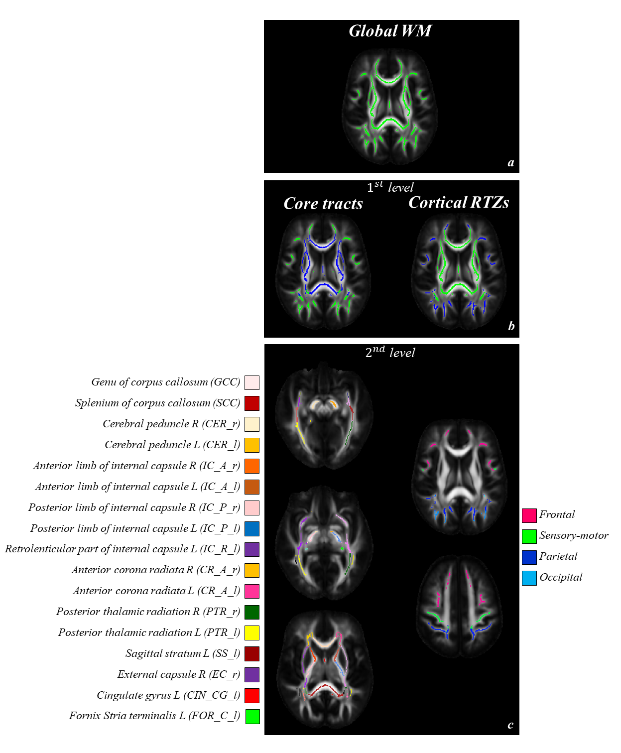
DW-images in 32 healthy subjects (19/13 men/women, age range 20-77y, Mean+/-SD=43.7+/-18.2y) were acquired at 3.0T with 12 b-shells up to 5000s/mm2 and 15 directions each. Figure.1 summarizes the pipeline: the images were pre-processed correcting for noise effects8, Gibbs ringing artifacts9, and eddy currents and subjects’ movements distortions10. Different subsets of the pre-processed data were used to compute DTI, DKI, NOODI and γ-imaging diffusion metrics. All the b-shells up to 1500s/mm2 were used to fit DTI11, obtaining FA,MD,D//,D⊥ maps. We used data up to 2500s/mm2 to fit both DKI12, obtaining MK, and NODDI13, obtaining νin,νfw,ODI. All the b-shells were used to fit the signal representation showing transient anomalous pseudo-superdiffusion, thus obtaining Mγ,γA,γ//,γ⊥1-5. Associations between diffusion metrics and subjects’ age were assessed using linear regression. All the maps were registered to a population-specific. Analysis of the correlations between diffusion metrics and subjects’ age was performed on a region of interest (ROI) basis using a hierarchical approach14. This is summarized in figure.2 for WM. As regard scGM, caudate, thalamus, putamen, and pallidum were considered in the study.Results
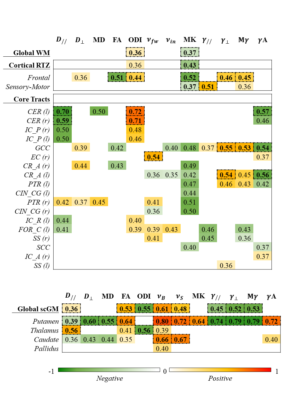
Figure.3 summarize the results. For each ROI and each diffusion metrics we reported the correlation coefficient when p<0.05. Red-yellow colors stand for positive correlations, while green colors stand for negative correlations. The correlation with a family-wise error corrected p-value (pfwe < 0.05) are highlighted in bold. On a global level ODI and MK were the only parameters showing a significant trend. The MK decrease seemed to be driven by a decrease within the cortical regional termination zones (RTZs) rather than in the core tracts. In particular, the tracts close to the frontal lobe showed the greatest number of significant differences. FA and MK decreased while ODI, γ⊥ and Mγ increased. MK decreased also in the tracts close to the sensory-motor lobe along with a parallel increase of γ//. As regard the core-tracts, the regions showing the strongest correlation were both sides of cerebral peduncle (CER). With a simultaneous decrease in D// and increase in ODI. Also, a significantly decreased γA was observed in the left side. γ-derived parameters showed a rather strong correlation within the genu of corpus callosum (GCC) and left anterior corona radiata (CR_A(l)). The increase in Mγ and the decrease in γA seemed to be driven by a decrease in γ⊥. A positive association was found in the left external capsule between νfw and age. An almost complete inversion of age-related trends was observed for all the parameters in the scGM: D⊥,MD,ODI,γ//,γ⊥,Mγ showed a decrease, while FA,νin,νfw,γA showed an increase with age. The putamen was, with no doubt the region showing the most widespread and strongest correlation with diffusion derived parameters. The thalamus showed a pattern like that found in the CER, although with inverted trends. Finally, the caudate showed a parallel increase of νin and νfw with aging.Discussion
Previous works highlighted how γ-metrics may reflect inhomogeneities due to susceptibility differences between various tissues and compartments, being potentially useful as an indirect measure of myelin integrity and iron content1-5. A decline in Mγ within the scGM and a complementary increase of γ⊥ in frontal WM, GCC and CR_A(l) with advancing age were found. We suggest that the increase of γ⊥ may reflect myelin density decline and Mγ decrease may mirror iron accumulation. An increase in D// and a decrease in ODI could be associated to axonal loss in the pyramidal tracts, while their inverted trends within the thalamus may reflect reduced architectural complexity of nerve fibers. γ-metrics together with conventional diffusion-metrics can more comprehensively characterize the complex mechanisms underlining age-related changes than conventional diffusion techniques alone.Acknowledgements
References
1. De Santis, S., Gabrielli, A., Bozzali, M., Maraviglia, B., Macaluso, E., and Capuani, S. (2011a). Anisotropic anomalous diffusion assessed in the human brain by scalar invariant indices. Magnetic resonance in medicine, 65(4):1043–1052.
2. Palombo, M., A. Gabrielli, S. De Santis, C. Cametti, G. Ruocco and S. Capuani (2011). Spatio-temporal anomalous diffusion in heterogeneous media by nuclear magnetic resonance. The Journal of chemical physics 135(3): 034504. doi:034510.031063/034501.3610367.
3. Palombo, M., A. Gabrielli, S. De Santis and S. Capuani (2012). The γ parameter of the stretched-exponential model is influenced by internal gradients: validation in phantoms. Journal of Magnetic Resonance 216: 28-36. doi:10.1016/j.jmr.2011.1012.1023.
4. Capuani, S., M. Palombo, A. Gabrielli, A. Orlandi, B. Maraviglia and F. S. Pastore (2013). Spatio-temporal anomalous diffusion imaging: results in controlled phantoms and in excised human meningiomas. Magnetic resonance imaging 31(3): 359-365. doi:310.1016/j.mri.2012.1008.1012.
5. Caporale, A., Palombo, M., Macaluso, E., Guerreri, M., Bozzali, M., and Capuani, S. (2017). The γ-parameter of anomalous diffusion quantified in human brain by mri depends on local magnetic susceptibility differences. Neuroimage, 147:619–631.
6. Peters, A. and Kemper, T. (2012). A review of the structural alterations in the cerebral hemispheres of the aging rhesus monkey. Neurobiology of aging, 33(10):2357–2372.
7. Acosta-Cabronero, J., Betts, M. J., Cardenas-Blanco, A., Yang, S., and Nestor, P. J. (2016). In vivo mri mapping of brain iron deposition across the adult lifespan. Journal of Neuroscience, 36(2):364–374.
8. Veraart, J., Novikov, D. S., Christiaens, D., Ades-Aron, B., Sijbers, J., and Fieremans, E. (2016b). Denoising of diffusion mri using random matrix theory. Neuroimage, 142:394–406.
9. Kellner, E., Dhital, B., Kiselev, V. G., and Reisert, M. (2016). Gibbs-ringing artifact removal based on local subvoxel-shifts. Magnetic resonance in medicine, 76(5):1574–1581.
10. Andersson, J. L. R. and Sotiropoulos, S. N. (2016). An integrated approach to correction for off-resonance effects and subject movement in diffusion MR imaging. NeuroImage, 125:1063-1078, 2016.
11. Basser, P. J., Mattiello, J., and LeBihan, D. (1994). Mr diffusion tensor spectroscopy and imaging. Biophysical journal, 66(1):259–267.
12. Jensen J.H., Helpern J.A., Ramani A., Lu H., Kaczynski K. (2005) Diffusional kurtosis imaging: the quantification of non-gaussian water diffusion by means of magnetic resonance imaging. Magn Reson Med. 53:1432–1440.
13. Zhang, H., Schneider, T., Wheeler-Kingshott, C. A., and Alexander, D. C. (2012). Noddi: practical in vivo neurite orientation dispersion and density imaging of the human brain. Neuroimage, 61(4):1000–1016.
14. Simmonds, D. J., Hallquist, M. N., Asato, M., and Luna, B. (2014). Developmental stages and sex differences of white matter and behavioral development through adolescence: a longitudinal diffusion tensor imaging (dti) study. Neuroimage, 92:356–368.
Figures