0315
Whole-brain 3D multi-parametric quantitative extraction at 7T using parallel transmission Universal Pulses1CEA - Neurospin, Gif-sur-Yvette, France, 2CRMBM/UMR 7339 CNRS, Aix Marseille Université, Marseille, France, 3CENIR, ICM, Paris, France, 4Inserm U 1127, CNRS UMR 7225, Sorbonne Universités, UPMC Univ Paris 06 UMR S 1127, Institut du Cerveau et de la Moelle épinière, ICM, Paris, France, 5Siemens Healthineers, Saint-Denis, France
Synopsis
Performing simultaneous quantitative MRI at ultra-high field is challenging, as B0 and B1 heterogeneities, and Specific Absorption Rate increase with field strength. In this work, Quantitative Imaging using Configuration States is successfully applied in vivo at 7T using calibration-free parallel transmission Universal Pulses to retrieve 3D whole-brain M0, flip angle, T1 and T2 maps in a clinically-relevant time. The method relies on the acquisition of multiple contrasts with spoiled SSFP sequence by varying flip angle and radiofrequency spoiling in a limited and optimized number of sets. Quantification of the physical parameters was then performed by fitting acquired data to the Bloch-Torrey equation.
Introduction
The simultaneous quantitative mapping of multiple parameters attracts considerable interest in the MR community1–5. However, existing techniques come with constraints, particularly at ultra-high field(UHF), where B0 and B1 inhomogeneities as well as Specific Absorption Rate(SAR) increase6. A method to perform Quantitative Imaging using Configuration States (QuICS) has been proposed7–9, allowing to retrieve 3D maps of magnetization(M0), flip angle(FA), T1 and T2. In this work, QuICS is applied in vivo at 7T using parallel transmission(pTx) universal pulses(UP)10 to achieve robust 3D whole-brain multi-parametric quantification over six volunteers.Materials & Methods
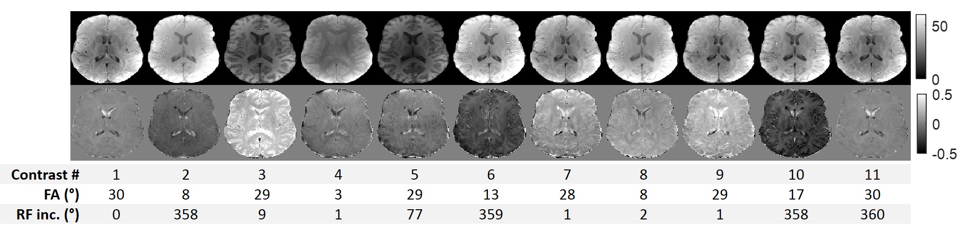
QuICS is based on a spoiled SSFP sequence, generating different contrasts by varying RF amplitude and phase cycling. To restrict the total acquisition time(TA), an optimization algorithm based on Fisher Information11 was used to select the 11 most informative contrasts, with a maximum FA set to 30° to obtain a low-SAR protocol. Resulting acquisition parameters and contrasts are shown in Figure 1.
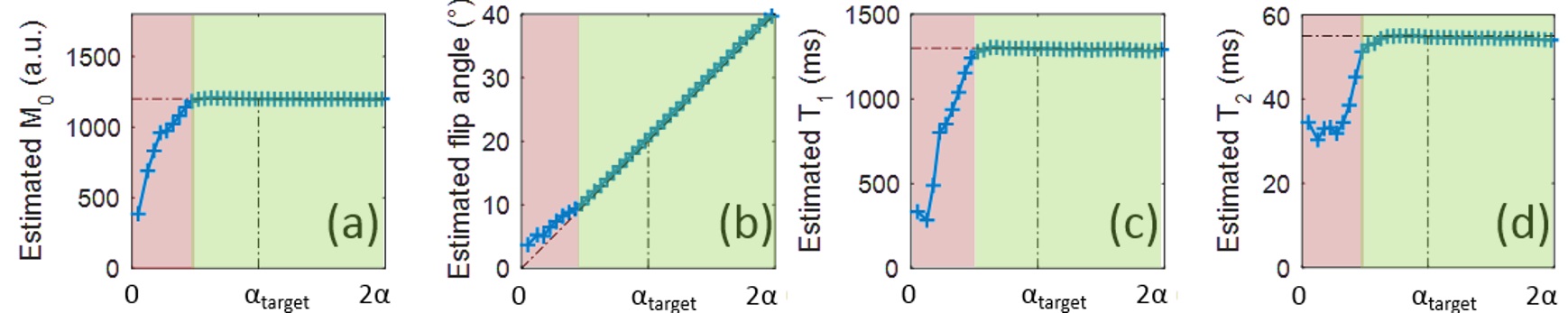
To determine the sensitivity of such acquisition protocol to FA variations encountered at UHF, in-silico Monte-Carlo simulations were performed. The setup was simulated, varying the FA from 0 to twice the targeted FA, with a precision of 1°, using 10,000 samples with randomized noise per FA respecting a fixed SNR similar to in vivo acquisitions. Results for white matter, Figure 2, show a relative immunity to effective FA down to 50% of the targeted FA. Below, NMR parameters will not be evaluated properly.
To mitigate RF inhomogeneity while incurring no time penalty for the user, we propose to employ pTx calibration-free universal pulses. In vivo acquisitions thus were performed at 7T using a 8Tx/32Rx head coil(Nova Medical, Wilmington, USA) with a kT-pointTM pulse for the UP, whose results were compared with the ones obtained in Circularly Polarized(CP) mode. The radiofrequency UP was designed offline on a database of 20 field maps from a prior study, to be robust to intersubject variability12. The duration of the UP was adapted to obtain the same mean input power of 1.8W in both acquisitions. Six healthy volunteers(3 females, age 22.6±2.7) were scanned. MRI acquisitions were performed in transverse orientation with FOV=256x160x168mm3, voxel size 1x1x3mm3, TR/TE 11ms/3.3ms, BW 650Hz/px, 500TR of dummy scan time, and a GRAPPA factor of 3. To avoid aliasing artefacts, an oversampling of 100% was added in the phase direction, leading to TA=16min36s.
The quantitative extraction was performed using Matlab(The Mathworks, Natick, USA) from DICOM images. Acquired data were fitted to the Bloch-Torrey equation to estimate voxel-wise M0, FA, T1 and T2 using a calculation approach based on configuration states formalism8,13 and Gauss-Newton non-linear least-squares11. Reference T1 and T2 maps were obtained separately using respectively a Variable FA technique corrected for incomplete spoiling and B1+ heterogeneities14, with FAs of 5 and 20°, TR/TE=14/3ms, at a 1x1x3mm3 resolution; and a multi-Spin-Echo acquisition with TE=10/30/50ms, TR=8s, resolution of 2x2x3mm3, and EPI factor of 3. The total scan time for B1 mapping, VFA and multi-spin-echo was 26 minutes.
Results
Exemplary in vivo obtained contrasts are shown in Figure 1, demonstrating the complex modulation of the signal.
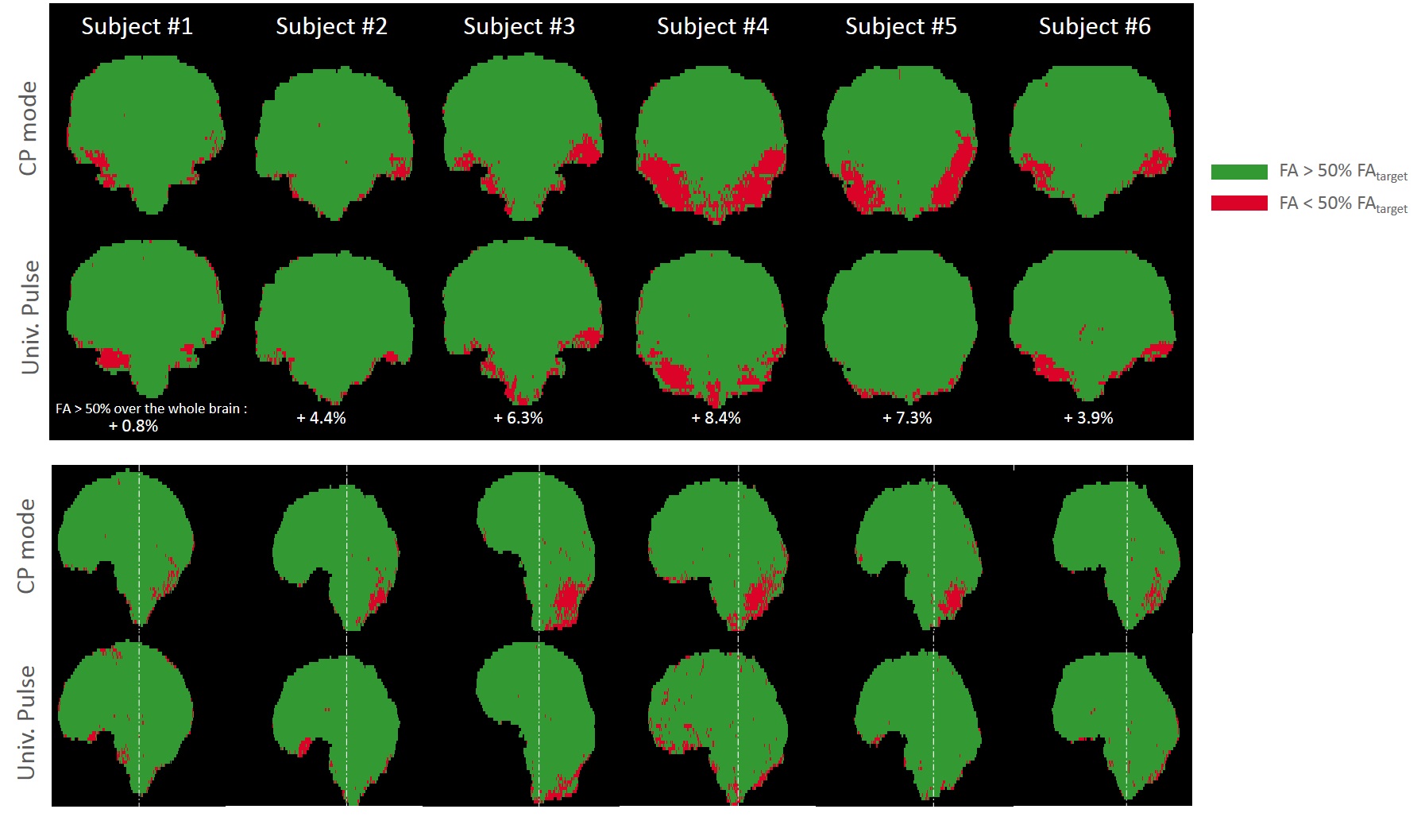
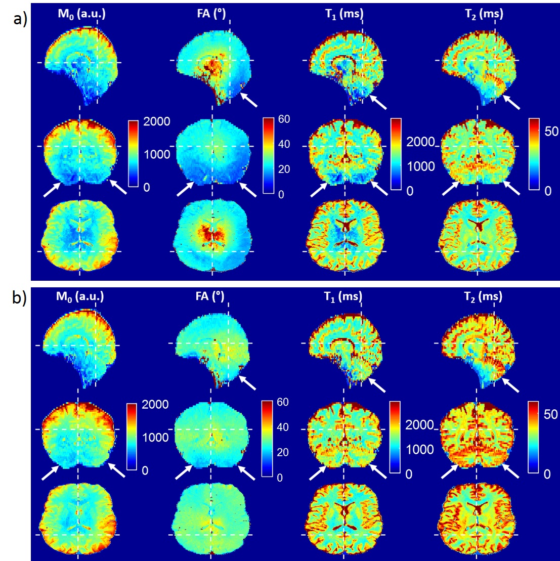
Figure 3 shows how the area where FA is lower than 50% of the target, in red, is greatly reduced(mean reduction of 54%) when applying UP over the six scanned subjects. The corresponding parametric maps, displayed in Figure 4, demonstrate a signal drop in cerebellum and temporal lobes for all the extracted parameters in CP-mode. When applying UP, a more homogeneous FA map is obtained, and quantitative results are acceptable over the whole brain.
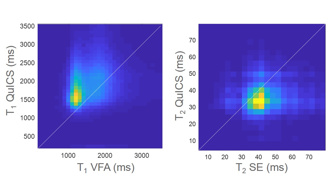
When comparing assessed parameters over the whole brain with reference methods, T1 values are higher than those obtained with VFA, as illustrated in Figure 5. This might be explained by partial volume or magnetization transfer effects, not considered in our model15,16. For T2, QuICS estimations are slightly lower than Spin-Echo values, but show an overall very good agreement.
Discussion/Conclusion
These results indicate that the use of UP improves the robustness of QuICS for in vivo relaxometry at 7Tesla, validating a low SAR quantitative protocol.
With the UP implementation, the SNR of the quantitative maps is improved compared to the CP-mode. Indeed, since the FA/RF increment schedule was chosen to maximize the Fisher information11, the proposed approach reduces biases and noise in the extraction, while reducing TA. Here, reference methods, for a longer TA, fail to achieve the same in-plane resolution of 1mm.
UP excitations could be replaced by universal slab-selective multispoke excitation12 to reduce the FOV dimension. The resulting time gain could be used to speed up the protocol or to retrieve an additional apparent diffusion coefficient map9,17.
Acknowledgements
This work received financial support from the French program ‘Investissement d’Avenir’ run by the ‘Agence Nationale pour la Recherche’, grant ‘Infrastructure d’avenir en Biologie Santé – ANR-11-INBS-0006’, from the ERPT equipment program of the Leducq Foundation, and from the European Union Horizon 2020 Research and Innovation program under Grant Agreement No. 736937.References
1. Ma D, Gulani V, Seiberlich N, Liu K, Sunshine JL, Duerk JL, Griswold MA. Magnetic resonance fingerprinting. Nature. 2013;495(7440):187–192. doi:10.1038/nature11971
2. Warntjes J b. m., Dahlqvist O, Lundberg P. Novel method for rapid, simultaneous T1, T*2, and proton density quantification. Magnetic Resonance in Medicine. 2007;57(3):528–537. doi:10.1002/mrm.21165
3. Heule R, Ganter C, Bieri O. Triple echo steady-state (TESS) relaxometry. Magnetic Resonance in Medicine. 2014;71(1):230–237. doi:10.1002/mrm.24659
4. Warntjes J b. m., Leinhard OD, West J, Lundberg P. Rapid magnetic resonance quantification on the brain: Optimization for clinical usage. Magnetic Resonance in Medicine. 2008;60(2):320–329. doi:10.1002/mrm.21635
5. Schmitt P, Griswold MA, Jakob PM, Kotas M, Gulani V, Flentje M, Haase A. Inversion recovery TrueFISP: Quantification of T1, T2, and spin density. Magnetic Resonance in Medicine. 2004;51(4):661–667. doi:10.1002/mrm.20058
6. Cloos MA, Knoll F, Zhao T, Block KT, Bruno M, Wiggins GC, Sodickson DK. Multiparametric imaging with heterogeneous radiofrequency fields. Nature Communications. 2016;7:12445. doi:10.1038/ncomms12445
7. de Rochefort L. Method and device for imaging by magnetic resonance. 2016 Nov 17;WO 2016/180947 A1.
8. de Rochefort L. Encoding with Radiofrequency Spoiling, Equilibrium States and Inverse Problem for Parametric Mapping. In: Proc. Intl. Soc. Mag. Reson. Med. 23. 2015. p. 445.
9. Leroi L, Coste A, de Rochefort L, Santin MD, Valabregue R, Mauconduit F, Giacomini E, Luong M, Chazel E, Valette J, et al. Simultaneous multi-parametric mapping of total sodium concentration, T1, T2 and ADC at 7 T using a multi-contrast unbalanced SSFP. Magnetic Resonance Imaging. 2018;53:156–163. doi:10.1016/j.mri.2018.07.012
10. Gras V, Vignaud A, Amadon A, Le Bihan D, Boulant N. Universal pulses: A new concept for calibration-free parallel transmission. Magnetic Resonance in Medicine. 2017;77(2):635–643. doi:10.1002/mrm.26148
11. Valabrègue R, de Rochefort L. Fisher Information Matrix for Optimizing the Acquisition Parameters in Multi-Parametric Mapping Based on Fast Steady-State Sequences. In: Proc. Intl. Soc. Mag. Reson. Med. 24. 2016. p. 1569.
12. Gras V, Boland M, Vignaud A, Ferrand G, Amadon A, Mauconduit F, Bihan DL, Stöcker T, Boulant N. Homogeneous non-selective and slice-selective parallel-transmit excitations at 7 Tesla with universal pulses: A validation study on two commercial RF coils. PLOS ONE. 2017;12(8):e0183562. doi:10.1371/journal.pone.0183562
13. Weigel M. Extended phase graphs: Dephasing, RF pulses, and echoes - pure and simple. Journal of Magnetic Resonance Imaging. 2015;41(2):266–295. doi:10.1002/jmri.24619
14. Preibisch C, Deichmann R. Influence of RF spoiling on the stability and accuracy of T1 mapping based on spoiled FLASH with varying flip angles. Magnetic Resonance in Medicine. 2009;61(1):125–135. doi:10.1002/mrm.21776
15. van Gelderen P, Jiang X, Duyn JH. Effects of Magnetization Transfer on T1 Contrast in Human Brain White Matter. NeuroImage. 2016;128:85–95. doi:10.1016/j.neuroimage.2015.12.032
16. Malik SJ, Teixeira RPAG, Hajnal JV. Extended phase graph formalism for systems with magnetization transfer and exchange. Magnetic Resonance in Medicine. 2018;80(2):767–779. doi:10.1002/mrm.27040
17. de Rochefort L, Guillot G, Dubuisson R-M, Valabrègue R. In Vivo Feasibility of Multi-Parametric Mapping Based on Fast Steady-State Sequences. In: Proc. Intl. Soc. Mag. Reson. Med. 24. 2016. p. 1823.
Figures