0312
T2-gSlider: rapid high resolution T2 mapping with generalized SLIce Dithered Enhanced Resolution and model-based reconstruction1Center for Brain Imaging Science and Technology, Department of Biomedical Engineering, Key Laboratory for Biomedical Engineering of Ministry of Education, Zhejiang University, Hangzhou, China, 2Department of Radiology, Harvard Medical School, Charlestown, MA, United States, 3Athinoula A. Martinos Center for Biomedical Imaging, Massachusetts General Hospital, Charlestown, MA, United States, 4Department of Electrical Engineering and Computer Science, Massachusetts Institute of Technology, Cambridge, MA, United States, 5Department of Imaging Sciences, University of Rochester, Rochester, NY, United States
Synopsis
To obtain rapid high isotropic resolution whole-brain T2 maps, a T2 generalized Slice-dithered enhanced resolution (T2-gSlider) acquisition/reconstruction framework is proposed. An accelerated RF-encoded multi-slab, multi-shot SE-EPI acquisition with variable TEs was developed to obtain high SNR acquisitions with reduced TR. A structured low-rank constraint was applied to reconstruct highly undersampled multi-shot data and achieve robust reconstruction for each slab. A Bloch simulated subspace shuffling model was utilized for T2 quantification and incorporated into gSlider reconstruction to further accelerate the acquisition. The proposed framework is demonstrated to enable whole-brain 1mm isotropic T2 mapping in ~40 seconds.
Introduction
High isotropic resolution T2 mapping has great potential in clinical and neuroscience applications, but is limited by low SNR and long acquisition time. Generalized Slice-dithered enhanced resolution (gSlider)1 uses self-navigated RF encoding to achieve higher SNR and shorter TR compared to conventional 2D-EPI, resulting in shortened acquisition time and enabling high resolution scans. Based on the gSlider acquisition, we propose a model-based reconstruction scheme which combines shuffling2 and gSlider super-resolution algorithms. To achieve high in-plane resolution with negligible geometric distortion, we extended the gSlider scheme to include in-plane multi-shot acquisition, where MUSSELS3 is applied to mitigate shot-to-shot phase variations and achieve high quality reconstructions. The proposed T2-gSlider enables high-quality whole-brain T2 mapping with 1-mm isotropic resolution within 40s.Methods
Figure1 shows the sequence diagram of proposed simultaneous RF-encoded multi-slab, multi-shot T2-gSlider acquisition, where five TEs are utilized to obtain different T2-weighted contrasts. Figure1b shows the designed 90° excitation pulses for sub-slab encoding and the 180° SLR pulse for refocusing4. To combine all sub-slab encodings, super slice-resolution images $$$\it x$$$ can be reconstructed from the acquired RF-encoded slab data $$$\it b$$$ by solving $$$\it b=\bf A\it x$$$ , where $$$\bf A$$$ is RF-encoding matrix calculated using Bloch simulation of the slab profiles. Herein, five RF-encodings with 5-mm thin-slabs were utilized to reconstruct images of 1-mm slice thickness. To reduce the TR and shorten the acquisition time, simultaneous multi-slab (SMS) imaging with blipped-CAIPI5 was utilized.
To obtain high in-plane resolution with negligible blurring and distortion, we extended gSlider to utilize in-plane multi-shot acquisition. For highly accelerated multi-shot, multi-slab data, a SMS-MUSSELS with FISTA iteration 3,6,7 was developed to enable high fidelity reconstructions by mitigating the shot-to-shot phase variations. After MUSSELS+FISTA reconstruction, data from all TE and RF-encoded slabs are employed for model-based shuffling and gSlider reconstructions.
As shown in Fig2a, three out of five different TEs were acquired in each RF-encoded slab, and used for fitting a signal evolution curve of a specific T2 value. Based on this curve, the missing two TEs’ data were synthesized using model-based matrix completion. Both sampled and synthetic images were then combined to expand along the TE-RF dimension of $$${\it b_{\tt exp}}$$$ and incorporated into the joint shuffling-gSlider model. By using the extended phase graph (EPG) algorithm8, a dictionary with signal evolution curves of T2’s from 1 to 1000 ms was build (temporal basis $$$\bf Φ$$$ shown in Figure2b) 2. Figure3c shows our joint shuffling-gSlider reconstruction. Thin slice-resolution temporal coefficient maps $$${\it c_{\tt exp}}$$$ of five slices (SL1 to SL5) were obtained by solving $$$ \min_{\it c_{\tt exp}}|| \it {b}_{\tt exp}-{\bf A}_{\tt exp} {\bf Φ}_{\tt exp}{\it c_{\tt exp}}||_2^2{\tt+R({\it c_{\tt exp}})}$$$ where $$${\bf A}_{\tt exp}$$$ is the expanded point spread function (PSF) matrix with RF encodings, $$${\tt R({\it c_{\tt exp}})}$$$ is Tikhonov regularization and $$${\bf Φ}_{\tt exp}$$$ is the expanded temporal basis. The weak temporal coefficients (c3 to c5) were truncated using low-rank approximation. Thin-slice images with different T2 contrast were recovered using $$${\bf Φ}_{\tt exp}^{\tt T}{\it c_{\tt exp}}$$$ and matched to the dictionary to obtain T2 maps (Figure2d).
Data acquisition: was performed using a Siemens Prisma scanner with 32ch head coil.
i) For high-quality T2 mapping with low distortion, multishot-EPI T2-gSlider with 3-shots at Rinplane=10 acceleration was acquired with: 5 RF-encodings, multi-band factors (MB) =1 and 2, FOV: 220×220×130mm3, 26 thin-slabs (slab-thickness=5mm), TR=2100 ms for MB2 and 3500 ms for MB1, 5 TEs=[56,71,86,101,116] ms. Acquisition times were 157.5s for MB1 and 94.5s for MB2.
ii) For faster T2 mapping at the cost of some distortion, a single-shot T2-gSlider with Rinplane=3 acceleration was acquired with: MB2, partial Fourier=6/8, FOV: 220×220×130mm3, 26 thin-slabs (slab-thickness=5mm), TR=2100 ms, 5 TEs=[56,71,86,101,116] ms. Acquisition time was 31.5s.
Results
Figure3 demonstrates high-quality MB1 and MB2 images of three specific RF-encoding and TE indices from different slabs using SMS-MUSSELS reconstruction. Figure4 shows whole-brain T2 maps at 1-mm3 resolution with/without undersampling along TE-RF dimension. The results with three acquired TEs per RF encoding were close to that with five TEs per RF encoding, while the former only needs 157.5 seconds for T2 mapping with negligible distortion. At MB2, the TR is further shortened to 2.1s (total acquisition=94.5s) without compromising image quality.
Figure5 shows T2 maps from single-shot T2-gSlider, demonstrating high-quality whole-brain maps from a 40s acquisition (31.5s for T2-gSlider and 9s for reference scan), albeit at the cost of some distortion.
Discussion and Conclusion
T2-gSlider enables rapid whole-brain T2 mapping with high isotropic resolution using SNR-efficient RF-encoded SE-EPI acquisition. Combined with gSlider and SMS for slice-direction acceleration, FISTA+MUSSELS reconstruction for in-plane undersampling and shuffling model for acceleration on RF-TE dimension, T2-gSlider can achieve high-quality, 1-mm isotropic whole-brain T2 maps in 94.5s with negligible distortion and in 40s with fast acquisition.Acknowledgements
This work was supported in part by NIH research grants: R01MH116173, R01EB020613, R01EB019437, U01EB025162, P41EB015896, and the shared instrumentation grants: S10RR023401, S10RR019307, S10RR019254, S10RR023043.References
1. Setsompop K, Fan Q, Stockmann J, Bilgic B, Huang S, Cauley SF, Nummenmaa A, Wang F, Rathi Y, Witzel T, Wald LL. High-resolution in vivo diffusion imaging of the human brain with generalized slice dithered enhanced resolution: Simultaneous multislice (gSlider-SMS). Magn Reson Med 2018;79(1):141-151.
2. Tamir JI, Uecker M, Chen W, Lai P, Alley MT, Vasanawala SS, Lustig M. T2 shuffling: Sharp, multicontrast, volumetric fast spin-echo imaging. Magn Reson Med 2017;77(1):180-195.
3. Mani M, Jacob M, Kelley D, Magnotta V. Multi-shot sensitivity-encoded diffusion data recovery using structured low-rank matrix completion (MUSSELS). Magn Reson Med 2017;78(2):494-507.
4. Pauly J, Roux PL, Nishimura D, Macovski A. Parameter relations for the Shinnar-Le Roux selective excitation pulse design algorithm (NMR imaging). IEEE Transactions on Medical Imaging 1991;10(1):53-65.
5. Setsompop K, Gagoski BA, Polimeni JR, Witzel T, Wedeen VJ, Wald LL. Blipped-controlled aliasing in parallel imaging for simultaneous multislice echo planar imaging with reduced g-factor penalty. Magn Reson Med 2012;67(5):1210-1224.
6. Beck A, Teboulle M. A Fast Iterative Shrinkage-Thresholding Algorithm for Linear Inverse Problems. SIAM Journal on Imaging Sciences 2009;2(1):183-202.
7. Bilgic B, Manhard M, Tian Q, Liao C, Feiweier T, Giri S, Cauley S, Huang S, Polimeni J, Wald L, Setsompop K. NEATR-SMS for highly accelerated multi-shot EPI. In Proceeding of ISMRM Workshop on Machine Learning, Capital Hilton, Washington, DC, USA 2018.
8. Weigel M. Extended phase graphs: dephasing, RF pulses, and echoes - pure and simple. J Magn Reson Imaging 2015;41(2):266-295.
Figures
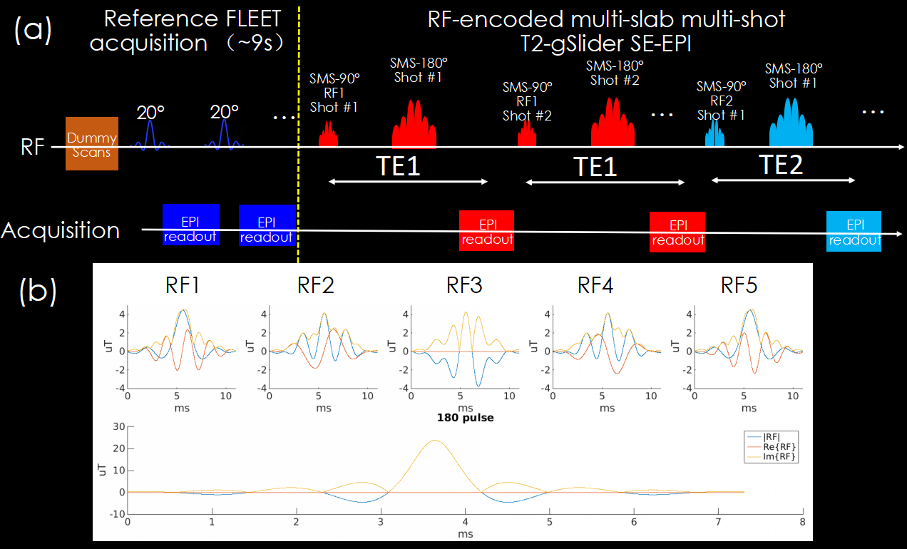
Figure 1
(a) Pulse sequence design.
(b) Profiles of 90° RF-encoding excitation pulse and the 180° SLR pulse.
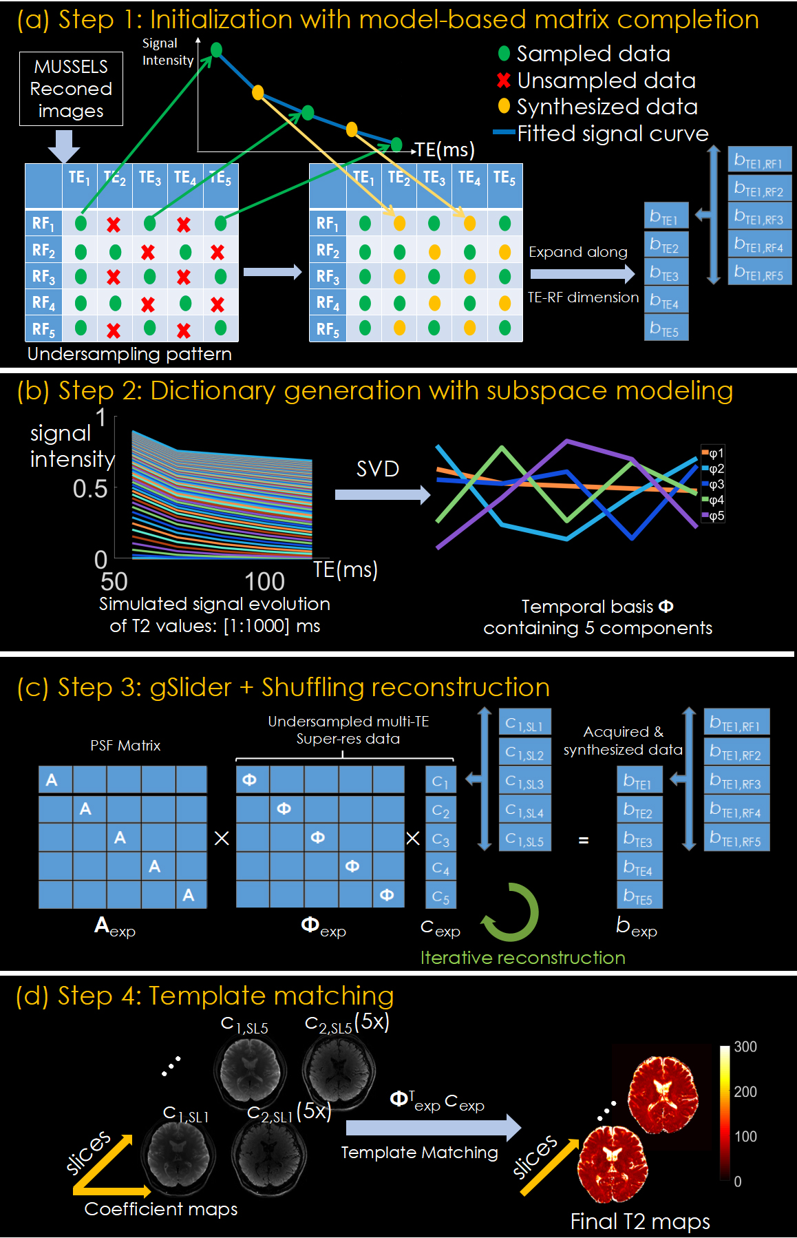
Figure 2
Reconstruction process including initialization (a), dictionary generation (b), gSlider-Shuffling joint reconstruction (c) and template match with a pre-calculated T2 dictionary (d). Due to the weak contributing rate of temporal basis from to (sum to about 0.2%), the corresponding coefficient maps C3 to C5 were truncated using low-rank approximation.
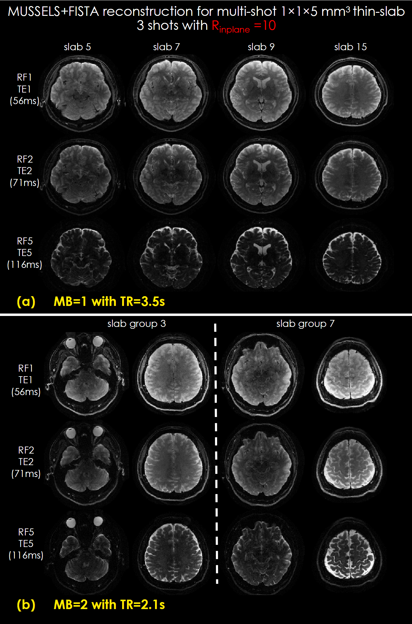
Figure 3
Reconstructed RF-encoded thin-slab images of different TEs and slabs with MB=1 (a) and MB=2 (b).
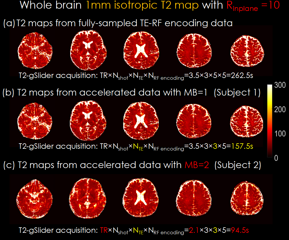
Figure 4
T2 maps in different slices of 1-mm3 isotropic resolution with MB=1 (a, b) and MB=2 (c). The MB=1 and MB=2 data were acquired from different subjects.
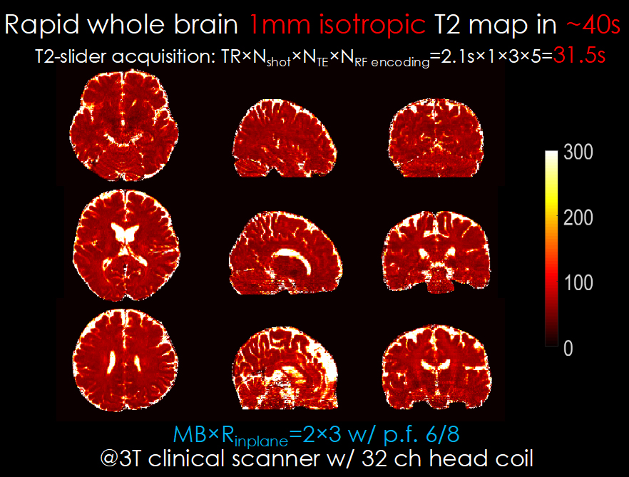
Figure 5
T2 maps in different slices of 1-mm3 isotropic resolution with single-shot acquisition and MB=2.