0311
Loss Adaptive Dipole Inversion (LADI): A novel data driven approach for quantitative susceptibility mapping1Radiology, University of Pennsylvania, Philadelphia, PA, United States, 2Bioengineering, University of Pennsylvania, Philadelphia, PA, United States, 3Biochemistry and Molecular Biophysics, University of Pennsylvania, Philadelphia, PA, United States
Synopsis
This abstract presents a novel data driven approach for high quality QSM reconstructions without the use of complex and computationally intensive reconstruction models. The purpose of this approach is to develop a reconstruction technique which does not depend on the use of spatial priors from the magnitude image to remove artifacts and reduce blurring of edges. In our proposed formulation, the data fidelity term is updated based on the deviation of the estimated susceptibility map from the measured local field. With the proposed fidelity-loss adaptive reconstruction formulation, removal of artifacts was achieved without causing smoothing of sharp features.
Introduction
Quantitative susceptibility mapping (QSM) is a technique that can quantify tissue magnetic properties and provide valuable information about hemorrhagic stroke [1,2] and impaired tissue oxygen consumption [2]. Reconstruction of magnetic susceptibility maps from the tissue field is a challenging ill-posed inverse problem. Most popular techniques use a combination of weighted least squares fidelity constraint and regularization constraints such as total variation (TV) [3] or total generalized variation (TGV) [4,5]. An exponential data fidelity model is sometimes used to further improve the quality of the reconstruction. This leads to slow and computationally complex reconstruction models. The purpose of this work was to develop a data driven QSM reconstruction algorithm that can faithfully reconstruct fine features and maintain sharp edges. The proposed Loss Adaptive Dipole Inversion (LADI) technique is a novel application of the constrained TV formulation that does not need to estimate the location of edges in the susceptibility maps or the nonuniform distribution of noise in the phase data. The advantage of this technique compared to algorithms that use a priori edge information is that our algorithm is unaffected by mismatch between the edge prior and estimated susceptibility map. We compare the performance of LADI to several CS based algorithms in for applications such as neuroimaging and hemorrhagic myocardial infarction.Methods
The constrained reconstruction formulation that we aim to minimize is $$_{min\chi}||\triangledown\chi||_{1}; S.T. ||M_{Bin}(F^H DFχ-ϕ)||^{2}\leq \sigma^{2}$$(1).
Here MBin is a binary mask, D is the magnetic dipole kernel represented in the frequency domain, χ is the susceptibility distribution, F is the Fourier operator, FH the inverse Fourier operator, ϕ is the tissue phase and σ is the noise standard deviation. By using the Bregman iterations [6], Eq. (1) can be reduced to $$_{min\chi}\lambda||\triangledown\chi||_{1}+\frac{\mu}{2} ||M_{Bin}(F^H DFχ-ϕ)||^{2}$$ and $$\phi^{k+1}=\phi^{k}+(ϕ-F^H DFχ^{k})$$The iterative update of ϕ is equivalent to the “adding-noise-back” step [6], which helps ensure better data fidelity to the measured local tissue field, while preventing smoothing of edges and fine features in the image due to data regularization. We compare the results from LADI to thresholded k-space division (TKD) [3], closed form L2 [3], morphology enabled dipole inversion (MEDI) [4,7] and a rapid implementation of non-linear MEDI with TGV constraints (FANSI) [4]. COSMOS phantom [5] and cardiac QSM data acquired in a large animal model of hemorrhagic myocardial infarction was used to compare the reconstructions. Visual inspection of images for presence of artifacts, metrics such as RMSE [5], SSIM [5], HFEN [5], mutual information [4] and quantification of mean susceptibilities from ROI’s were used for to compare the different reconstructions.
Results
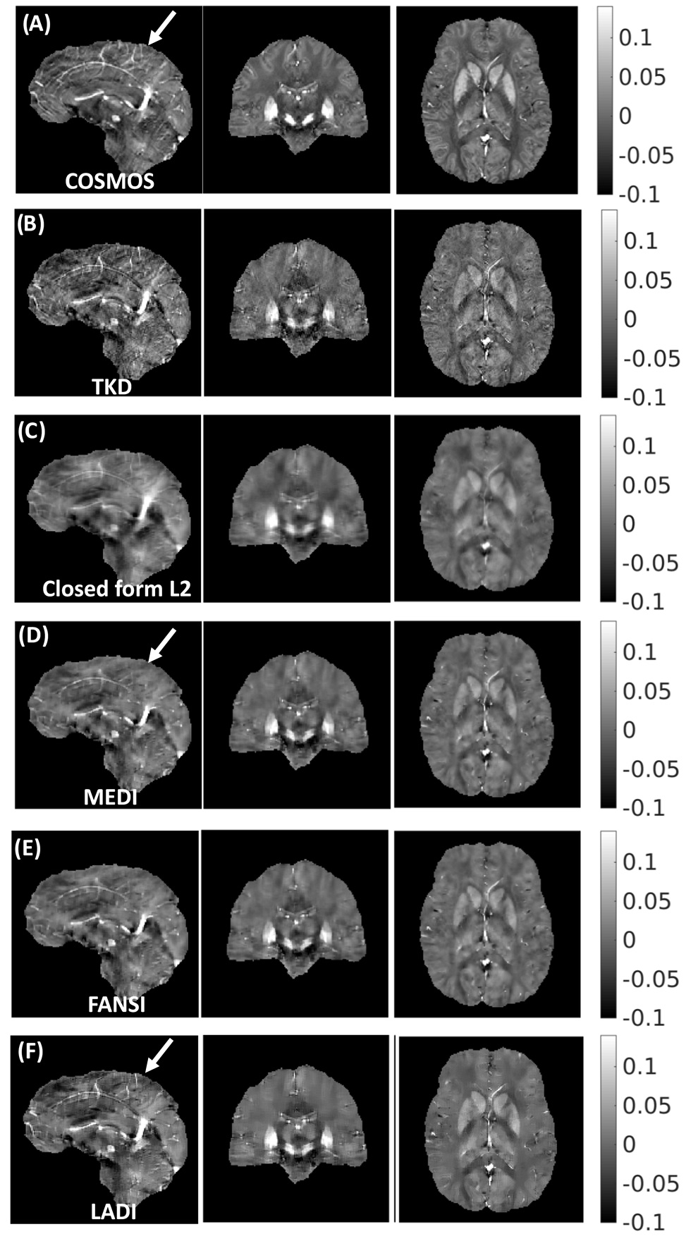
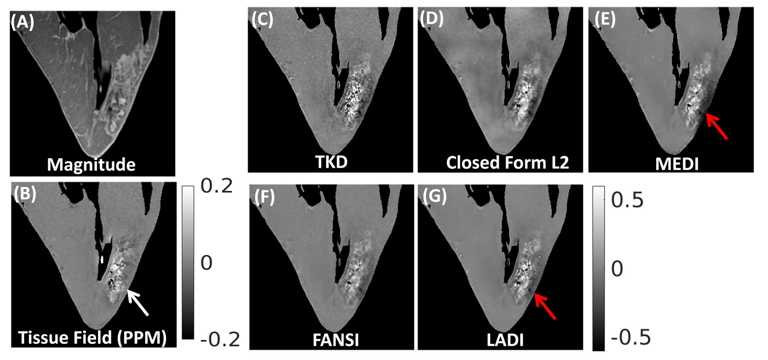
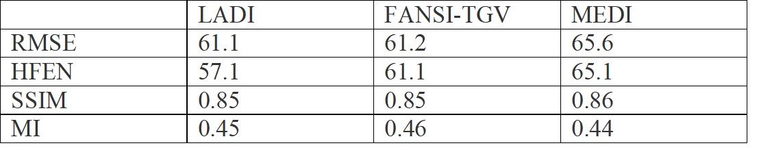
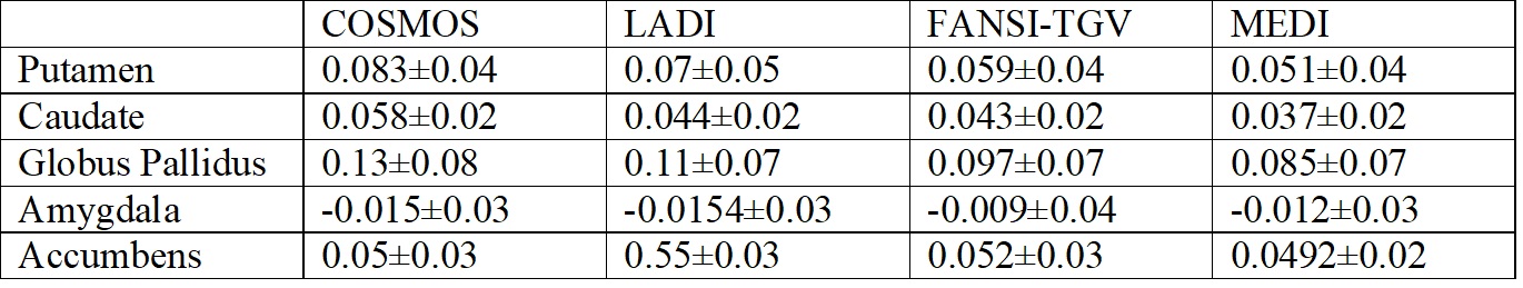
A comparison of the reconstructions from COSMOS phantom are shown in Fig (1). TKD reconstructions suffered from large streaking artifacts (SSIM=0.75,RMSE=73.1,HFEN=66.7) while in closed form L2 reconstructions, removal of artifacts was achieved at the cost of smoothing of edges (SSIM=0.81,RMSE=70.1,HFEN=68.3). FANSI and LADI had slightly better performance with respect to global error quality metrics(Table I); though sharp features such as veins were better preserved in LADI (location shown by arrow in Fig 1). Overall, LADI (corr coeff=0.81) and FANSI (corr coeff=0.79) performed better at matching COSMOS (corr coeff=0.77). ROI based comparison from five different regions in the brain (Table II) showed that FANSI and LADI were able to match COSMOS better than MEDI reconstructions. A comparison of the reconstructions from the hemorrhagic myocardial infarction data is shown in Fig 2. LADI and FANSI reconstructions had a good balance of feature preservation and artifacts removal, and the dark artifacts seen in MEDI, TKD and closed form L2 was well mitigated.Discussion
The proposed LADI technique does not need to estimate the location of edges in the susceptibility maps or the nonuniform distribution of noise in the phase data. The major advantage of LADI algorithm compared to algorithms that use a priori edge information is that our algorithm is unaffected by mismatch between the edge prior and estimated susceptibility map. Hence blurring of edges that could occur in existing MEDI and non-linear MEDI based formulation is avoided. The results from the COSMOS phantom and the cardiac QSM data showed that the proposed model is robust to variations in data quality. LADI formulation can be easily accelerated by incorporating the Split Bregman based variable substitution techniques in [3,4].Conclusion
We developed a data driven approach for high quality QSM reconstructions. LADI had better image quality metrics compared to MEDI and performed as well as FANSI-TGV, one of the current state-of-the-art reconstruction models. Ease of implementation, high image quality and robustness to different data types are the important features of the proposed LADI technique.Acknowledgements
This work is supported by R00-HL108157, McCabe Foundation, and W.W. Smith Foundation.References
1.Chang S, Zhang J, Liu T, Tsiouris A J, Shou J, Nguyen T and Kovanlikaya I. (2016). Quantitative Susceptibility Mapping of Intracerebral Hemorrhages at Various Stages. JMRI, 44(2), 420–425.
2. Wang Y and Liu T. (2015), Quantitative susceptibility mapping (QSM): Decoding MRI data for a tissue magnetic biomarker. Magn. Reson. Med., 73: 82–101. doi:10.1002/mrm.25358
3. Bilgic B, Chatnuntawech I, Fan A P, Setsompop K, Cauley S F, Wald L L and Adalsteinsson E. (2014), Fast image reconstruction with L2-regularization. J. Magn. Reson. Imaging, 40: 181–191. doi:10.1002/jmri.24365.
4. Milovic C, Bilgic B, Zhao B, Acosta-Cabronero J and Cristian Tejos, Fast nonlinear susceptibility inversion with variational regularization, Magn. Reson. Med. 2018: 80(2):814-821, doi: 10.1002/mrm.27073.
5. Langkammer C, Schweser F, Shmueli K, Kames C, Li X, Guo L, Milovic C, Kim J., Wei H, Bredies K, Buch S, Guo Y, Liu Z, Meineke J, Rauscher, A., Marques, J. P. and Bilgic, B. (2017), Quantitative susceptibility mapping: Report from the 2016 reconstruction challenge. Magn. Reson. Med. doi:10.1002/mrm.26830.
6. Goldstein T and Osher S, The Split Bregman Method for L1-Regularized Problems, SIAM Journal on Imaging Sciences 2009 2:2, 323-343
7. Liu T, Liu J, de Rochefort L, Spincemaille P, Khalidov I, Ledoux JR, Wang Y. Morphology enabled dipole inversion (MEDI) from a single-angle acquisition: comparison with COSMOS in human brain imaging. Magnetic Resonance in Medicine. 2011; 66(3):777–783.
Figures