0310
Echo Planar Time-Resolved Imaging (EPTI) with subspace constraint and optimized k-t trajectory1A. A. Martinos Center for Biomedical Imaging, Massachusetts General Hospital, Charlestown, MA, United States, 2Electrical Engineering and Computer Science, MIT, Cambridge, MA, United States, 3Harvard-MIT Health Sciences and Technology, MIT, Cambridge, MA, United States
Synopsis
Echo planar time-resolved imaging (EPTI) is a multi-contrast quantitative imaging technique, which achieved fast acquisition of distortion- and blurring-free images at multiple echo times (TE). To improve the SNR and accuracy of EPTI at high-accelerations, in this study, we developed a subspace-constrained reconstruction for EPTI and proposed new k-t sampling trajectories to take advantage of this reconstruction. The subspace reconstruction is also augmented with phase-cycling to extract high-resolution phase data, without need of high-resolution B0 calibration scan. Using the proposed approach, whole-brain 1.1mm-isotropic multi-echo images, and T2* and B0 maps are reconstructed from 3D-EPTI data acquired within 50 seconds.
Purpose
Echo planar time-resolved imaging[1] is a multi-contrast quantitative imaging technique, which achieved rapid acquisition of distortion- and blurring-free images at multiple echo times (TE). In EPTI, a spatio-temporal CAIPI sampling[1-3] is used to cover k-t space efficiently through customized multishot-EPI trajectory. The highly-undersampled ky-t data is then recovered by exploiting signal correlation across the time and coil dimensions through a GRAPPA-like reconstruction. In such reconstruction, the edge of ky-t space cannot be recovered accurately through kernel-based interpolation, reducing the number of echo images that can be reconstructed. Moreover, the SNR of the reconstruction can be low for high spatial resolution cases with high-undersampling. To address these issues, we develop a new reconstruction using the subspace-constrained approach[4,5], and validated its improved performance on both 2D- and 3D-EPTI acquisitions. New EPTI sampling patterns were also designed and combined with subspace reconstruction to further improve highly-accelerated 3D-EPTI.Methods
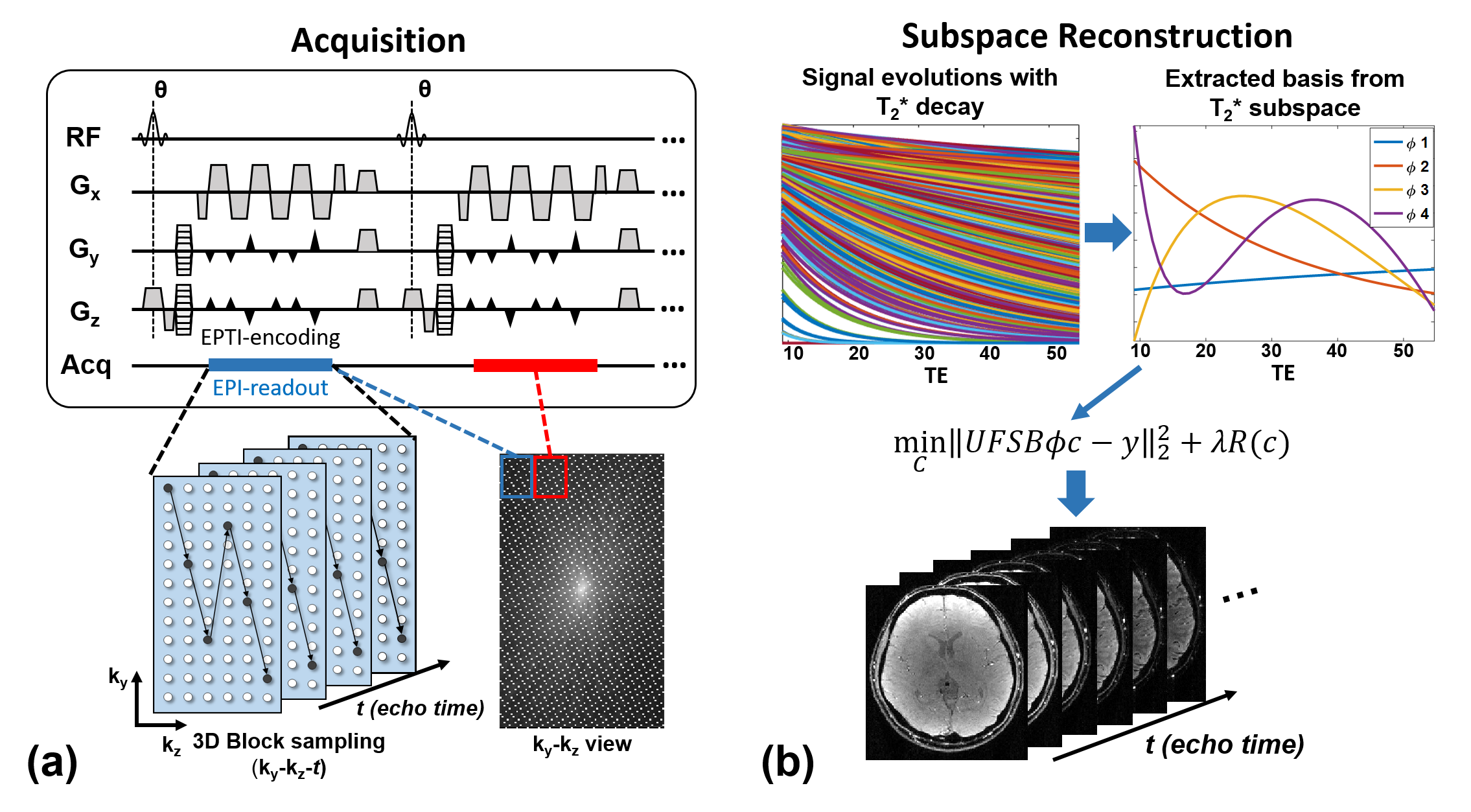
In EPTI, continuous EPI readouts are performed with spatio-temporal CAIPI-sampling to efficiently sample the desired signal evolution in k-t space. For instance, a 3D gradient-echo (GE) EPTI sequence shown in Fig.1a was used to acquire the T2* signal decay. After each RF excitation, an EPTI readout is used to cover a small ky-kz block using highly-accelerated zigzag trajectory. Such spatio-temporal CAIPI trajectory has been demonstrated to enable effective use of coil sensitivity in recovering highly undersampled ky-kz-t data within each sampling block[1]. Acquisition across k-space blocks are performed across TRs to fill ky-kz space. To recover the signal evolution from undersampled data, subspace reconstruction is developed for EPTI (Fig.1b). First, the desired signal evolution (T2* decay in this study) within the possible parameter range is simulated based on the acquisition parameters. Then, several bases are extracted through PCA as Φ, which form a low-dimensional subspace used to approximate the signal space. Using these bases, the temporal image series can be calculated by Φc, where c is the coefficient map of the bases that needs to be estimated. Using this approach, the degrees of freedom of reconstruction is reduced from the number of time points to the number of bases, which improves conditioning of the reconstruction and image-SNR[5]. The subspace-constrained reconstruction is solved by:
$$min_{c}\parallel UFSB\phi c-y \parallel_2^2+\lambda R(x)$$
where B is temporal B0 phase-evolution, S is coil sensitivity, F is Fourier transform, U is undersampling mask, and y is the acquired undersampled kx-ky-kz-t data. Regularization R(c) can be used to improve the conditioning and SNR for higher undersampling. After estimating c, we can generate the time-series images/volumes by computing Φc. The B0 phase evolution and coil sensitivity in the forward model are both estimated using a fast-low-resolution calibration data with 6 GE echoes. High-resolution B0 maps can be estimated by the phase-cycling approach[6] using the reconstructed magnitude images and acquired signals.The following brain data were acquired at 3T using a 32-channel coil.
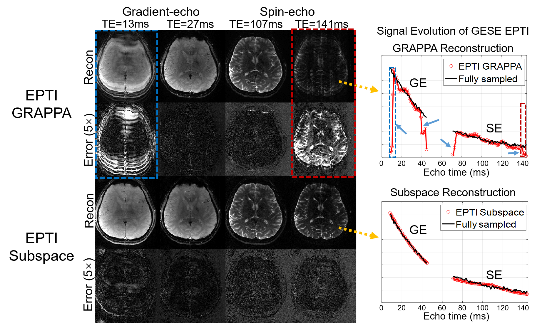
2D GE-SE EPTI acquisition was used to compare GRAPPA-like method and subspace reconstruction. Gradient- and spin-echo k-t data with FOVxy = 220x220mm2 and resxyz =1.1×1.1×3mm3 were acquired with full 216 phase-encodings across 40 gradient-echo and 80 spin-echo time-points. The data were retrospectively undersampled along ky-t by 24x to synthesize a 9-shot 2D-EPTI acquisition used for reconstruction comparison.
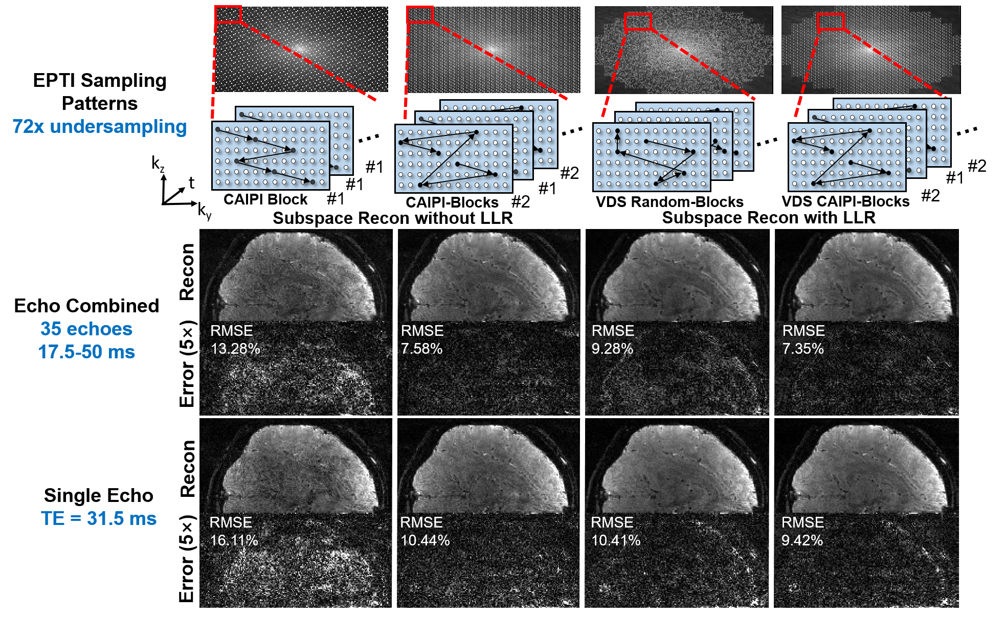
3D GE EPTI acquisition was used to evaluate subspace reconstruction under four different EPTI sampling strategies: i)&ii) regular CAIPI-like EPTI samplings, iii) variable density (VDS) random-sampling, and iv) VDS CAIPI-sampling. For VDS cases (iii&iv), locally low-rank (LLR) constraint was employed. GE k-t data at 1.1mm isotropic resolution were fully acquired across 50 echo-time points, with FOVxyz = 220x220x110mm3. The data were retrospectively undersampled along ky-kz-t by 72x with a block size of Ryblock x Rzblock=12×6 for all sampling strategies to reduce acquisition time from 26-mins to 22s.
Results
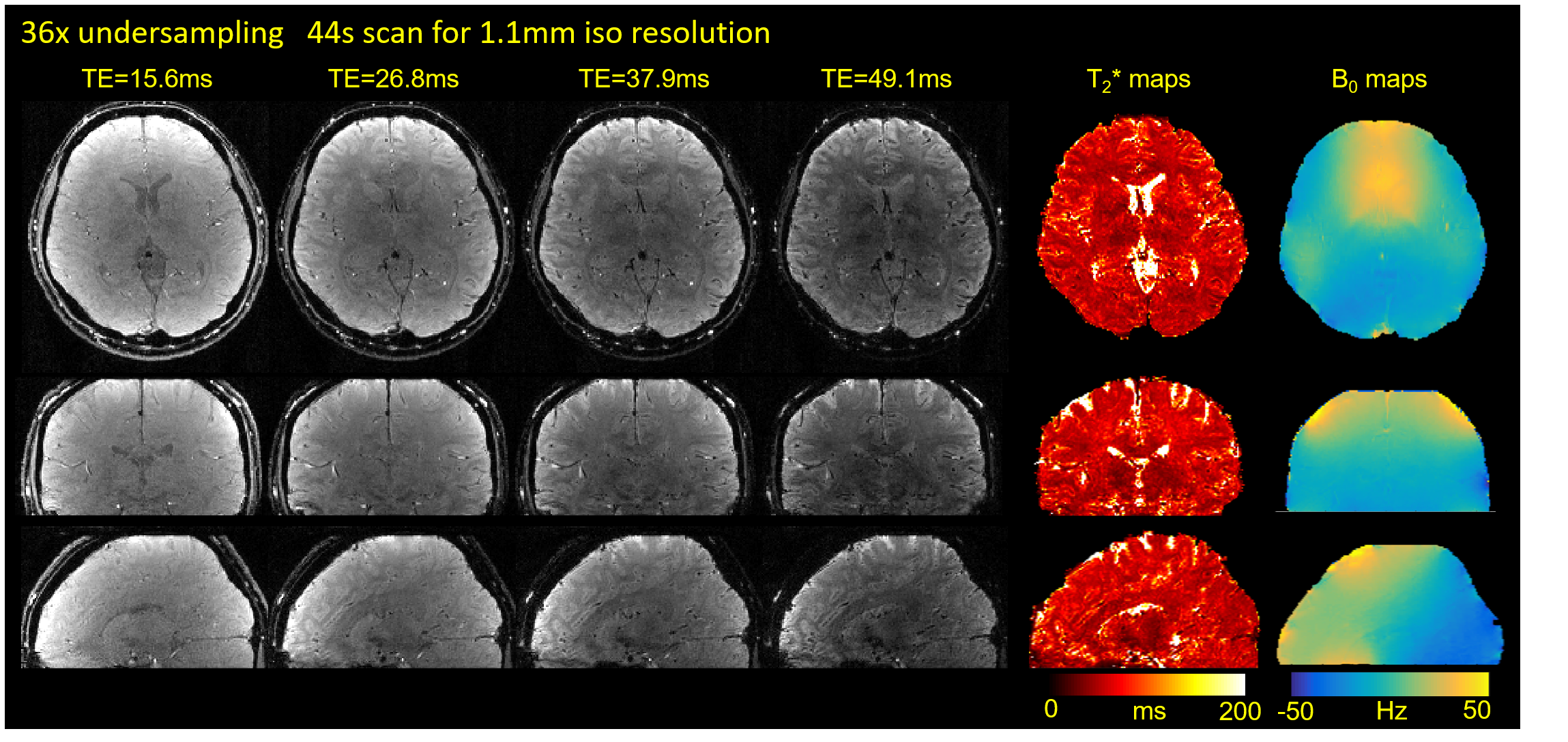
Fig.2 shows the comparison between GRAPPA-like reconstruction and subspace reconstruction for 2D GE-SE EPTI. The edge echoes which are corrupted in GRAPPA reconstruction are well-recovered by the subspace reconstruction. Fig.3 shows the results of different 3D EPTI sampling strategies combined with subspace reconstruction. Through 72× acceleration, 3D brain volumes at 1.1mm isotropic resolution across 50 echoes time-points can be obtained in 22 seconds. Here, VDS CAIPI-sampling with LLR-subspace reconstruction achieves the best results with low errors. The reconstructed multi-echo images, T2* and B0 maps of 3D EPTI data at 36× acceleration with VDS CAIPI-sampling (44s scan) and LLR-constrained subspace reconstruction are shown in Fig.4.Conclusion
Subspace reconstruction improves the performance of EPTI for better signal evolution recovery, and the designed 3D sampling strategy enables efficient high-resolution EPTI, providing a powerful technique for fast multi-contrast and quantitative imaging.Acknowledgements
This work was supported in part by NIH research grants: R01MH116173, R01EB020613, R01EB019437, U01EB025162, P41EB015896, and the shared instrumentation grants: S10RR023401, S10RR019307, S10RR019254, S10RR023043.References
1. Wang F, Dong Z, Reese T, Wald L, Setsompop K. Echo Planar Time-resolved Imaging. In Proceedings of the 26th Annual Meeting of ISMRM, Paris, France, 2018. p 0217.
2. Breuer FA, Blaimer M, Mueller MF, Seiberlich N, Heidemann RM, Griswold MA, Jakob PM. Controlled Aliasing in Volumetric Parallel Imaging (2D CAIPIRINHA). Magn Reson Med 2006; 55:549–556.
3. Setsompop K, Gagoski BA, Polimeni JR, Witzel T, Wedeen VJ, Wald LL. Blipped‐controlled aliasing in parallel imaging for simultaneous multislice echo planar imaging with reduced g‐factor penalty. Magn Reson Med 2012;67:1210-1224.
4. Liang Z P. Spatiotemporal imaging with partially separable functions. Biomedical Imaging: From Nano to Macro, 2007. ISBI 2007. 4th IEEE International Symposium on. IEEE, 2007: 988-991.
5. Tamir JI, Uecker M, Chen W, Lai P, Alley MT, Vasanawala SS, Lustig M. T2 shuffling: Sharp, multicontrast, volumetric fast spin‐echo imaging. Magn Reson Med. 2017;77:180-195.
6. Ong F, Cheng J Y, Lustig M. General phase regularized reconstruction using phase cycling. Magn Reson Med. 2018, 80: 112-125.
Figures