0301
Intra-Voxel Incoherent Motion at 7T to quantify human spinal cord microperfusion: pitfalls and promises1Faculty of Medicine, Aix-Marseille Univ, CNRS, CRMBM, Marseille, France, 2APHM, Hopital Universitaire Timone, CEMEREM, Marseille, France, 3Faculty of Medicine, Aix-Marseille Univ, IFSTTAR, LBA, Marseille, France, 4iLab-Spine International Associated Laboratory, France-Canada, Marseille-Montreal, France, 5Siemens Healthcare GmbH, Erlangen, Germany, 6Siemens Healthcare SAS, Saint-Denis, France
Synopsis
Spinal cord microperfusion assessment in human is challenging but would greatly help characterize tissue integrity and surgery decision-making. Intra-Voxel Incoherent Motion (IVIM) microperfusion measurement is promising but remains highly Signal-to-Noise ratio (SNR) demanding. Monte-Carlo simulations show that IVIM two-step segmented fitting approach is less accurate than directly fitting the bi-exponential representation to all b-values. Simulations also help quantify required SNR and estimation errors to measure IVIM parameters in the context of low perfusion. Exploiting 7T SNR gain, large number of repetitions and group average, IVIM was able to unveil the gray matter higher microperfusion-related pattern, compared to white matter, in agreement with brain studies.
Introduction
Being able to assess spinal cord (SC) microperfusion in-vivo would help characterize SC tissue integrity and hence managing surgery decisions, such as in compressive injuries where tissue damage originates from perfusion alteration1,2. Non-invasive MRI-based techniques have been successfully applied to measure microperfusion in several human organs (e.g. brain, kidney, liver) and mouse SC3,4. However, microperfusion has never been quantified, and even less mapped, in human SC. The challenge arises from an expected low microperfusion, pulsatile cord movements and magnetic field inhomogeneities. Besides, the inner SC vascular system is complex and shows interindividual variability, complicating blood-tagging strategies such as for Arterial Spin Labeling. Given the major progress in diffusion MRI of human SC5–12, Intra-Voxel Incoherent Motion (IVIM)13 seems promising. IVIM aims at quantifying the signal decrease at low b-values supposedly induced by blood water circulation through capillaries mimicking a Brownian motion random-walk at large scale. To overcome low expected signal, the technique can be deployed at ultra-high field. In this view, we got on with optimizing an IVIM 7T acquisition protocol, data post-processing and tailored fitting pipelines to propose the first maps of microperfusion-related parameters within the human SC.Methods
Data acquisition. Axial acquisitions were performed on a 7T Siemens Magnetom system, based on a prototype 2D single-shot EPI diffusion-prepared sequence6. Parameters (GRAPPA, partial Fourier, TE, BW, repetitions, reconstruction algorithm) were optimized for SNR. Acquisition was triggered on pulse oxymeter to minimize effects of cord motion and cerebrospinal fluid (CSF) pulses. Acquisition was split into forward and reverse phase-encoding blips to further correct for susceptibility-induced distortions. Such data were acquired for 16 b-values in three orthogonal diffusion-encoding directions (~14min/direction) within 8 healthy volunteers (aged 25.8±3.3, 2 females).
Data processing. Data were denoised using local PCA filter14, Gibbs artefacts were removed with Unring15 and distortions were corrected using Topup16,17.
Data fitting. Fig.1 describes IVIM parameters estimation pipelines, implemented using LMFIT 0.9.11 python module (lmfit.github.io/lmfit-py). Accuracy of two conventional approaches was assessed through Monte-Carlo simulations: the “two-step segmented”18,19 (1) and the “full”18 (2) fitting approaches. To do so, range of $$$f_{IVIM}$$$, $$$D^*$$$ and $$$D$$$ values were defined based on IVIM literature in brain20–27. First, data were generated using the signal representation without noise for the defined parameter ranges and b-values of 0,5,10,15,20,30,50,75,100,125,150,200,250,600,700,800 s/mm2. Estimation error on each parameter was assessed as $$estimation\ error(\%)=100\times\frac{estimated\ value-true\ value}{true\ value}$$Secondly, random gaussian noise was added to synthetic data so as to match in-vivo measured Signal-to-Noise Ratio (SNR) – mean voxel-wise SNR across subjects in cord for single diffusion-encoding directions was 116 [min=61,max=187] – and errors were averaged across 100 random draws. Thirdly, the minimum SNR required to get less than 10% error on $$$f_{IVIM}D^*$$$ with approach (2) was computed for the defined parameter ranges.
Finally, real data were fitted voxel-wise using approach (2). IVIM parameters maps were registered to high-resolution anatomical images (MEDIC28) to include gray matter (GM) shape interindividual variability, and then to PAM50 template29 – using the SpinalCordToolbox (SCT)30, to finally be averaged across slices (mostly spanning C4) and subjects.
Results & Discussion
The developed algorithm yielded perfect estimation of parameters when no noise was added with approach (2) while approach (1) showed errors up to 44%, especially for low $$$D^*$$$ (Fig.2-left). With realistic SNR, errors dramatically increased for both approaches – mainly at low $$$f_{IVIM}$$$ and $$$D^*$$$ values, in agreement with previous studies18,31,32 – nevertheless approach (2) yielded better estimation as supported by median error, especially for $$$D$$$ (Fig.2-right). Higher $$$D$$$ yielded larger errors, suggesting that IVIM parameters might be more difficult to estimate along SC axis.
To reach an error$$$\leq$$$10% on $$$f_{IVIM}D^*$$$ in individual maps for a single diffusion-encoding direction given the distribution of chosen b-values, median SNRs of 157 and 212 are required for low and high $$$D$$$ respectively (Fig.3). If $$$f_{IVIM}≥0.042$$$ and $$$D^*\geq6.6\times10^{-3}mm^2/s$$$, median required SNRs are 126 and 200, which stands close to observed in-vivo SNR. Note that these values do not account for the SNR gain obtained through maps averaging across diffusion-encoding directions.
Still, IVIM maps computed from individual in-vivo
datasets (Fig.4-top) hardly discriminate between GM and white matter (WM). However,
when averaging maps (Fig.4-bottom) across slices and subjects, higher values
are revealed within GM compared to WM, in agreement with brain microperfusion studies22,23,33. Quantitative analyses (Fig.5) suggest that most
vascularized ($$$f_{IVIM}$$$) regions are intermediate and anterior GM while highest
blood flow ($$$f_{IVIM}D^*$$$)34 would occur within intermediate GM.
Conclusion
This
work reports the first high-resolution IVIM parameters maps of human SC.
Provided high SNR, mainly obtained here through 7T MRI, relatively long acquisition time
and group average, IVIM seems able to provide new knowledge on SC microperfusion
topography. Further work will aim to refine those results with more subjects
and to shorten acquisitions.
Acknowledgements
The authors would like to particularly thank Olivier Girard and Ludovic de Rochefort for useful discussions.
This project has received funding from the European Union’s Horizon 2020 research and innovation program under the Marie Skłodowska-Curie grant agreement No713750. Also, it has been carried out with the financial support of the Regional Council of Provence-Alpes-Côte d’Azur and with the financial support of the A*MIDEX (n° ANR- 11-IDEX-0001-02), funded by the Investissements d'Avenir project funded by the French Government, managed by the French National Research Agency (ANR).
References
1. Breig, A., Turnbull, I. & Hassler, O. Effects of mechanical stresses on the spinal cord in cervical spondylosis: a study on fresh cadaver material. Journal of neurosurgery 25, 45–56 (1966).
2. Fehlings, M. G. & Skaf, G. A review of the pathophysiology of cervical spondylotic myelopathy with insights for potential novel mechanisms drawn from traumatic spinal cord injury. Spine 23, 2730–2737 (1998).
3. Duhamel, G., Callot, V., Cozzone, P. J. & Kober, F. Spinal cord blood flow measurement by arterial spin labeling. Magnetic Resonance in Medicine 59, 846–854 (2008).
4. Duhamel, G. et al. Mouse lumbar and cervical spinal cord blood flow measurements by arterial spin labeling: Sensitivity optimization and first application. Magnetic Resonance in Medicine 62, 430–439 (2009).
5. Taso, M. et al. Tract-specific and age-related variations of the spinal cord microstructure: a multi-parametric MRI study using diffusion tensor imaging (DTI) and inhomogeneous magnetization transfer (ihMT). NMR in Biomedicine 29, 817–832 (2016).
6. Massire, A. et al. Feasibility of single‐shot multi‐level multi‐angle diffusion tensor imaging of the human cervical spinal cord at 7T. Magnetic Resonance in Medicine (2018). doi:10.1002/mrm.27087
7. Massire, A. et al. High-resolution multi-parametric quantitative magnetic resonance imaging of the human cervical spinal cord at 7T. NeuroImage 143, 58–69 (2016).
8. Li, D. C. et al. The role of diffusion tensor imaging in spinal pathology: A review. Neurology India 65, 982 (2017).
9. Rasoanandrianina, H. et al. Region-specific impairment of the cervical spinal cord (SC) in amyotrophic lateral sclerosis: A preliminary study using SC templates and quantitative MRI (diffusion tensor imaging/inhomogeneous magnetization transfer). NMR in Biomedicine 30, e3801 (2017).
10. Grussu, F., Schneider, T., Zhang, H., Alexander, D. C. & Wheeler–Kingshott, C. A. M. Neurite orientation dispersion and density imaging of the healthy cervical spinal cord in vivo. NeuroImage 111, 590–601 (2015).
11. Duval, T. et al. g-Ratio weighted imaging of the human spinal cord in vivo. NeuroImage 145, Part A, 11–23 (2017).
12. Duval, T. et al. In vivo mapping of human spinal cord microstructure at 300mT/m. NeuroImage 118, 494–507 (2015).
13. Le Bihan, D. et al. Separation of diffusion and perfusion in intravoxel incoherent motion MR imaging. Radiology 168, 497–505 (1988).
14. Manjón, J. V. et al. Diffusion Weighted Image Denoising Using Overcomplete Local PCA. PLOS ONE 8, e73021 (2013).
15. Kellner, E., Dhital, B., Kiselev, V. G. & Reisert, M. Gibbs-ringing artifact removal based on local subvoxel-shifts. Magnetic Resonance in Medicine 76, 1574–1581 (2016).
16. Andersson, J. L. R., Skare, S. & Ashburner, J. How to correct susceptibility distortions in spin-echo echo-planar images: application to diffusion tensor imaging. NeuroImage 20, 870–888 (2003).
17. Smith, S. M. et al. Advances in functional and structural MR image analysis and implementation as FSL. NeuroImage 23, S208–S219 (2004).
18. Pekar, J., Moonen, C. T. W. & van Zijl, P. C. M. On the precision of diffusion/perfusion imaging by gradient sensitization. Magnetic Resonance in Medicine 23, 122–129 (1992).
19. Jalnefjord, O. et al. Comparison of methods for estimation of the intravoxel incoherent motion (IVIM) diffusion coefficient (D) and perfusion fraction (f). Magn Reson Mater Phy (2018). doi:10.1007/s10334-018-0697-5
20. Federau, C., O’Brien, K., Meuli, R., Hagmann, P. & Maeder, P. Measuring brain perfusion with intravoxel incoherent motion (IVIM): Initial clinical experience. Journal of Magnetic Resonance Imaging 39, 624–632 (2014).
21. Federau, C. et al. Quantitative Measurement of Brain Perfusion with Intravoxel Incoherent Motion MR Imaging. Radiology 265, 874–881 (2012).
22. Wang, C. et al. Distribution of intravoxel incoherent motion MRI-related parameters in the brain: evidence of interhemispheric asymmetry. Clinical Radiology 72, 94.e1-94.e6 (2017).
23. Bisdas, S. & Klose, U. IVIM analysis of brain tumors: an investigation of the relaxation effects of CSF, blood, and tumor tissue on the estimated perfusion fraction. Magn Reson Mater Phy 28, 377–383 (2015).
24. Bertleff, M. et al. Diffusion parameter mapping with the combined intravoxel incoherent motion and kurtosis model using artificial neural networks at 3 T. NMR in Biomedicine 30, e3833 (2017).
25. Suo, S. et al. Stroke assessment with intravoxel incoherent motion diffusion-weighted MRI. NMR in Biomedicine 29, 320–328 (2016).
26. Bisdas, S. et al. Intravoxel incoherent motion diffusion-weighted MR imaging of gliomas: feasibility of the method and initial results. Neuroradiology 55, 1189–1196 (2013).
27. Federau, C., Meuli, R., O’Brien, K., Maeder, P. & Hagmann, P. Perfusion Measurement in Brain Gliomas with Intravoxel Incoherent Motion MRI. American Journal of Neuroradiology 35, 256–262 (2014).
28. Held, P., Dorenbeck, U., Seitz, J., Fründ, R. & Albrich, H. MRI of the abnormal cervical spinal cord using 2D spoiled gradient echo multiecho sequence (MEDIC) with magnetization transfer saturation pulse. A T2* weighted feasibility study. J Neuroradiol 30, 83–90 (2003).
29. De Leener, B. et al. PAM50: Unbiased multimodal template of the brainstem and spinal cord aligned with the ICBM152 space. NeuroImage (2017). doi:https://doi.org/10.1016/j.neuroimage.2017.10.041
30. De Leener, B. et al. SCT: Spinal Cord Toolbox, an open-source software for processing spinal cord MRI data. NeuroImage 145, 24–43 (2017).
31. Federau, C. et al. Functional Mapping of the Human Visual Cortex with Intravoxel Incoherent Motion MRI. PLOS ONE 10, e0117706 (2015).
32. Qinwei Zhang, Yi-Xiang Wang, Ma, H. T. & Jing Yuan. Cramer-Rao bound for Intravoxel Incoherent Motion Diffusion Weighted Imaging fitting. in 2013 35th Annual International Conference of the IEEE Engineering in Medicine and Biology Society (EMBC) 511–514 (IEEE, 2013). doi:10.1109/EMBC.2013.6609549
33. Parkes Laura M., Rashid Waqar, Chard Declan T. & Tofts Paul S. Normal cerebral perfusion measurements using arterial spin labeling: Reproducibility, stability, and age and gender effects. Magnetic Resonance in Medicine 51, 736–743 (2004).
34. Bihan, D. L. & Turner, R. The capillary network: a link between ivim and classical perfusion. Magnetic Resonance in Medicine 27, 171–178 (1992).
35. Storn, R. & Price, K. Differential Evolution – A Simple and Efficient Heuristic for global Optimization over Continuous Spaces. Journal of Global Optimization 11, 341–359 (1997).
36. Lévy, S. et al. White matter atlas of the human spinal cord with estimation of partial volume effect. NeuroImage 119, 262–271 (2015).
Figures
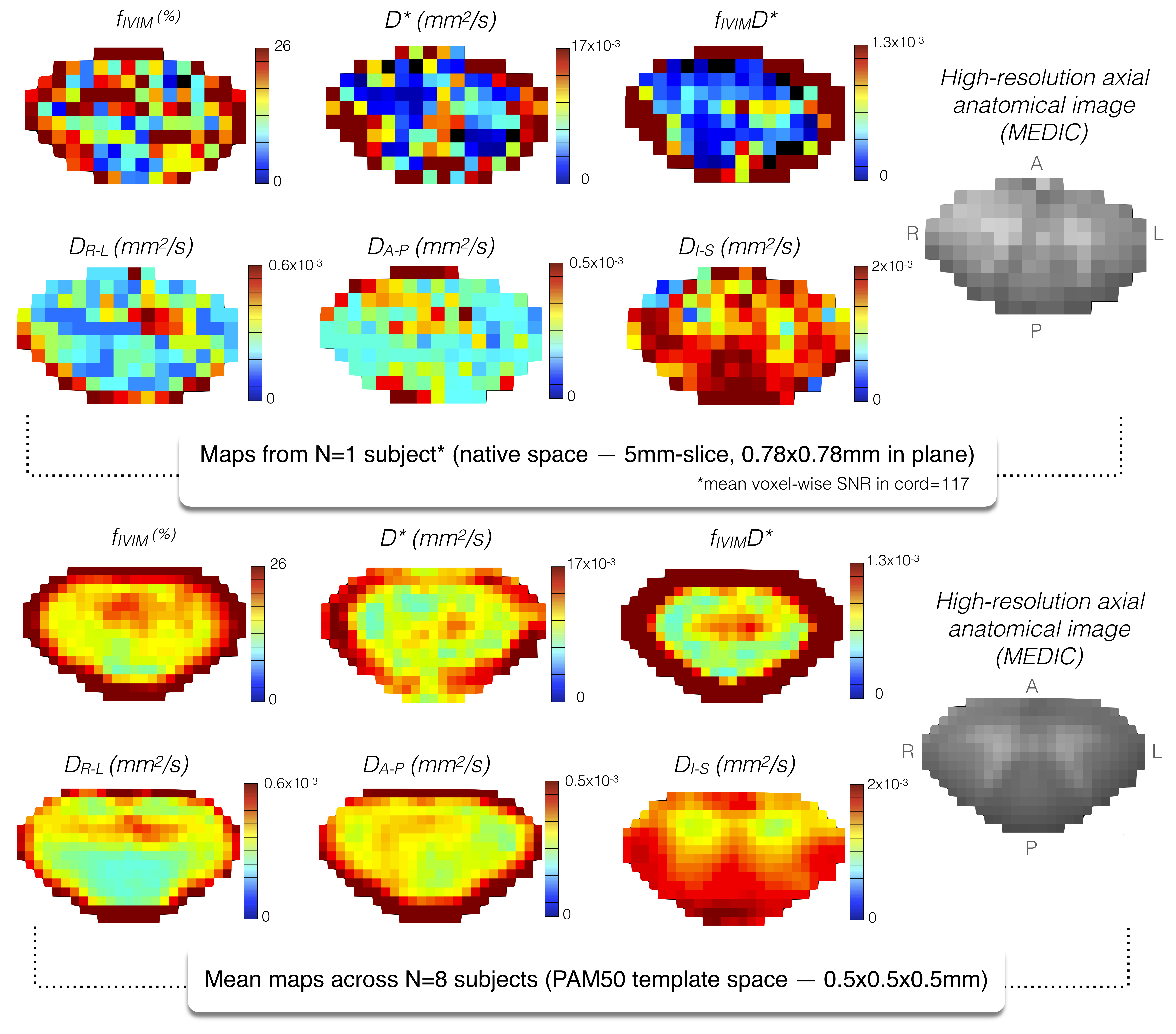
Fig.4 Top: High-resolution individual IVIM maps averaged across diffusion-encoding directions from single 5mm-slice, and corresponding anatomical image in diffusion-weighted images space. Expected higher perfusion values within GM are difficult to discern. Bottom: IVIM maps (and anatomical image) averaged across volunteers and slices (at C4) registered to PAM50 template29. After averaging, higher values show up within GM. Note that location of hot spots differs between $$$f_{IVIM}$$$, $$$D^*$$$ and $$$f_{IVIM}D^*$$$. High parameter values at the cord periphery might be due to partial voluming with CSF as can be also seen on maps estimated from individual dataset (top).
$$$D_{R-L}, D_{A-P}, D_{I-S}$$$: diffusion coefficients in right-left, anterior-posterior, inferior-superior directions.