0290
High Risk Characteristics of Cervicocranial Artery Dissection Associated with Ischemic Stroke: A Head and Neck Combined Vessel Wall MR Study1Xuanwu Hospital Capital Medical University, Beijing, China
Synopsis
The aim of this study was to investigate high risk characteristics of dissected artery using VWMRI. A total of 114 Patients with CeAD were prospectively recruited and 139 dissected vessels were analyzed. Dissected arteries in the stroke group showed a significantly higher prevalence of irregular surface, intraluminal thrombus and severe stenosis (>70%) compared with that of the non-stroke group. Logistic regression analysis showed that the presence of irregular surface and intraluminal thrombus were independently associated with ischemic stroke in CeAD. Our results provide insights into the vascular pathophysiology of symptomatic CeAD and may reveal important predictor of stroke in CeAD.
Introduction/Purpose
Cervicocranial Artery Dissection(CeAD) is one of the most important etiologies of ischemic stroke in younger patients1. However, the association of vessel wall magnetic resonance imaging (VWMRI) characteristics of CeAD and ischemic stroke remains unclear. Thus, the aim of the study was to investigate the characteristics of CeAD on head-neck Methods:combined VWMRI that are associated with ischemic stroke.Methods
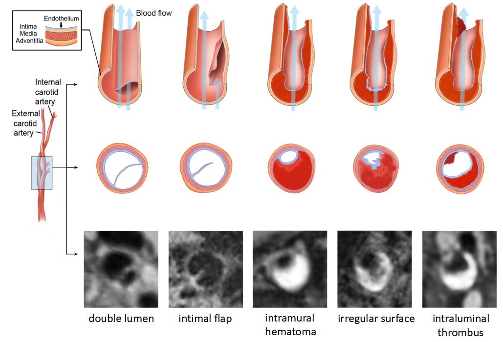
CeAD patients were scanned on a 3-tesla MRI scanner (Magnetom Verio; Siemens, Erlangen, Germany) with a 32-channel integrated head/neck coil. All patients underwent diffusion weighted imaging(DWI) and head-neck combined VWMRI within 1 month after symptom onset. Parameters for VWMRI were: 3D T1w-SPACE sequence, repetition time = 900 ms, echo time = 14 ms, FOV = 230 mm, matrix = 288×384, slices number = 224, slice thickness = 0.6 mm, voxel size = 0.6×0.6×0.6 mm3, acquisition time = 7 minutes2,3. The following types of patients were excluded: 1) accompanied with other cerebral vasculopathies (e.g. Moyamoya disease, vasculitis, intracranial or carotid arteriosclerosis with≥50% stenosis; 2) evidence of cardioembolism; 3) previous strokes or transient ischemic attacks; 4) history of transluminal intervention. Patients with divided into stroke and non-stroke groups. A diagnosis of CeAD was made if any pathognomonic signs of dissection (double lumen, intimal flap and intramural hematoma) were identified on VWMRI4,5. The VWMRI characteristics of dissected arteries including lesion location, the presence of double lumen, intimal flap, intramural hematoma, irregular surface, intraluminal thrombus and other quantitative parameters were compared between the two groups. The association among CeAD characteristics and stroke was analyzed using multivariate logistic regression.Results
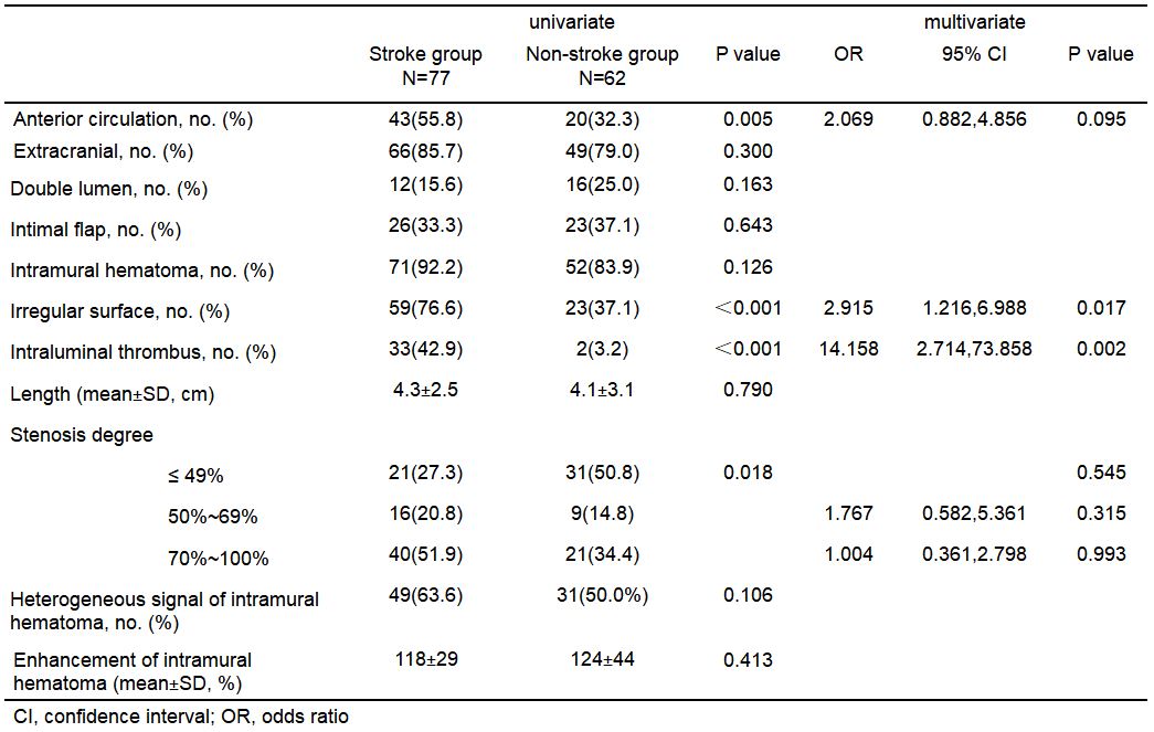
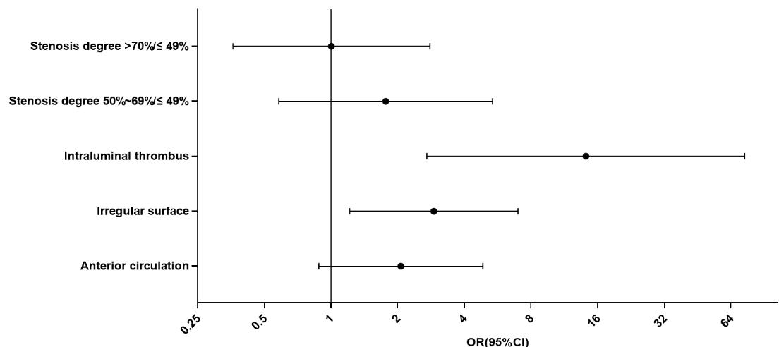
One hundred and fourteen CeAD patients were finally included. A total of 139 dissected vessels were analyzed, including 77 (55.4%) in the stroke group and 62 (44.6%) in the non-stroke group. Dissected arteries in the stroke group showed a significantly higher prevalence of an irregular surface (76.6% vs. 37.1%; P<0.001), intraluminal thrombus (42.9% vs. 3.2%; P<0.001), and severe stenosis (>70%) (51.9% vs. 34.4%; p=0.018) compared with that of the non-stroke group. Logistic regression analysis showed that the presence of an irregular surface and intraluminal thrombus were independently associated with ischemic stroke in CeAD with an odds ratio of 2.915 (95% confidence interval, 1.216-6.988, P=0.017) and 14.158 (95% confidence interval, 2.714-73.858, P=0.002).Discussion
In this study, we found a higher prevalence of irregular surface, intraluminal thrombus and severe stenosis in CEAD patients with stroke. Multivariate analysis showed that the presence of irregular surface and intraluminal thrombus were independently associated with the occurrence of ischemic stroke. Therefore, it is understandable that these high risk VWMRI features may help to better understand the relationship between unstable CEAD, formation of blood clots, and embolic stroke.
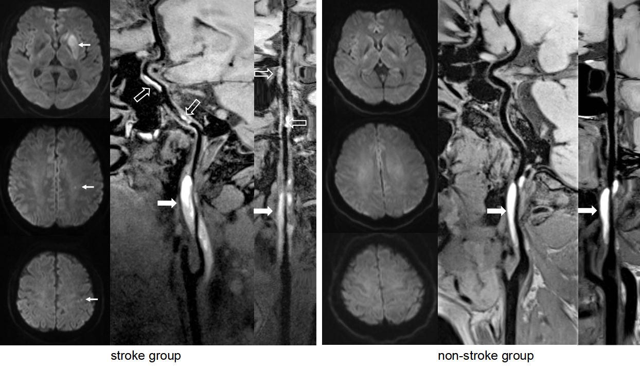
3D-VWMRI technique with an integrated head-neck coil which provided larger vessel coverage and high spatial resolution, enabling a more comprehensive evaluation of the involved artery in CeAD. In our study, we found intraluminal thrombus usually form in the distal segments of the dissected vessels on VWMRI. The presence of irregular surface may be explained by the intimal damage which may increase the risk of the formation of surface thrombus.
Conclusion
An irregular luminal surface and intraluminal thrombus are related to stroke occurrence in CeAD patients and may be useful for individual stroke stratification early after clinical presentation. Our results provide insights into the vascular pathophysiology of CeAD.Acknowledgements
No acknowledgement found.References
1. Putaala J, Metso A J, Metso T M, et al. Analysis of 1008 Consecutive Patients Aged 15 to 49 With First-Ever Ischemic Stroke: The Helsinki Young Stroke Registry. Stroke. 2009; 40(4): 1195-1203.
2. Fan Z, Yang Q, Deng Z, et al. Whole-brain intracranial vessel wall imaging at 3 Tesla using cerebrospinal fluid-attenuated T1-weighted 3D turbo spin echo. Magn Reson Med. 2017; 77(3): 1142-1150.
3. Yang Q, Deng Z, Bi X, et al. Whole-brain vessel wall MRI: A parameter tune-up solution to improve the scan efficiency of three-dimensional variable flip-angle turbo spin-echo. J Magn Reson Imaging. 2017; 46(3): 751-757.
4. Debette S M L D. Cervical-artery dissections: predisposing factors, diagnosis, and outcome. Lancet Neurol. 2009; 8(7): 668-678.
5. Debette S, Compter A, Labeyrie M A, et al. Epidemiology, pathophysiology, diagnosis, and management of intracranial artery dissection. Lancet Neurol. 2015; 14(6): 640-654.
Figures