0284
Independent Validation of U-Net Based Breast and Fibroglandular Tissue Segmentation Method on MRI Datasets Acquired Using Different ScannersYang Zhang1, Jeon-Hor Chen1,2, Kai-Ting Chang1, Vivian Youngjean Park3, Min Jung Kim3, Siwa Chan4, Peter Chang1, Daniel Chow1, Alex Luk1, Tiffany Kwong1, and Min-Ying Lydia Su1
1Department of Radiological Sciences, University of California, Irvine, CA, United States, 2Department of Radiology, E-Da Hospital and I-Shou University, Kaohsiung, Taiwan, 3Department of Radiology and Research Institute of Radiological Science, Severance Hospital, Yonsei University College of Medicine, Seoul, Korea, Democratic People's Republic of, 4Department of Medical Imaging, Taichung Tzu-Chi Hospital, Taichung, Taiwan
Synopsis
Segmentation of breast and fibroglandular tissue (FGT) using the U-net architecture was implemented using training MRI from 286 patients, and the developed model was tested in independent validation datasets from 28 healthy women acquired using 4 different MR scanners. The dice similarity coefficient was 0.86 for breast, 0.83 for FGT; and the accuracy was 0.94 for breast and 0.93 for FGT. The results on MRI acquired using different MR scanners were similar. U-net provides a fully automatic, efficient, segmentation method in large MRI datasets for evaluating its role on breast cancer risk assessment and hormonal therapy response prediction.
Introduction
Breast MRI is an established clinical imaging modality for high-risk screening, diagnosis, pre-operative staging and neoadjuvant therapy response evaluation. The most common clinical indication was diagnostic evaluation (40.3%), followed by screening (31.7%) [1]. Passage of the breast density notification law has had a major impact on MRI utilization, showing increases from 8.5% to 21.1% in non-high-risk women after the law in California went into effect [2]. The increasing popularity of breast MRI has led to the fast accumulation of large breast MRI database. This offers a great opportunity to address unanswered clinical questions regarding the use of breast density, e.g. whether the volumetric density can be incorporated into risk models to improve the prediction accuracy [3], or be used as a surrogate biomarker to predict hormonal treatment efficacy [4,5]. Since MRI is a three-dimensional (3D) imaging modality with distinctive tissue contrast, it can be used to measure the fibroglandular tissue (FGT) volume. However, because many imaging slices are acquired in one MRI, an efficient, objective, and reliable segmentation method is needed. In a previous study we have developed an automatic segmentation method using the Fully-Convolutional Residual Neural Network (FC-RNN), commonly noted as U-net, with non-fat-sat T1-weighted MRI of 286 patients [6]. In this study we tested the segmentation performance of this developed method in 56 breasts of 28 healthy volunteers, each woman acquired using 4 different scanners.Methods
The initial dataset used for training included 286 patients with unilateral invasive breast cancer (median age, 49 years; range, 30–80 years), and only the contralateral normal breast was analyzed. MRI was performed on a 3T Siemens Trio-Tim scanner, and the pre-contrast T1-weighted images without fat suppression were used for segmentation. The ground truth was generated using a template-based segmentation method [7]. Deep learning segmentation was performed using the U-net, which is a fully connected convolutional residual network [8], and consists of convolution and max-pooling layers at the descending part (the left component of U), and convolution and up-sampling layers at ascending part (the right component of U). The final segmentation model was developed using 10-fold cross-validation. The independent validation dataset included 28 healthy volunteers (age 20–64, mean 35 years old). Each subject was scanned using four different MR scanners in two institutions, including GE Signa-HDx 1.5T, GE Signa-HDx 3T (GE Healthcare, Milwaukee, WI), Philips Achieva 3.0T TX (Philips Medical Systems, Eindhoven, Netherlands) and Siemens Symphony 1.5T TIM (Siemens, Erlangen, Germany). Non-contrast T1-weighted images without fat suppression were used for segmentation. Since both left and right breasts were normal, they were analyzed separately, so there was a total of 56 breasts. The validation was done using the 56 breasts acquired by each scanner first, and then using all 224 breasts acquired by all 4 scanners together. The ground truth was generated using the template-based method for comparison. The segmentation performance was evaluated using the Dice Similarity Coefficient (DSC) and the overall accuracy based on all pixels. In addition, the Pearson’s correlation was applied to evaluate the correlation between the U-net prediction output and the ground truth.Results
The DSC and accuracy for each scanner was calculated separately, and then combined for all 4 scanners together. The results are shown in Table 1. Figures 1 and 2 illustrate the segmentation results of two women with different breast morphology. The correlation between the U-net prediction output and ground truth for breast volume is shown in Figure 3. The obtained results for four different scanners were similar. The correlation coefficient r was high, in the range of 0.96-0.98. In each figure, the fitted line was very close to the unity line, and the slope was close to 1. The segmentation result for FGT volume is shown in Figure 4. The FGT segmentation results for MRI acquired using 4 different scanners were similar. The correlation coefficient r was very high, in the range of 0.97-0.99. However, using the unity line as reference, the U-net segmented FGT volume was lower compared to the ground truth, as in the two case examples demonstrated in Figures 1 and 2.Discussion
The results suggest that deep learning segmentation using U-net is feasible to perform fully automatic segmentation for the breast and FGT and yield reasonable accuracy compared to the ground truth segmented by using a template-based method verified by a radiologist. Over the last decade, segmentation of the breast and FGT on MRI has been studied using semiautomatic to automatic approaches with some operator inputs. The processing time for these methods varies from minutes to more than half an hour, which is partially due to the need for the post-segmentation manual correction. The testing in this study using independent validation datasets allows us to evaluate whether the developed segmentation method can be applied widely to other MRI datasets acquired using different imaging sequences on different MR scanners. This fully-automatic deep learning-based segmentation method may provide an accurate and efficient means to quantify FGT volume for evaluation of breast density in very large datasets. The results can potentially be used for breast cancer risk assessment, and for evaluating the response in women receiving hormonal therapy or chemoprevention.Acknowledgements
This study is supported in part by NIH R01 CA127927, R21 CA208938, and a Basic Science Research Program through the National Research Foundation of Korea (NRF) funded by the Ministry of Education (NRF-2017R1D1A1B03035995).References
[1] Wernli et al. Patterns of breast magnetic resonance imaging use in community practice. JAMA internal medicine. 2014;174(1):125-132. [2] Ram et al. Impact of the California Breast Density Law on Screening Breast MR Utilization, Provider Ordering Practices, and Patient Demographics. Journal of the American College of Radiology. 2018;15(4):594-600. [3] Kerlikowske et al. Combining quantitative and qualitative breast density measures to assess breast cancer risk. Breast Cancer Research. 2017;19(1):97. [4] Lundberg et al. Association of infertility and fertility treatment with mammographic density in a large screening-based cohort of women: a cross-sectional study. Breast Cancer Research. 2016;18(1):36. [5] Chen et al. Reduction of breast density following tamoxifen treatment evaluated by 3-D MRI: preliminary study. Magnetic resonance imaging. 2011;29(1):91-98. [6] Zhang et al. Automatic Breast and Fibroglandular Tissue Segmentation Using Deep Learning by A Fully-Convolutional Residual Neural Network. Presented at the Joint Annual Meeting ISMRM-ESMRMB, Paris, France, June 16-21, 2018; Program Number: 2420. [7] Lin et al. Template‐based automatic breast segmentation on MRI by excluding the chest region. Medical physics. 2013;40(12). [8] Ronneberger et al. U-net: Convolutional networks for biomedical image segmentation. Paper presented at: International Conference on Medical image computing and computer-assisted intervention 2015.Figures
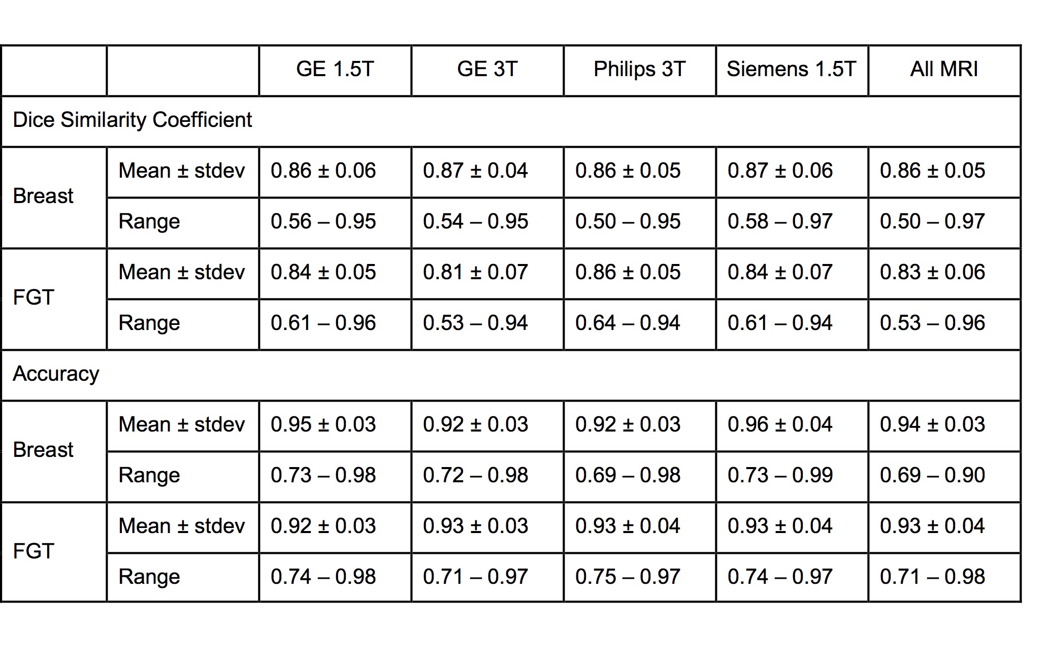
Table 1. The dice similarity
coefficient (DSC) and the accuracy for the segmentation of breast and FGT in
different MR scanners.
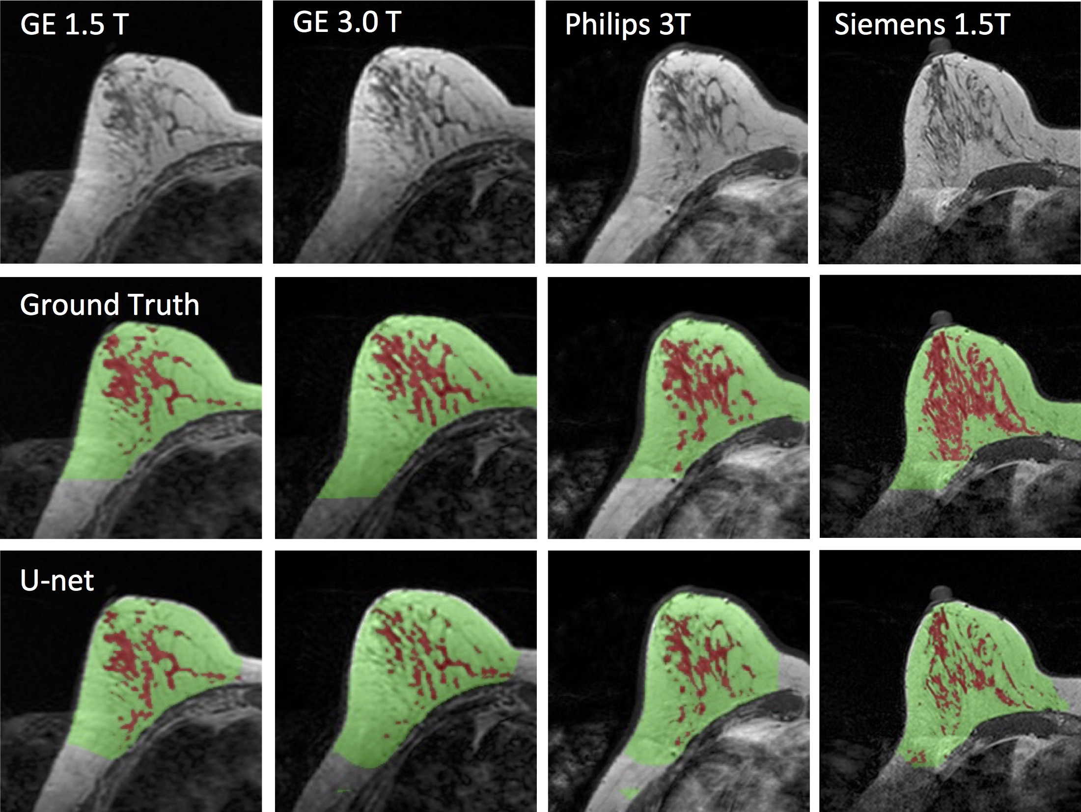
Figure 1. Images of a 43-year-old woman with
heterogeneous breast morphology acquired using the GE 1.5T, GE 3.0T, Philips
3.0T, and Siemens 1.5T systems. The top row shows the original images. The
center row shows the ground truth obtained by using the template-based segmentation
method. The bottom row shows the U-net prediction results. The FGT volume
segmented by U-net is smaller compared to the ground truth.
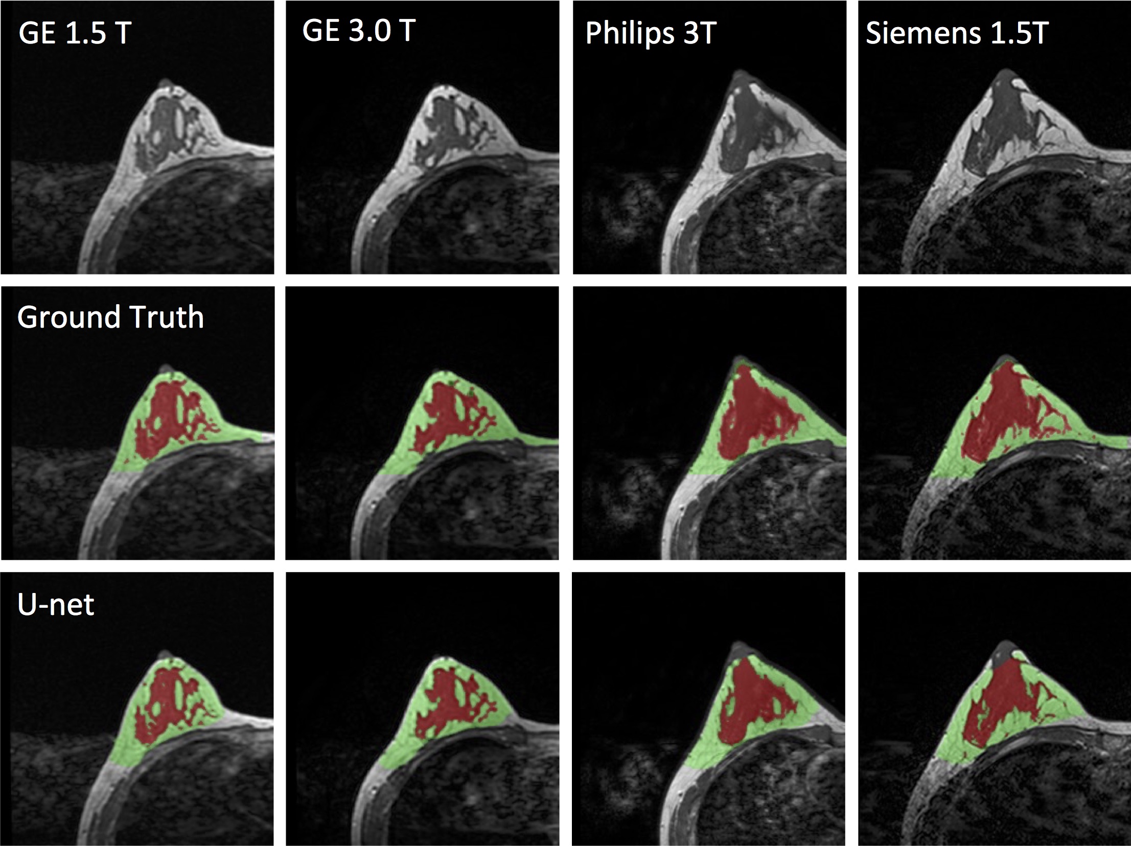
Figure 2. Images of a 29-year-old
woman with dense breast acquired using the GE 1.5T, GE 3.0T, Philips 3.0T, and Siemens
1.5T systems. The top row shows the original images. The center row shows the
ground truth obtained by using the template-based segmentation method. The
bottom row shows the U-net prediction results.
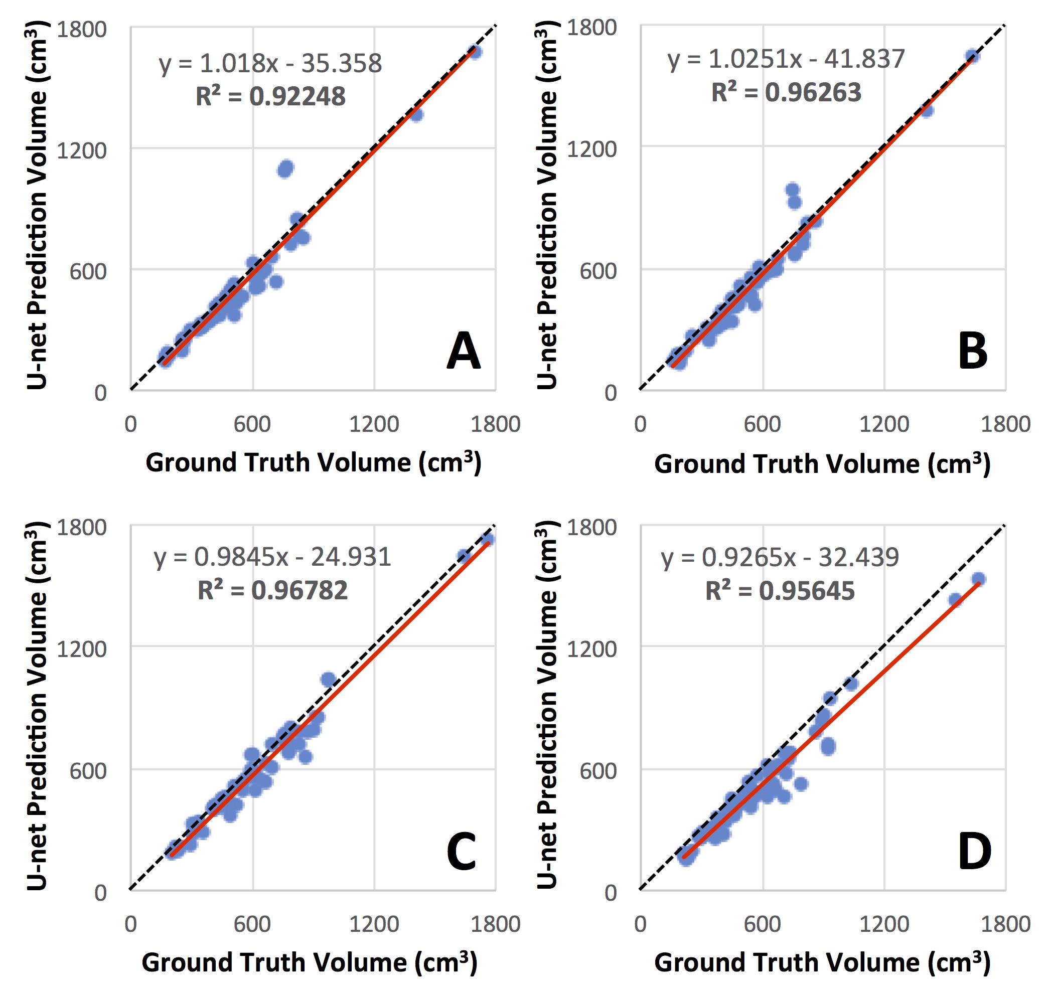
Figure 3. Correlation of breast volume between the
ground truth obtained from the template-based segmentation method and the U-net
prediction. (A) GE 1.5 T, (B) GE 3T, (C) Philips 3T, (D) Siemens 1.5T. The red
line is the trend line, and the dashed black line is the unity line as
reference.
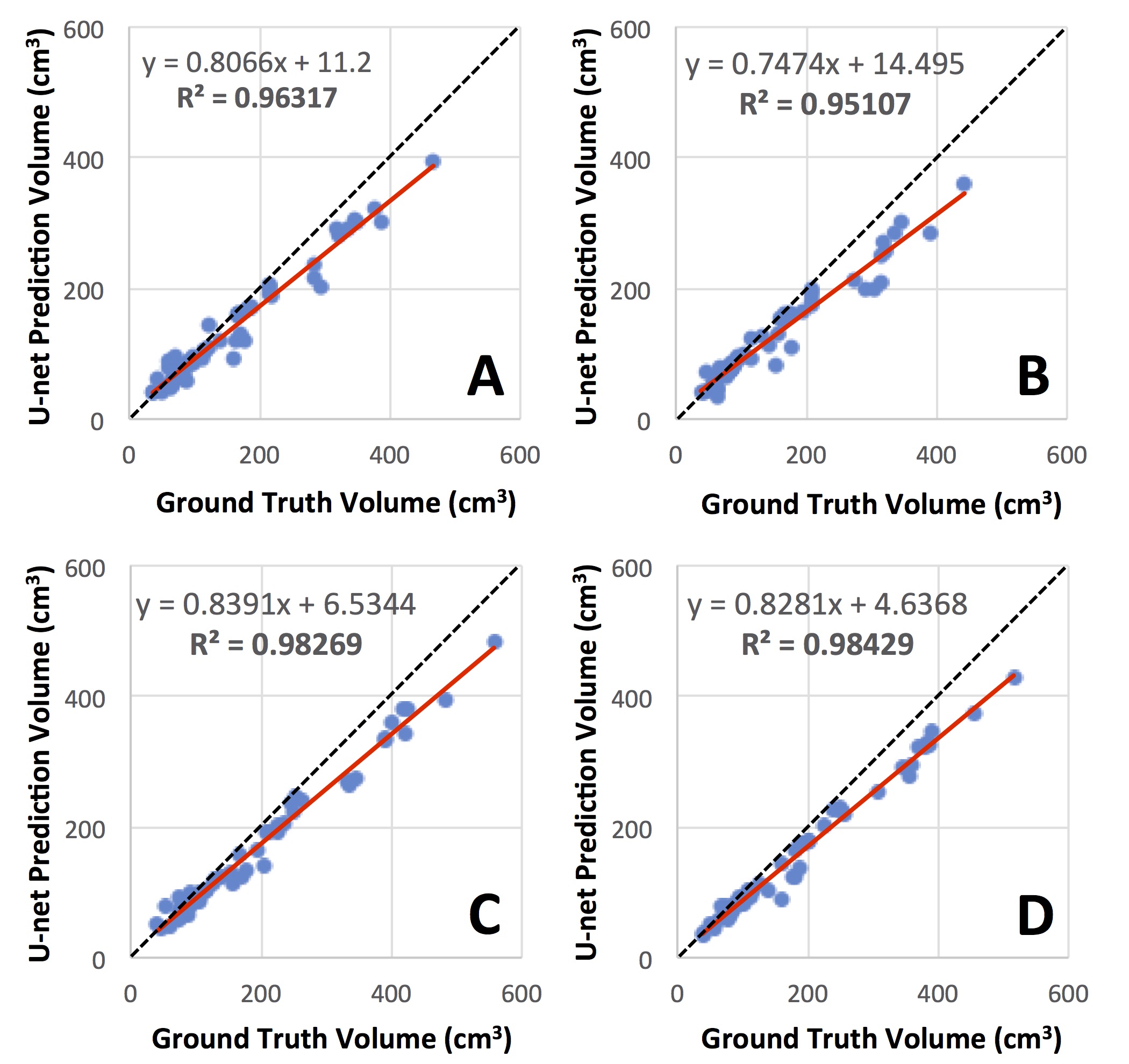
Figure 4. Correlation of FGT volume
between the ground truth obtained from the template-based segmentation method
and the U-net prediction. (A) GE 1.5 T, (B) GE 3T, (C) Philips 3T, (D) Siemens
1.5T. The red line is the trend line, and the dashed black line is the unity
line as reference. The volume segmented by U-net is smaller compared to the
ground truth.