0280
Co-registration of Breast Diffusion MR Images Across Multiple Time Points in a Longitudinal Study to Evaluate the Response to Neoadjuvant Chemotherapy1Center for MR Research, University of Illinois at Chicago, Chicago, IL, United States, 2Department of Electrical and Computer Engineering, University of Illinois at Chicago, Chicago, IL, United States, 3Department of Bioengineering, University of Illinois at Chicago, Chicago, IL, United States, 4Department of Diagnostic Radiology, National Cancer Center and National Clinical Research Center for Cancer, Cancer Hospital, Chinese Academy of Medical Sciences and Peking Union Medical College, Beijing, China, 5Departments of Radiology and Neurosurgery, University of Illinois at Chicago, Chicago, IL, United States
Synopsis
Breast cancer is one of the most common cancers among women. Recently, several diffusion models have been proposed to characterize breast cancer. To extend these models for the assessment or prediction of treatment response of breast cancer, image co-registration throughout the time course of treatment is a significant challenge, particularly considering the vulnerability to deformation of the breast tissue. In this study, we demonstrate a 3D non-rigid co-registration method, and apply it to diffusion weighted (DW) images acquired in a longitudinal study during neoadjuvant chemotherapy.
Introduction
Neoadjuvant chemotherapy has been considered as the standard of care for the treatment of breast cancer. An accurate and non-invasive prediction of treatment response to neoadjuvant therapy is of great importance for optimizing the treatment strategies. To overcome the limitations of the morphological imaging techniques in assessing microstructural changes in breast tumor tissue during treatment, diffusion-weighted MRI (DWI) has been proposed for predicting pathological response1. Lately, advanced non-Gaussian diffusion models have shown promise in predicting breast cancer’s response to treatment2-4. Although these models provide voxel-level spatial information on tumor characteristics, changes in the quantitative parameters are typically performed in the manually drawn regions of interests (ROIs) across multiple treatment time points. Efforts in co-registering breast images in longitudinal studies have been hampered by soft tissue deformation, tumor-shrinking, and inconsistency in patient positions. In this study, we employ a 3D non-rigid registration technique based on a demon algorithm5-8 to co-register the diffusion-weighted (DW) images at different time points during neoadjuvant chemotherapy. This technique is demonstrated through a collection of DWI parameters obtained from both Gaussian and non-Gaussian DWI models, including mono-exponential, stretched-exponential9, intra-voxel incoherent motion (IVIM)10, and continuous-time random-walk (CTRW) models11.Methods
Image Acquisition: DWI was performed on twenty-five breast cancer patients receiving neoadjuvant chemotherapy on a 3T MRI scanner (GE Healthcare, Discovery MR750) with 12 b-values from 0 to 3000 sec/mm2. The other acquisition parameters were: TR/TE = 3378/79.3ms, slice thickness = 5mm, matrix size = 256x256. Images were obtained at three time points: before treatment (T1), during treatment (T2), and after treatment (T3).
Longitudinal Co-registration of Images: To perform a 3D affine and non-rigid registration, the demon algorithm5-8 was employed by defining a "pixel velocity" with the use of intensity differences and gradient information between time points, smoothing this velocity field by a Gaussian kernel, and using it to transform the images at T2 or T3 to co-register with the reference images at T1, iteratively.
DWI Analysis: The co-registered DW images were analyzed with four DWI models: 1) mono-exponential model to estimate apparent diffusion coefficient (ADC) by using b-values of 0 and 1000 sec/mm2. 2) stretched-exponential model to estimate distributed diffusion coefficient (DDC) and αSE. 3) IVIM model to estimate f, Dperf, and Ddiff, and 4) CTRW model12: S/S0 = (Eα(-bDm)β), to estimate the anomalous diffusion coefficient, Dm,and temporal and spatial diffusion heterogeneity parameters αCTRW and βCTRW, respectively. Twelve b-values (0-3000 sec/mm2) were used in the stretched-exponential and CTRW models, while 5 b-values (0-750 sec/mm2) were used in the IVIM model.
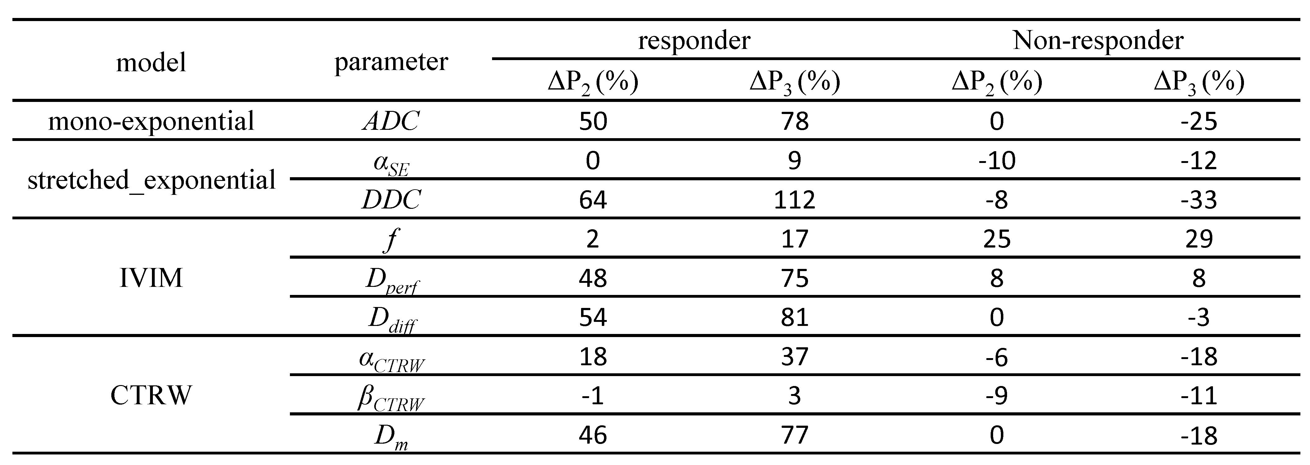
Statistical Analysis: With co-registration, the percentage changes in the mean parameter values at T2 (ΔP2) and T3 (ΔP3) were calculated over the tumor ROIs drawn on the images at T1 with the following formula: ΔPi = (mean value at Ti - mean value at T1) / (mean value at T1) x100. The descriptive statistics of the parameter changes in all models were computed for one representative responder and one representative non-responder breast cancer patient.
Results
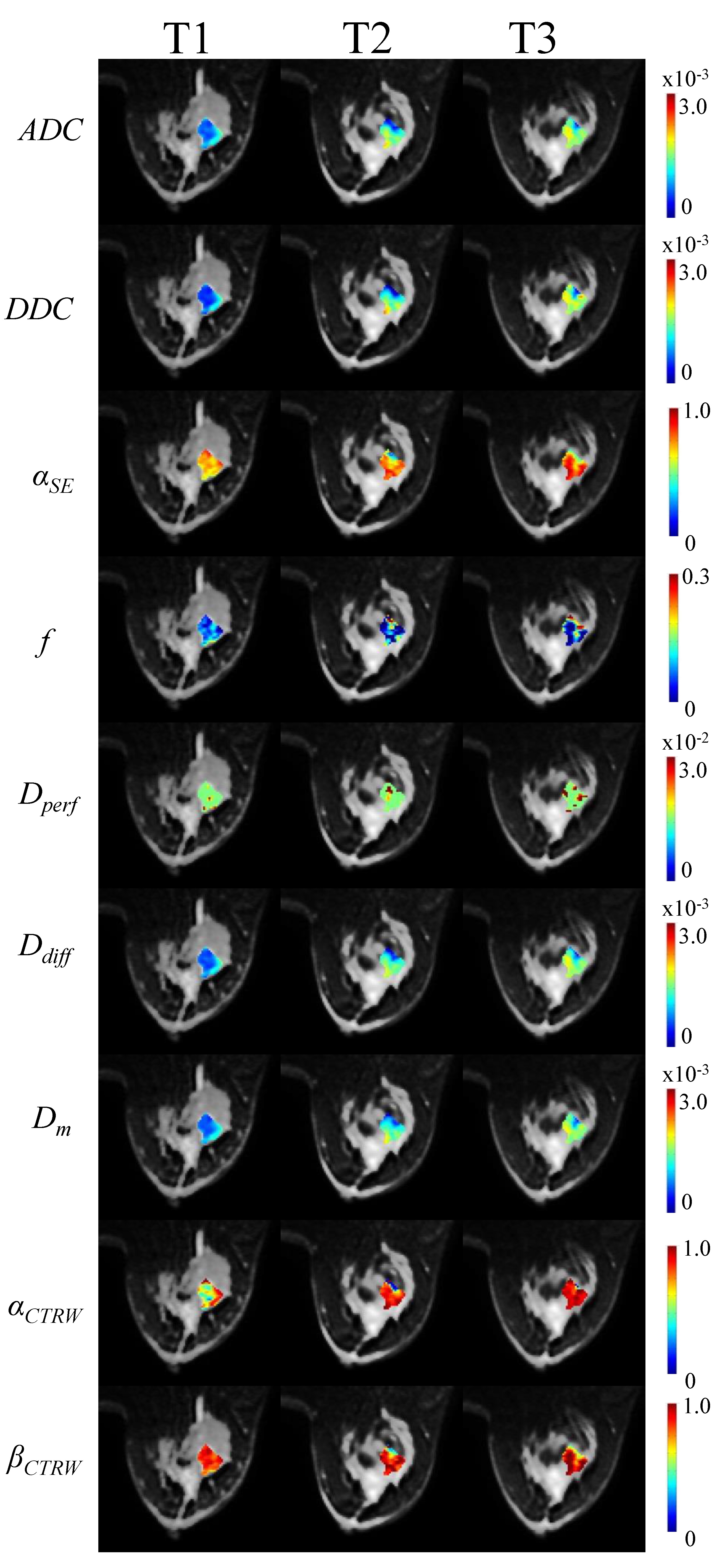
The top and bottom rows of Fig. 1 display the original and co-registered DW images, respectively, with the first three columns corresponding to T1, T2, and T3 from a representative patient. After co-registration, the images at T2 and T3 are well-aligned with the reference image at T1. The effectiveness of co-registration is further illustrated in the right two columns as indicated by the arrows. Fig. 2 shows a panel of parameter maps from a responder throughout the time course of treatment. Substantial changes in many parameters were evident. This observation was quantified in Fig. 3 by summarizing the mean parameter value changes, ΔP2 and ΔP3, computed from one representative responder and one representative non-responder. The mean changes in diffusion coefficients, ADC, DDC, Dperf, Ddiff, and Dm, were all positive and substantially higher in the responder than the non-responder. The parameters, αSE, αCTRW , and βCTRW, were also higher while f was lower in the responder patient. Within the non-diffusion coefficient parameters, αCTRW exhibited more pronounced change as compared to others.Discussion and Conclusion
We have demonstrated the use of a 3D non-rigid and affine co-registration technique for co-registering longitudinal DW images throughout the course of neoadjuvant chemotherapy on breast cancer patients. We have also shown that changes in the tumor in response to treatment can be well-reflected in a set of diffusion parameters computed from several DWI models on the co-registered DW images. Enabling a quantitative comparison between DWI parameters at different time points in a rigorous way, this approach contributes to the ongoing efforts of exploring various DWI models to assess breast cancer’s response to treatment.Acknowledgements
This work was supported in part by General Electric Healthcare in China.References
- Virostko J, Hainline A, Kang H, et al. Dynamic contrast-enhanced magnetic resonance imaging and diffusion-weighted magnetic resonance imaging for predicting the response of locally advanced breast cancer to neoadjuvant therapy: a meta-analysis. Journal of Medical Imaging. 2017; 5(1): 011011.
- Che S, Zhao X, Ou Y, et al. Role of the intravoxel incoherent motion diffusion weighted imaging in the pre-treatment prediction and early response monitoring to neoadjuvant chemotherapy in locally advanced breast cancer. Medicine. 2016; 95(4): e2420.
- Kim Y, Kim SH, Lee HW, et al. Intravoxel incoherent motion diffusion-weighted MRI for predicting response to neoadjuvant chemotherapy in breast cancer. Magnetic Resonance Imaging. 2018; 48: 27-33.
- Cho GY, Gennaro L, Sutton EJ, et al. Intravoxel incoherent motion (IVIM) histogram biomarkers for prediction of neoadjuvant treatment response in breast cancer patients. Eur J Radiol Open. 2017; 4: 101-107.
- Kroon DJ and Cornelis HS. MRI modality transformation in demon registration. Biomedical Imaging: From Nano to Macro. ISBI'09. IEEE International Symposium on. IEEE, 2009.
- Cachier P, Xavier P, Ayache N, et al. Fast non rigid matching by gradient descent: Study and improvements of the "demons" algorithm. Diss. INRIA, 1999.
- Thirion, JP. Image matching as a diffusion process: an analogy with Maxwell's demons. Medical Image Analysis. 1998; 2(3): 243-260.
- Wang H, Lei D, O'Daniel J, et al. Validation of an accelerated "demons" algorithm for deformable image registration in radiation therapy. Physics in Medicine & Biology. 2005; 50(12): 2887.
- Bennett KM, Schmainda KM, Bennett RT, et al. Characterization of continuously distributed cortical water diffusion rates with a stretched‐exponential model. Magn Reson Med. 2003; 50(4): 727-734.
- Le Bihan D. Intravoxel incoherent motion perfusion MR imaging: a wake-up call. Radiology. 2008; 249(3): 748-752.
- Ingo C, Magin RL, Colon-Perez L, et al. On random walks and entropy in diffusion‐weighted magnetic resonance imaging studies of neural tissue. Magn Reson Med. 2014; 71(2): 617-627.
- Karaman MM, Sui Y, Wang H, et al. Differentiating low- and high-grade pediatric brain tumors using a continuous-time random-walk diffusion model at high b-values. Magn Reson Med. 2016; 76 (4): 1149-1157.
Figures