0275
Equivalent-Charge-Based Optimization of Spokes-and-Hub Magnets for Hand-Held and Classroom MR Imaging1Department of Electrical Engineering and Computer Science, Massachusetts Institute of Technology, Cambridge, MA, United States, 2Athinoula A. Martinos Center for Biomedical Imaging, Massachusetts General Hospital, Charlestown, MA, United States, 3Harvard Medical School, Boston, MA, United States, 4Institute for Medical Engineering and Science, Massachusetts Institute of Technology, Cambridge, MA, United States
Synopsis
Differentiating the potential from end-cap equivalent charges is so efficient at computing fields from bar magnets, that in less than minute, a laptop running MATLAB can computationally-optimize field uniformity in hundred-bar wagon-wheel (or spokes-and-hub) magnets. And optimized spokes-and-hub magnets have several advantages for hand-held and classroom low-field single-slice MR imaging (50-200 mT). Their frame is open, and they are easily assembled and scaled. We demonstrate several such magnets, from finger to wrist to infant size, and match magnetic field measurements to quadrature-based equivalent-charge simulation. We also demonstrate generating spin echoes using a spokes-and-hub magnet in a 200 mT tabletop imager.
Introduction
The cost, size, and safety of the magnets used in clinical MRI scanners disqualify them from point-of-care diagnostics. And even in educational MR systems, the magnets are expensive and dangerous to handle. As an alternative, we propose a spoke-and-hub permanent magnet topology that is easy to assemble, safe to handle, and efficiently simulated and optimized.1,2 Below we show several “spoke-and-hub” magnets designed using simulation-based optimization, and compare the measured and simulated fields for designs of varying size (1.3”-6” diameter openings), field-strengths (50-200 mT), and application type (classroom to clinical systems).Methods
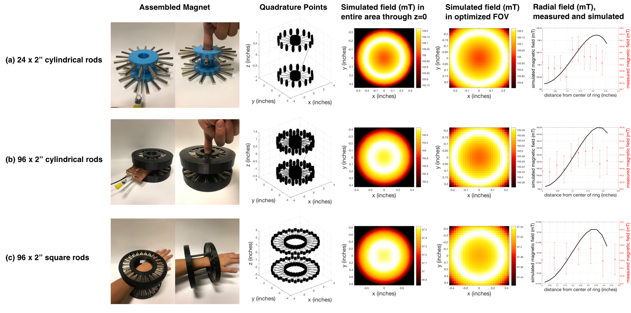
Gauss-Legendre-quadrature based discretization of the equivalent-charge method was used to compute magnetic fields from axially-magnetized, cylindrical and square rods arranged in two large rings with opposite magnet directions (Fig. 1).3 Because the fields are so efficiently computed, optimization techniques can be used to quickly tune parameters for magnet design. Levenberg-Marquardt nonlinear least squares optimization was used to minimize the variation in field about a desired FOV at the center of the magnet, and obtain an optimal z-axis separation distance between the two sets of magnet rings.
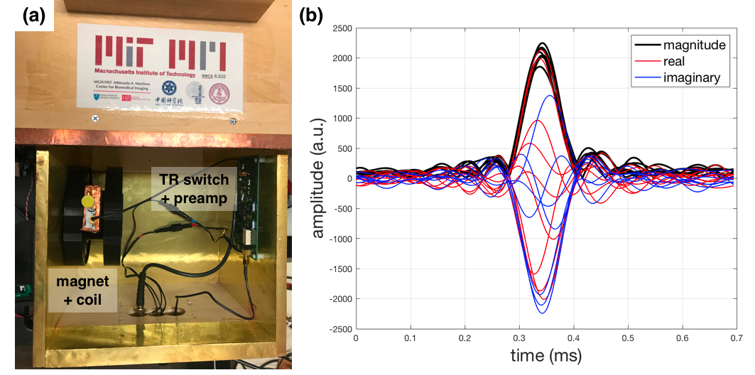
From these simulations, we built three spokes-and-hub magnets by arranging cylindrical- and square-shaped bar magnets (K&J Magnetics, Plumsteadville, PA) into slots in 3D-printed enclosures. As shown in Fig. 1a, b, and c, the B0 fields of the three magnets with increasing ring size were 105, 195, and 60 mT, respectively. Since the z-directed field is assumed to be radially symmetric about the z = 0 plane, we took field measurements along the inner radius of the magnet within the optimized FOV of interest using an ALS31300 3D Linear Hall-Effect Sensor (Allegro Microsystems, Manchester, NH). Additionally, spin echoes were generated for samples placed inside a 10 mm NMR tube using the TR switch, preamplifier, and coil from the MIT/Martino’s tabletop scanners.4
Results
We obtained MATLAB-simulated maps and measurements for the z-directed static magnetic field of the three magnets (Fig. 1). The comparison plot of radial fields shows that, within the precision of of the Hall-Effect probe, the magnitude and trend of the measured and simulated field inhomogeneities match quite closely, though there is a slight (<1 mT) absolute offset bias due to the weighting . These three low-field magnet prototypes validate the scalability of spokes-and-hub design, from a classroom scanner capable of imaging a small 10 mm test tube to a larger hand-held clinical design with an imaging area of a wrist. Additionally, using the MIT/Martino’s tabletop scanner signal chain, we generated spin echoes using the 195 mT magnet shown in Fig. 1b (Fig. 2).4
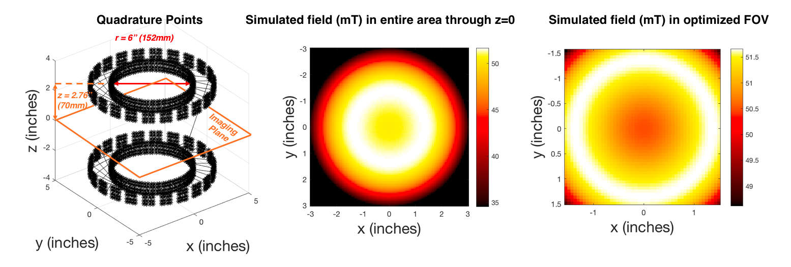
Furthermore, we confirmed in simulation that reasonable 50 mT fields for an imaging plane capable of encompassing limbs or even a newborn baby’s brain can be obtained with a 6” wide spokes-and-hub ring opening and constructed with easily-obtainable 0.75” x 0.75” x 1.5” axially-magnetized bar magnets similar to the magnet prototypes we have built (Fig. 3).
Discussion and Conclusion
We characterized and demonstrated feasibility of several, easy-to-assemble spokes-and-hub low-field permanent magnet structures for MRI. In simulation, we have a computationally-efficient method for accurately modeling the B0 field across multiple planes within the magnet bore. With the ability to efficiently simulate the fields, we can further optimize the fields within the magnet to also allow for built-in linear gradients in the static magnetic field by introducing unequal numbers of bar magnets within each ring. Regarding the design of the magnet, its open structure allows for imaging along all three axes, as well as potential for utilization of rotation for gradient encoding and projection-based encodings.5 The spoke rings can also be easily taken apart to allow for novel coil geometries. Of particular interest to us, the simulation results for a 6” wide spokes-and-hub magnet demonstrates its promise as a low-cost, hand-held tool for clinical scanning of pediatric brains most notably with applications towards hydrocephalus detection in newborns. Moving further, we plan to apply our spokes-and-hub topology to an imaging system capable of these clinical applications with fields optimized to capitalize on the magnet’s inherent inhomogeneities.Acknowledgements
The authors graciously acknowledge support from NIH NIBIB R01EB018976, MIT-MGH seed grant, and Skolkovo Institute of Technology Next Generation Program.References
- Moresi, G., Magin, R. Miniature Permanent Magnet for Table-top NMR. Concepts Magn. Res. B. 2003, p. 35.
- Raich, H., Blumler, P. Design and Construction of a Dipolar Halbach Array with a Homogeneous Field from Identical Bar Magnets: NMR Mandhalas. Concepts Magn. Res. B. 2004, p. 16.
- Curti M., Paulides JJH., Lomonova, EA. An Overview of Analytical Methods for Magnetic Field Computation. Conference on Ecological Vehicles and Renewable Energies, 2015.
- Cooley CZ, Stockmann JP, LaPierre C, Witzel T, Jia F, Zaitsev M, Stang P, Scott G, Wenhui Y, Zheng W, Wald LL. Implementation of low-cost, instructional tabletop MRI scanners. Int. Soc. Magn. Res. Med. 2014, p. 4819.
- Cooley CZ, Stockmann JP, Armstrong BD, Sarracanie M, Lev MH, Rosen MS, Wald LL. 2D Imaging in a Lightweight Portable MRI Scanner without Gradient Coils. Magn. Res. Med. 2015, p. 1.
Figures