0270
MRI Powered and Triggered Current Stimulator for Concurrent Stimulation and MRI1Weldon School of Biomedical Engineering, Purdue University, West Lafayette, IN, United States, 2MR-LINK LLC, West Lafayette, IN, United States, 3Electrical and Computer Engineering, Purdue University, West Lafayette, IN, United States
Synopsis
The integration of stimulation, recording and high-field MRI has significant potential to evaluate different organs (brain, heart, gut etc.). However, externally powered devices consisting of cables and connectors for powering and synchronization may perturb the magnetic field within the MRI system and create additional safety concerns at high-fields. Here we present an MRI powered and triggered system, namely X-ON, to deliver electrical stimulation during MRI operation. The MR-compatible system can harvest wireless energy from varying gradient fields and provide programmable current stimulation in synchronization with MRI or fMRI acquisition.
Introduction
The integration of stimulation, recording and high-field MRI has opened new avenues for understanding functions of various organs (brain, heart, gut etc.). However, strong, varying magnetic fields introduced during MR imaging poses a hostile environment for any externally powered device (for recording and stimulation) to be integrated to the imaging platform. Furthermore, additional powering and synchronizing cables can perturb the highly uniform magnetic field, thus degrading image acquisition. To address these issues, here we present an MRI powered and triggered system (X-ON) to deliver electrical stimulation during MRI operation. The system can harvest wireless energy from varying gradient fields and provide programmable current stimulation in synchronization with MRI or fMRI acquisition.Methods
The device (X-ON) consisted of two major parts: (a) a power harvesting and triggering unit and, (b) a programmable stimulation unit (Fig. 2). The whole system fitted inside a custom enclosure (Fig. 1a) that clamped on to the animal holder tray (Fig. 1c) of a 7-tesla horizontal-bore small animal MRI system (BioSpec 70/30; Bruker Instruments, Billerica, USA). The power harvesting unit utilized a low frequency (LF) machine wound copper coil (14mm diameter, Fig. 1b) to pick up gradient changes during MR imaging. This signal was rectified and passed through a DC/DC converter to generate two voltage rails: VHigh (11V) and VLow (3.3V) for operating the stimulation circuit and the on-board micro-controller, respectively (Fig. 2). Additionally, the pickup signal was used to generate imaging triggers for synchronization of the stimulation pulses with MR imaging1,2,3. The high voltage compliant (±11V), bi-phasic current stimulator (Fig. 2) provided tunable stimulation profiles for a wide range of stimulation paradigms applicable to tissue, brain, and nerves, to name a few.
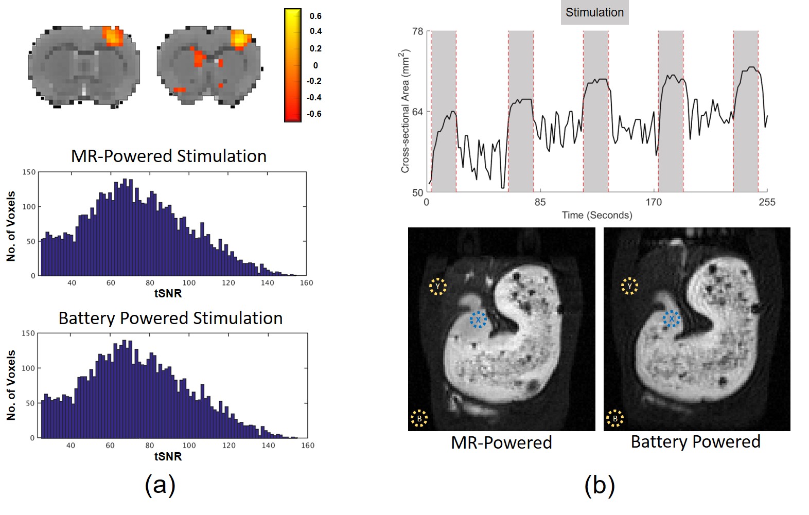
The power-harvesting capability of the system was tested by evaluating the load power characteristics of the system during two widely used MRI sequences: (a) echo-planar imaging (EPI) and (b) fast low angle shot (FLASH). Furthermore, the low power stimulator (12mW) was operated simultaneously during these imaging sessions to provide MRI powered and triggered stimulation. Efficacy of the wireless stimulator was analyzed through two experimental setups (a) BOLD response evoked by forepaw stimulation (EPI) and (b) gastric motility in response to VNS (multi-slice 2D FLASH scans). Four Sprague-Dawley rats (male, 228-330 g) were used for these studies in accordance with procedures approved by Institutional Animal Care and Use Committee (IACUC). Lastly, the MR-compatibility of the system was further quantitatively analyzed by obtaining the temporal SNR (tSNR) and contrast-to-noise ratio (CNR) for EPI and FLASH, respectively, during which the stimulator harvested power and provided stimulation.
BOLD fMRI: 2-D single-shot gradient echo EPI sequence (TR = 1 s, TE = 15 ms, FA = 55°, in-plane resolution about 0.6×0.6 mm2, slice thickness = 1 mm. Stimulation Setting: Monophasic, 10Hz (frequency), 2 mA (amplitude), 2 ms (pulse width), 30s (ON) and 30s (OFF) block design.
Gastric MRI: a 2-D FLASH sequence (TR = 11.784 ms, TE = 1.09ms, FA = 25°, in-plane resolution about 0.47×0.47 mm2, slice thickness =1.5 mm). Stimulation Setting: Monophasic, 20Hz (frequency), 0.4 mA (amplitude), 0.5 ms (pulse width), 20s (ON) and 40s (OFF) block design.
Results
As load resistance was varied during the gradient echo sequences, a continuous peak power of 72mw and 22mw was harvested for the EPI and FLASH sequence respectively (Fig. 2c). Both peak powers were sufficient for the operation of the stimulator system. For the fMRI study, a clear stimulation-evoked response was observed in the somatosensory cortex forelimb region (Fig. 3a). During the VNS experiment, monophasic, efferent stimulation of the vagal nerve with small stimulation dose (20Hz, 0.4mA, 0.5ms) relaxed antral contractions and produced secretion in the gastric antrum (Fig. 3b). Finally, MR-compatibility experiments for EPI study shows no significant changes in tSNR and image quality. Although CNR in the FLASH scans decreased by 5dB (36db during power harvesting compared to 31dB when the device was powered through battery), no qualitative degradation of the MR-images was observed. This decreased CNR was attributed to an increase of SD (1.68 to 3.3) of the image background noise while the mean signal intensity of both the stomach (~120) and muscle tissues (~20) remained constant.Conclusions
In this study, we have presented a miniaturized stimulation system, that is tailored for simultaneous operation alongside MR imaging. The unique plug-and-play system wirelessly synchronizes with the imaging system and does not require any additional cable connection. The power harvesting unit described herein, can scavenge up to 72 mW power from the varying gradients present during fMRI. This wireless power harvesting technique can be further expanded to support a wide range of MRI integrated systems or wireless receiver coils to enhance MRI capabilities for animal and human applications.Acknowledgements
No acknowledgement found.References
- R. Mandal et al., "Adaptive and Wireless Recordings of Electrophysiological Signals during Concurrent Magnetic Resonance Imaging," in IEEE Transactions on Biomedical Engineering.doi: 10.1109/TBME.2018.2877640
- R. Mandal et al., “Multimodal imaging: MR-compatible, gradient artifact free, wireless recording system integrated with MR-scanner for simultaneous EEG and fMRI acquisition,” in Proc. Int. Soc. Mag. Reson. Med., 2017 vol. 25, paper. 4072.
- R. Mandal et al., “Recording electrophysiological signals through MR receiver coils during concurrent fMRI,” Proc. Int. Soc. Mag. Reson. Med., 2018 vol. 26, paper. 4660.
- J. Hofflin et al., “Energy harvesting with a figure-8 coil - towards energy autonomous MRI detection,” in Proc. Exp. Nucl. Magn. Resonance Conf., 2016.
Figures