0257
Hyperpolarized [1-13C]pyruvate MRS in a large animal model of partial renal obstruction supports clinical use in patients1MR Research Centre, Aarhus University, Aarhus N, Denmark, 2Department of Cardiology, Aarhus University Hospital, Skejby, Denmark, 3GE Healthcare, Munich, Germany
Synopsis
Hyperpolarized [1-13C]pyruvate was used in this study to explorer the capability of detecting ischemic injury in the kidneys and to present the heterogeneity from the different kidney compartments. This was done using Michaelis–Menten kinetics by obtaining data in
Purpose
Previous studies in rodents have demonstrated altered hemodynamic and metabolic status in renal ischemia-reperfusion models (1-3). These models, however, expose the kidney to complete ischemia and do not allow for partial obstruction of the kidneys. Here, we evaluate the potential of a spectral-spatial saturation-recovery imaging approach (4) to investigate the potential intra-renal heterogeneities associated with a local occlusion. We aimed to show the extent and localization of hemodynamic and metabolic changes associated with partial obstruction of the kidney. We hypothesize that a spectral-spatial saturation recovery imaging approach can accurately determine the intra-renal hemodynamic and metabolic fates of [1-13C]pyruvate, in a large animal model of partial obstruction of the kidney.Methods
Four healthy, female, domestic pigs weighing 30 kg were included in this study. During general anaesthesia, a branch of the left renal artery was occluded with a balloon introduced through the femoral artery to obtain local ischemia for 60 min. After 90 minutes of reperfusion, hyperpolarized measurements were performed with and without a low dose (2.5 uq/kg/hr) of continuous dopamine infusion. The MR experiments were performed on a clinical 3T scanner (MR750, GE Healthcare, Milwaukee, WI, USA), equipped with a 13C Helmholtz loop coil (PulseTeq, UK) (ø=20cm) covering both kidneys. Proton scout images were acquired with a modified 1H 8-channel heart array coil. Spectral-spatial saturation recovery spiral imaging (4) was used to image the renal distribution of [1-13C]pyruvate and its metabolic conversion. [1-13C]pyruvate was polarized in a SpinLab polarizer (GE Healthcare, Milwaukee, WI, USA) for ~2.5 hours to ~40% polarization. Hyperpolarized [1-13C]pyruvate images: 2D spectral-spatial excitation (80 Hz bandwidth) with spiral read-out in a saturation recovery approach, flip angle 8° for pyruvate and 90° for metabolites, in-plane resolution 8 x 8 mm2, 20 mm slice over 2 min. Metabolic-exchange rate images from hyperpolarized [1-13C]pyruvate were calculated in an in-house MATLAB script using Michaelis–Menten kinetics. Kidney perfusion was measured using a 3D 1H dual-echo 3D SPGR sequence following a bolus of Dotarem (Guerbet, 279.32mg/ml, 0.4ml/kg).Results
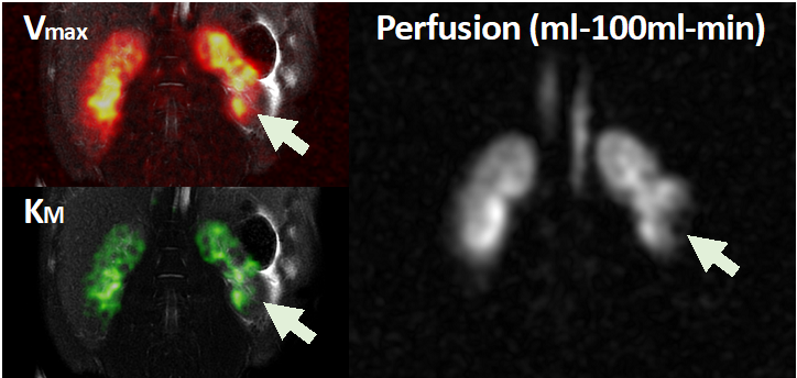
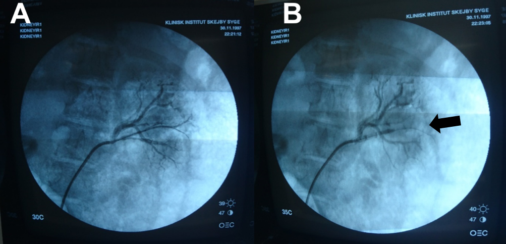
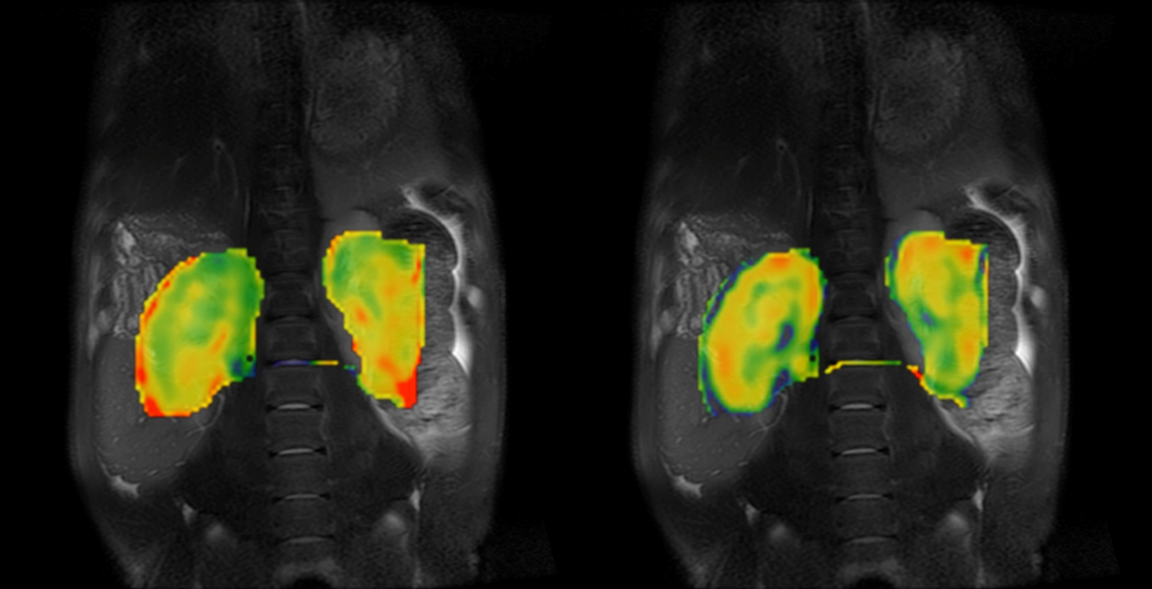
In Figure 1, one pig is shown with the resulting Vmax and Km maps for lactate and the area highlighted by arrows showing a great correlation between Michealis-Menten maps and the DCE-MRI perfusion map. Figure 2 show the X-ray image from the same pig before and after the locale balloon occlusion. A intra-renal heterogeneity was observed in both perfusion and metabolite accumulation (Michaelis–Menten kinetic Vmax and Km maps). Figure 3 is an example of this heterogeneity from another pig where Km maps from alanine is found dominantly in the medulla tissue and lactate dominantly in the cortex.Discussion
The principal finding of this study is that hyperpolarized [1-13C]-pyruvate spectral-spatial saturation recovery MRI can effectively identify intra-renal accumulation of pyruvate and its metabolites in a large animal model similar to human physiology. Ischemic injury could be assessed by Michealis-Menten maps and heterogeneity caused by the two major functional tissue compartments was observed.Conclusion
This work demonstrates, for the first time, in vivo intra-renal heterogeneity in hemodynamics and metabolism using hyperpolarized 13C-pyruvate MR imaging in multi-papillary kidneys, highlighting the potential for human translation. Future work aims to link perfusion measures both obtained from Dotarem and [1-13C]pyruvate to describe the ischemic injury further.Acknowledgements
No acknowledgement found.References
1. Nielsen PM, Eldirdiri A, Bertelsen LB, Jorgensen HS, Ardenkjaer-Larsen JH, Laustsen C. Fumarase activity: an in vivo and in vitro biomarker for acute kidney injury. Sci Rep 2017;7:40812.
2. Nielsen PM, Laustsen C, Bertelsen LB, Qi H, Mikkelsen E, Kristensen ML, Norregaard R, Stodkilde-Jorgensen H. In situ lactate dehydrogenase activity: a novel renal cortical imaging biomarker of tubular injury? Am J Physiol Renal Physiol 2017;312(3):F465-F473.
3. Nielsen PM, Szocska Hansen ES, Norlinger TS, Norregaard R, Bonde Bertelsen L, Stodkilde Jorgensen H, Laustsen C. Renal ischemia and reperfusion assessment with three-dimensional hyperpolarized 13 C,15 N2-urea. Magnetic resonance in medicine 2016.
4. Schulte RF, Sperl JI, Weidl E, Menzel MI, Janich MA, Khegai O, Durst M, Ardenkjaer-Larsen JH, Glaser SJ, Haase A, Schwaiger M, Wiesinger F. Saturation-recovery metabolic-exchange rate imaging with hyperpolarized [1-13C] pyruvate using spectral-spatial excitation. Magnetic resonance in medicine 2013;69(5):1209-1216.
Figures