0252
Probing Cerebral Lactate Compartmentalization with Hyperpolarized Diffusion Weighted 13C MRI1UCSF, San Francisco, CA, United States
Synopsis
Hyperpolarized 13C MRI has been used to non-invasively measure metabolism in real-time. However, perfusion and transporter expression can impact the compartmentalization of metabolites. In this work, we investigated the feasibility of diffusion weighted imaging of lactate generated from HP [1-13C]pyruvate in the human brain to assess lactate efflux and compartmentalization in a healthy volunteer. Whole brain lactate ADC values were 0.37ⅹ10-3 mm2/s, 0.29ⅹ10-3 mm2/s, and 0.41ⅹ10-3 mm2/s when diffusion gradients were applied in the X, Y, and Z direction, respectively, demonstrating the feasibility of diffusion weighted HP 13C MRI in a clinical setting.
Introduction
Dissolution DNP provides more than a four orders of magnitude enhancement to carbon-13 nuclei. Coupled with the ability of MRI to resolve both substrate and metabolites, dissolution DNP of 13C substrates has been used extensively for metabolic imaging in both pre-clinical1 and proof-of-concept clinical studies2 to non-invasively assess metabolic conversion. In addition to the Warburg Effect, many cancers - such as malignant renal cell carcinoma3 and prostate cancer4 - overexpress MCT4, the monocarboxylate transporter primarily responsible for lactate efflux. Moreover, lactate can also be used as an energy source in the brain, with increased MCT4 expression in astrocytes and increased MCT2 expression for lactate uptake in neuronal cells5.
Because of structural differences in the intra- and extra-cellular microenvironments, diffusion weighted imaging (DWI) of hyperpolarized lactate (generated intracellularly via LDH catalyzed conversion from HP pyruvate) could provide unique information on lactate efflux and microstructure. This potentially could provide insight into MCT4 expression and lactate transport in a rapid, non-invasive manner, and has been explored previously in pre-clinical imaging of prostate cancer6. In this work, we investigated the feasibility of DWI of lactate generated from HP [1-13C]pyruvate in the human brain to assess lactate efflux and compartmentalization in a healthy volunteer.
Methods
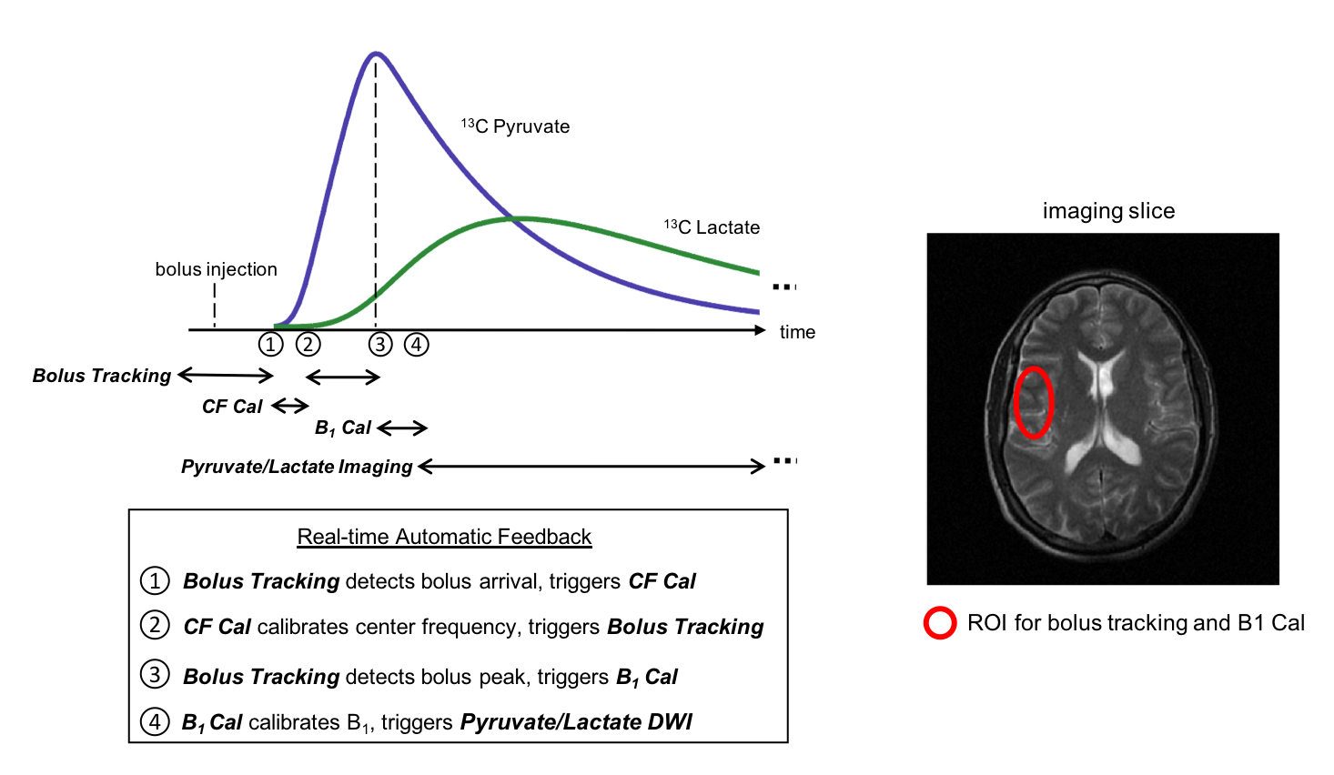
Hyperpolarized [1-13C]pyruvate was generated in a SPINlab polarizer operating at 5T and 0.8K (GE Healthcare). The sample was polarized for 3 hours and then rapidly dissolved and neutralized to yield 42mL of 220mM pyruvate with 43.8% polarization. The sample was transferred to the scan room and injected at a rate of 0.43mL/kg followed by a 20mL flush, both at 5mL/s. The acquisition was triggered at bolus arrival within the brain using an integrated RT-Hawk platform (HeartVista) for real-time frequency and B1 calibration7 (Fig. 1).
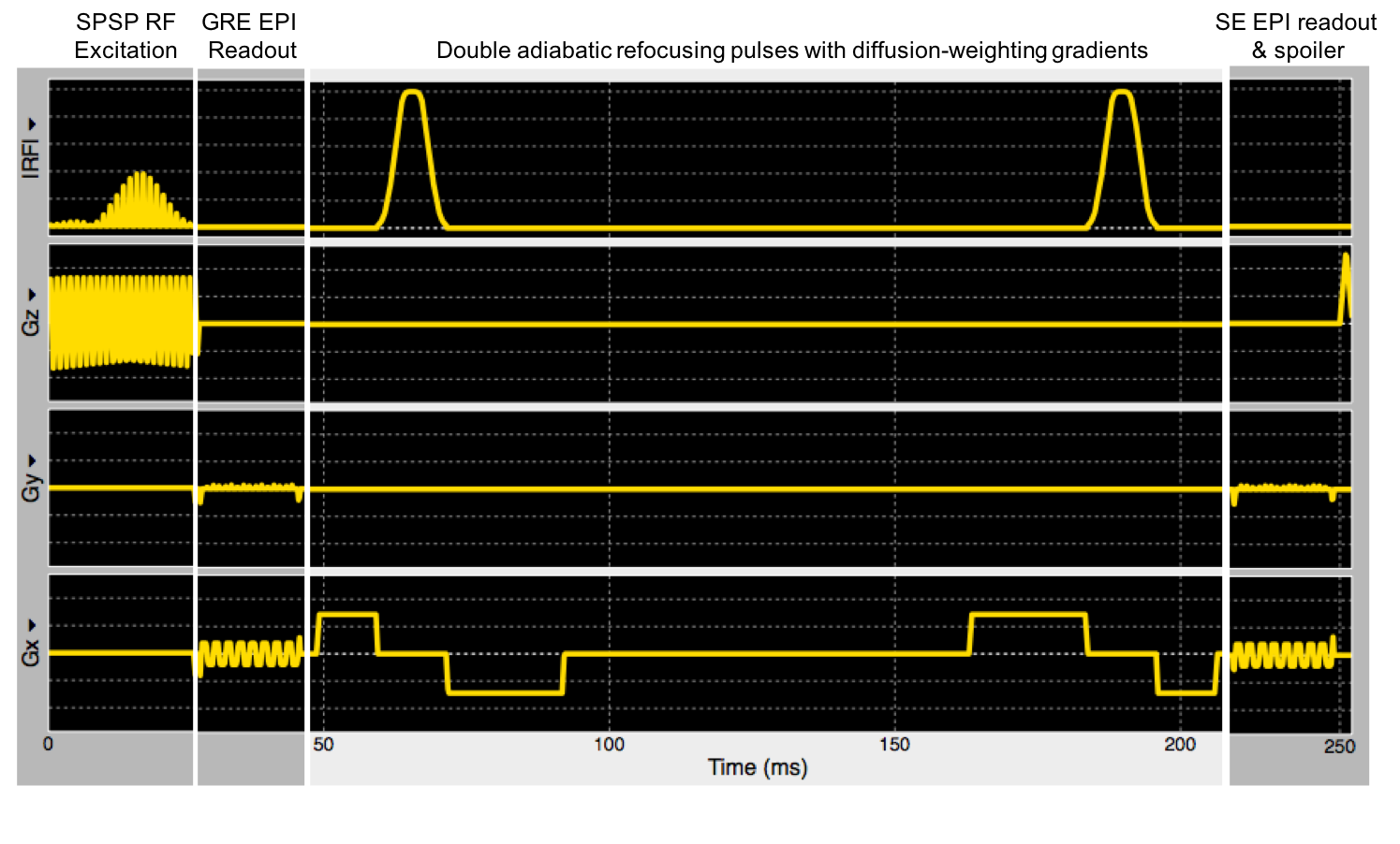
Hyperpolarized data were acquired using a double spin-echo diffusion weighted EPI sequence (Fig. 2). A gradient echo EPI module was inserted before the first refocusing pulse to provide an internal reference for signal normalization to account for T1 and RF utilization. Scan parameters were 250ms TR, 10ms (GRE) and 142ms (SE) TE, 1.5ⅹ1.5cm2 in-plane resolution, one 40mm thick slice, 20o pyruvate flip angle, 45o lactate flip angle, a low b-value of 51 s/mm2 applied in the Z direction and a high b-value of 319 s/mm2 applied in either the X, Y, or Z direction. A twice-refocused spin echo was employed to reduce eddy-current induced distortion in the high b-value images8. Each timepoint had one gradient-echo pyruvate image and four spin-echo lactate diffusion-weighted images, followed by a 2.25s delay, yielding a temporal resolution of 3.5s. To isolate the effects of diffusion weighting, spin-echo signal was normalized to the gradient echo readout for each b-value, with the ADC then calculated over the whole brain as a two-point fit: $$ADC = \frac{-ln \left( \frac{SE_{high-b} / GRE_{high-b}}{SE_{low-b} / GRE_{low-b}} \right)}{b_{high}-b_{low}} $$
Results & Discussion
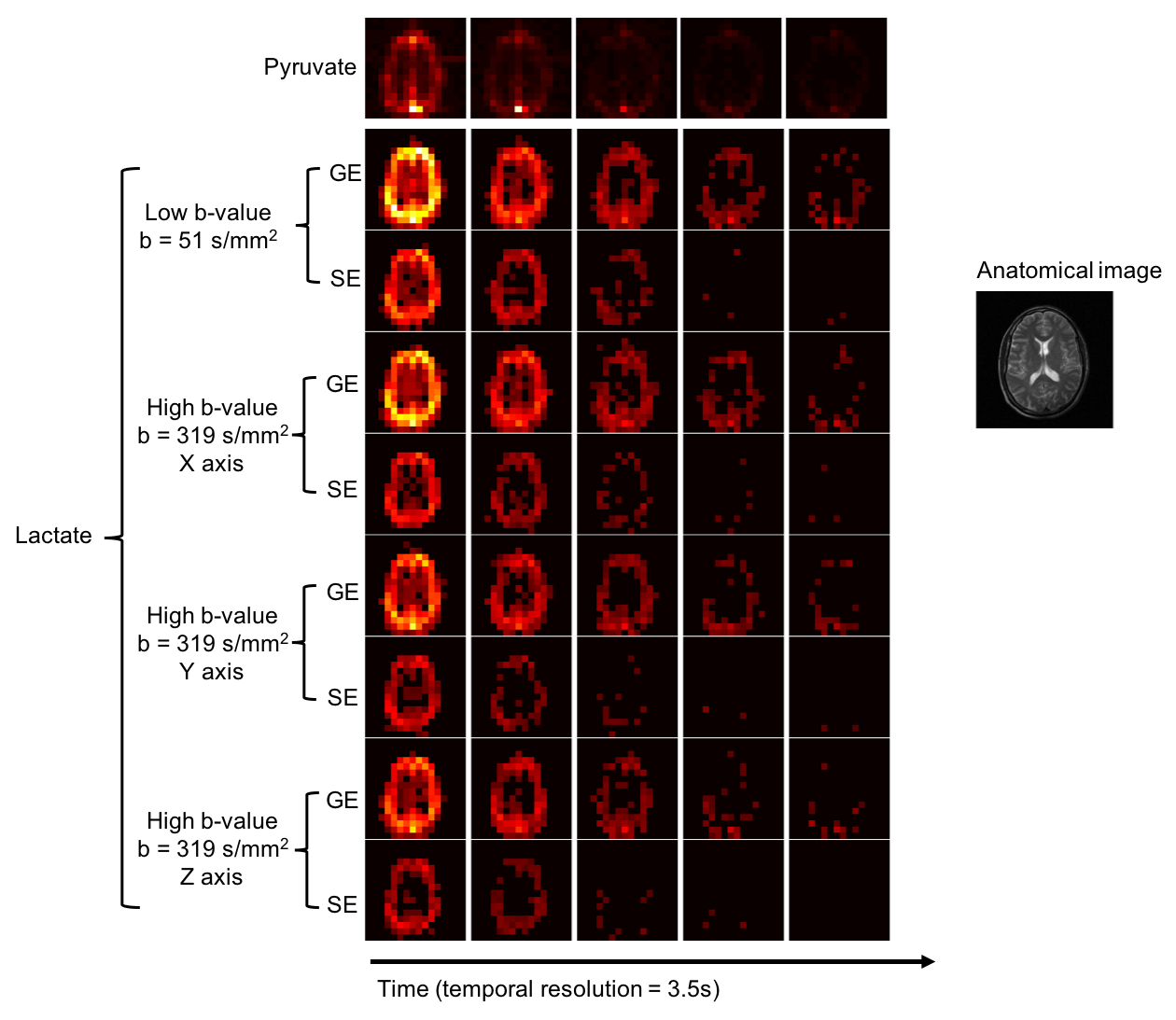
Fig. 3 shows the dynamic hyperpolarized 13C pyruvate and lactate images acquired using the sequence seen in Fig. 2. The shape and location of the brain is consistent among all lactate images, regardless of b-value or direction, indicating no apparent eddy-current or EPI artifacts are observed. Qualitatively, the signal for both pyruvate and lactate decayed faster compared to a conventional gradient echo acquisition, where no refocusing was applied9. This could be a result of imperfect inversion of the adiabatic RF pulse, as well as saturation of inflowing pyruvate and lactate at the coil boundary since the refocusing pulse was not frequency selective. Future work will explore the use of slice-selective adiabatic refocusing pulses to control out-of-slice saturation and improve signal at later timepoints6. A frequency-selective refocusing pulse could also be used to minimize saturation of inflowing pyruvate and may enable dynamic imaging over the entire time course. Whole brain lactate ADC values were 0.37ⅹ10-3 mm2/s, 0.29ⅹ10-3 mm2/s, and 0.41ⅹ10-3 mm2/s when diffusion gradients were applied in the X, Y, and Z direction, respectively, reasonable values given previously reported pre-clinical ADC values of lactate10,11. Future work will focus on using a variable flip angle scheme to improve SNR in the diffusion weighted data to calculate the ADC throughout the time course, as well as acquiring data in glioma patients to characterize lactate ADC in healthy and cancerous tissue.Conclusion
This research demonstrated the feasibility of diffusion weighted HP 13C MRI and showed that HP [1-13C]lactate ADC can be acquired in a clinical setting. Future developments will work on improved RF pulse design and flip angle schedules to increase SNR, enabling more b-values and improved volumetric coverageAcknowledgements
P41EB013598, R01EB016741, P41EB013598, and American Cancer Society Research Scholar Grant 131715‐RSG‐18‐005‐01‐CCE.References
1.) Albers, M.J., et al., Hyperpolarized 13C Lactate, Pyruvate, and Alanine: Noninvasive Biomarkers for Prostate Cancer Detection and Grading. Cancer Res., 2008. 68(20): p. 8607-8615.
2.) Nelson, S.J., et al., Metabolic Imaging of Patients with Prostate Cancer Using Hyperpolarized [1-13C]Pyruvate. Science Translational Medicine, 2013. 5(198): p. 198ra108.
3.) Keshari, K.R., et al., Hyperpolarized 13C-Pyruvate Magnetic Resonance Reveals Rapid Lactate Export in Metastatic Renal Cell Carcinomas. Cancer Research, 2013. 73(2): p. 529-538.
4.) Pertega-Gomes, N., et al., Monocarboxylate transporter 4 (MCT4) and CD147 overexpression is associated with poor prognosis in prostate cancer. BMC Cancer, 2011. 11(1): p. 312.
5.) Pérez-Escuredo, Jhudit, Vincent F. Van Hée, Martina Sboarina, Jorge Falces, Valéry L. Payen, Luc Pellerin, and Pierre Sonveaux. Monocarboxylate transporters in the brain and in cancer. Biochimica et Biophysica Acta (BBA)-Molecular Cell Research, 2016. 1863(10): 2481-2497.
6.) Zhu X, Gordon JW, Bok RA, Kurhanewicz J, Larson PEZ. Dynamic diffusion-weighted hyperpolarized 13C imaging based on a slice-selective double spin echo sequence for measurements of cellular transport. Magnetic Resonance in Medicine 2018. https://doi.org/10.1002/mrm.27501
7.) Tang S, Milshteyn E, Reed G, Gordon J, Bok R, Zhu X, Zhu Z, Vigneron DB, Larson PEZ. A regional bolus tracking and real-time B1 calibration method for hyperpolarized 13C MRI. Magnetic Resonance in Medicine 2018. https://doi.org/10.1002/mrm.27391.
8.) Reese TG, Heid O, Weisskoff RM, Wedeen VJ. Reduction of eddy-current-induced distortion in diffusion MRI using a twice-refocused spin echo. Magnetic Resonance in Medicine 2003;49(1):177-182.
9.) Gordon JW, Chen H-Y, Autry A, Park I, Van Criekinge M, Mammoli D, Milshteyn E, Bok R, Xu D, Li Y, Aggarwal R, Chang S, Slater JB, Ferrone M, Nelson S, Kurhanewicz J, Larson PEZ, Vigneron DB. Translation of Carbon-13 EPI for hyperpolarized MR molecular imaging of prostate and brain cancer patients. Magnetic Resonance in Medicine 2018. https://doi.org/10.1002/mrm.27549.
10.) Pfeuffer J, Lin JC, DelaBarre L, Ugurbil K, Garwood M. Detection of intracellular lactate with localized diffusion {1H–13C}-spectroscopy in rat glioma in vivo. Journal of Magnetic Resonance 2005;177(1):129-138.
11.) Koelsch BL, Reed GD, Keshari KR, Chaumeil MM, Bok R, Ronen SM, Vigneron DB, Kurhanewicz J, Larson PEZ. Rapid in vivo apparent diffusion coefficient ping of hyperpolarized 13C metabolites. Magnetic Resonance in Medicine 2015;74(3):622-633.
Figures