0250
High-resolution MR imaging of human brain with multi-echo integrated SSFP1State Key Laboratory of Brain and Cognitive Science, Beijing MRI Center for Brain Research, Institute of Biophysics, Chinese Academy of Sciences, Beijing, China, 2University of Chinese Academy of Sciences, Beijing, China, 3CAS Center for Excellence in Brain Science and Intelligence Technology, Chinese Academy of Sciences, Beijing, China, 4Siemens Shenzhen Magnetic Resonance Ltd, Shenzhen, China, 5Stevens Neuroimaging and Informatics Institute, University of Southern California, Los Angeles, CA, United States, 6Beijing Institute for Brain Disorders, Beijing, China
Synopsis
Balanced SSFP (bSSFP) has been used in structural and functional MRI, but always suffered from banding artifacts. While phase cycling was widely used to reduce banding artifact, it would take more scan time. Recently, integrated SSFP (iSSFP) was introduced to acquire banding-free images in shorter scan time than phased-cycled bSSFP. In this work, multi-echo iSSFP was further developed to improve SNR and acquire ultrahigh-resolution images with moderate scan time at 7T. Phantom and in vivo experiments demonstrated that the combined image produced by weighted averaging of multi-echo iSSFP showed obvious SNR and contrast improvement and inherited the characteristics of bSSFP.
Introduction
In the past decade, bSSFP has been used in research for structural and functional MR imaging and routine clinical applications for pathology detection due to its unique T2/T1 contrast and high SNR per unit time1. However, it always exhibits characteristic banding artifacts in the presence of B0 field inhomogeneity, especially at ultrahigh-field MRI systems as 7T. A previous study realized ultrahigh-resolution imaging of the human brain by averaging 8 increments of phase-cycled bSSFP at the cost of dramatically increased scan time2. Recently, integrated SSFP (iSSFP), was introduced to eliminate banding artifacts in high field by adding an extra 2pi dephasing gradient to compress the bSSFP signal profile into a single voxel3. iSSFP can acquire banding-free images that inherit the advantages of bSSFP without lengthening scan time, whereas it shows relatively low SNR compared with bSSFP. In this work, we developed a multi-echo iSSFP method to acquire high-resolution MR images of human brain with comparable SNR with bSSFP in moderate scan time which may have potential in structural and fMRI studies to depict microarchitecture or detect abnormalities for intact human brain.Methods
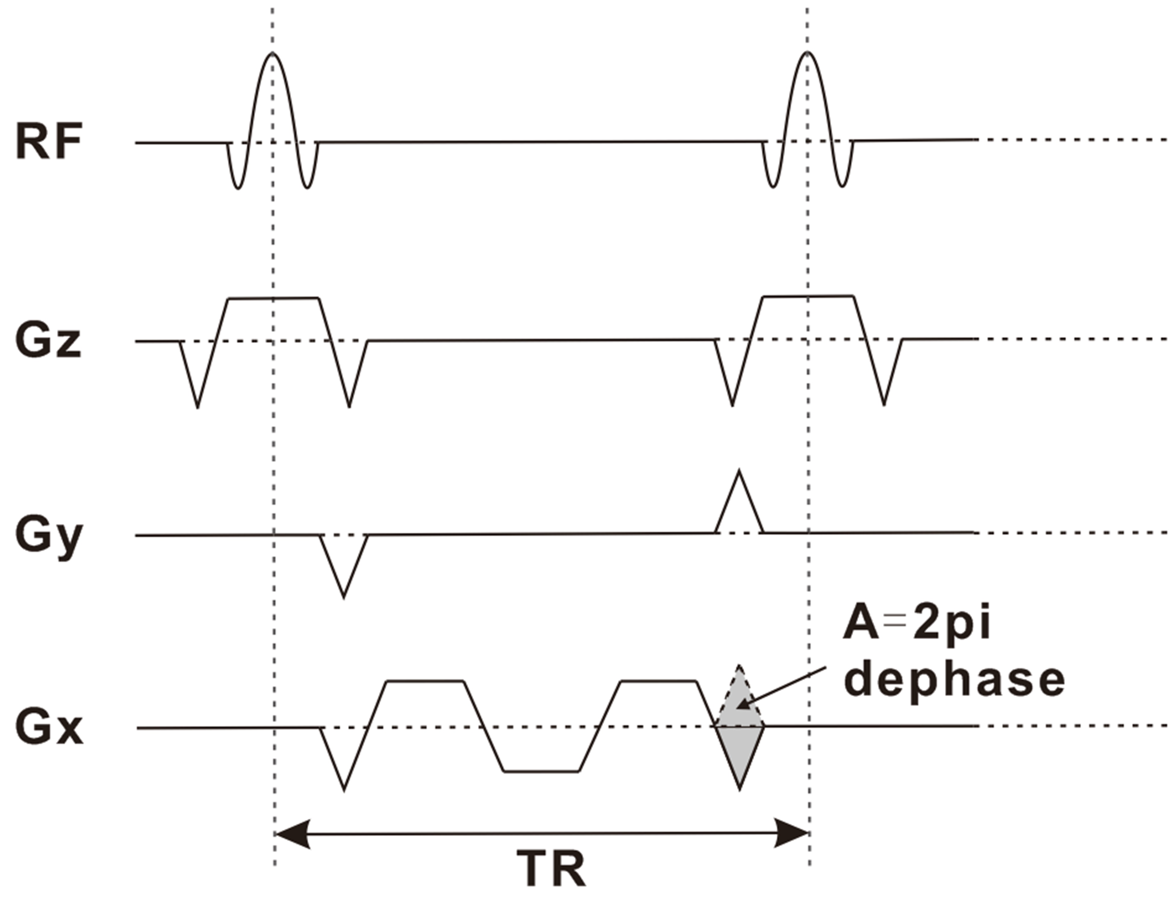
This study was performed at a 7T research system (Siemens Healthcare, Erlangen, Germany) with a 1TX/32RX head coil (Nova Medical). The study was approved by the Institutional Review of Beijing MRI Center for Brain Research. Image post-processing was performed in MATLAB 2016 (The MathWorks, Natick, MA). As shown in Fig.1, the multi-echo iSSFP sequence was modified from bSSFP by applying bipolar readout gradients to acquire three echoes and adding an extra dephasing gradient in the readout direction, which caused a 2pi dephasing of the spins within a voxel during a TR period. iSSFP integrated a 2pi cycle of the bSSFP signal profile within a voxel which eliminated its sensitivity to B0 inhomogeneity. Images of three echoes were averaged with different weighting factors to get a composite image with higher SNR. Comparative phantom experiments were performed using bSSFP and multi-echo iSSFP sequence respectively to validate the signal behavior of both sequences with following parameters: flip angle=30°, field of view=200*200mm2, spatial resolution=0.5*0.5*3.0mm3, number of slices=60, echo time of bSSFP=2.58ms, 1st-, 2nd- and 3rd- echo times of multi-echo iSSFP=2.58/4.78/6.98ms, bandwidth=568Hz/Px, repetition time of one slice for bSSFP=2123.46ms, repetition time of one slice for three-echo iSSFP=3929.66ms, scan time of bSSFP=2:07 mins,scan time for three-echo iSSFP=3:56 mins. No GRAPPA acceleration. Transverse and coronal high-resolution whole-brain images were acquired from a healthy volunteer with careful shimming. We drew regions of interest (ROI) in images and chose noise regions at four corners of an image to calculate the SNR.Results
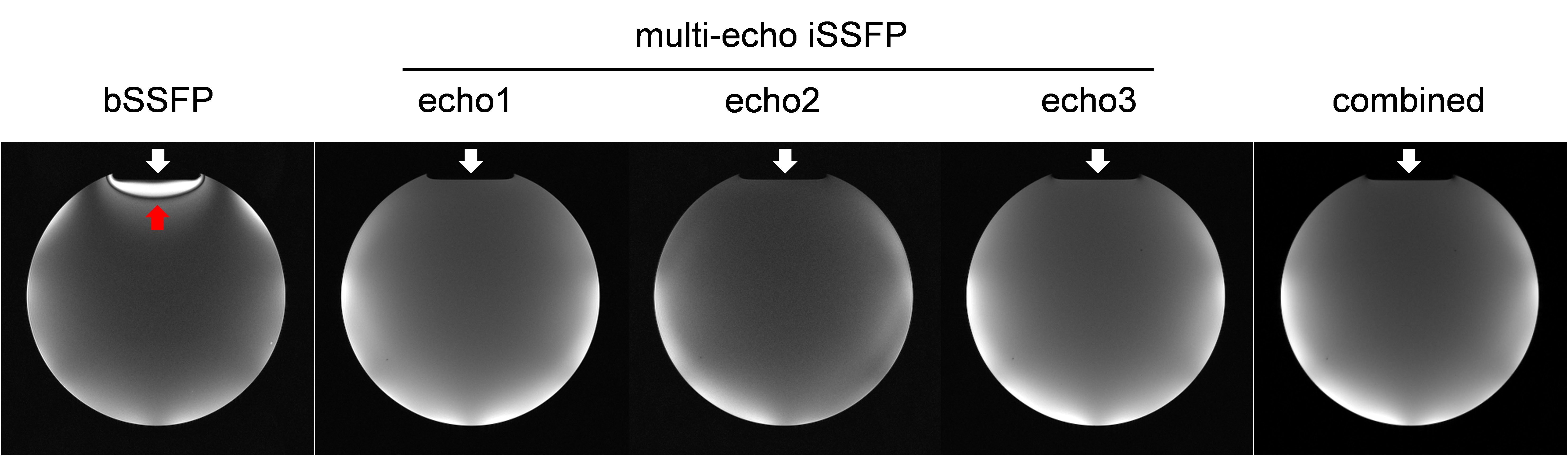
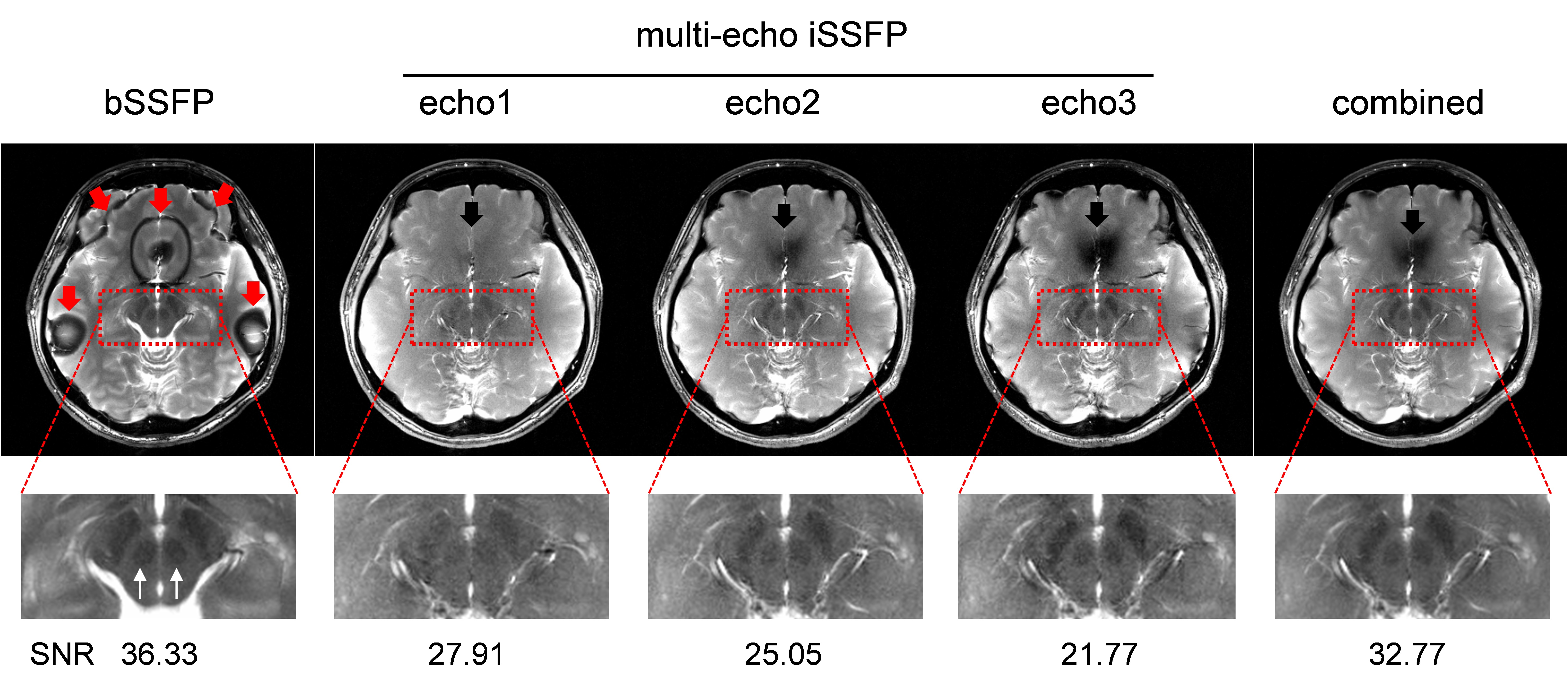
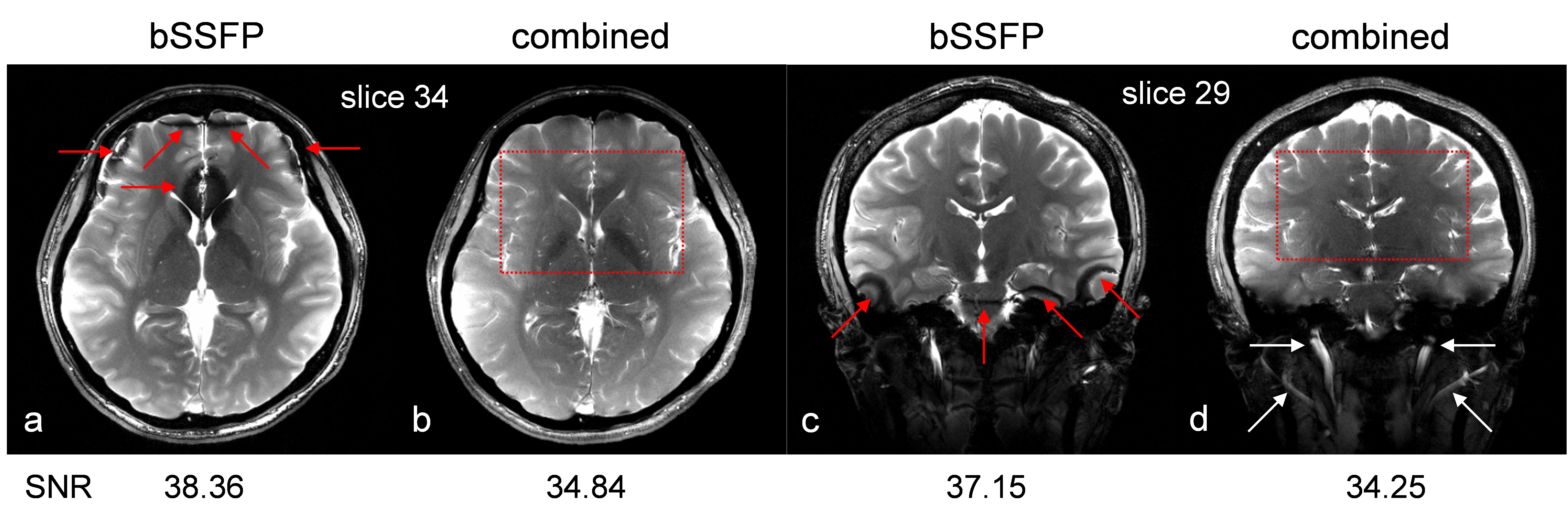
The phantom experiment demonstrated that multi-echo iSSFP was insensitive to B0 field inhomogeneity and straightforward arithmetic averaging of three echoes, i.e., combined image=(echo1+echo2+ehco3)/3, can improve the SNR of iSSFP. In vivo experiments also validated the feasibility of multi-echo iSSFP used for 2D ultrahigh in-plane resolution of human brain, especially in areas with B0 inhomogeneity where banding artifacts were observed with bSSFP. We found the strategy of weighted averaging of three echoes could improve the SNR of selected ROI in human brain images further. As shown in Fig. 3, the combined image by weighted averaging manifested about 18% SNR improvement and can better depict microstructures in midbrain such as red nuclei and substantia nigra, which benefited from better contrast and good SNR. Fig. 4 further demonstrated that the combined image of multi-echo iSSFP was insensitive to B0 field inhomogeneity, and inherited the advantages of bSSFP with comparable SNR.Discussion and Conclusion
In this study, we developed a multi-echo iSSFP sequence, modified from bSSFP, and applied a weighted averaging method to get a composite image with improved SNR, which was insensitive to B0 inhomogeneity and inherited the characteristics of bSSFP. In practice, key parameters, such as receiver bandwidth and flip angle can be optimized to realize further SNR improvement. In addition, we will further investigate 3D multi-echo iSSFP sequence combined with acceleration methods to achieve ultrahigh resolution imaging with isotropic voxels in shortened scan time. Averaged multi-echo iSSFP images with ultrahigh resolution and improved SNR can better depict microstructures and have great potential to detect abnormalities of the human brain.Acknowledgements
This work was supported in part by the Ministry of Science and Technology of China (MOST) grants (2015CB351701), National Nature Science Foundation of China grants (81871350, 31730039), and Chinese Academy of Sciences grant (XDBS01000000).References
1. Bieri O, Scheffler K. Fundamentals of balanced steady state free precession MRI[J]. Journal of Magnetic Resonance Imaging, 2013, 38(1): 2-11.
2. Zeineh M, Parekh M, Zaharchuk G, et al. Ultra-High Resolution Imaging of the Human Brain with Phase-Cycled Balanced Steady State Free Precession at 7.0 T[J]. Investigative radiology, 2014, 49(5): 278-289.
3. Sun K, Xue R, Zhang P, et al. Integrated SSFP for functional brain mapping at 7 T with reduced susceptibility artifact[J]. Journal of Magnetic Resonance, 2017, 276: 22-30.
Figures