0247
Whole brain sub-mm resolution T2* weighted anatomy imaging in less than 2 minutes1Department of Radiology, University Medical Center Utrecht, Utrecht, Netherlands, 2Department of Radiotherapy, University Medical Center Utrecht, Utrecht, Netherlands, 3Department of Neuroscience, University of Turin, Turin, Italy, 4Philips Healthcare, Best, Netherlands
Synopsis
T2* weighted imaging can be used to study both normal and pathological tissue. These images are commonly obtained using traditional gradient echo sequences which can lead to long scan times that are problematic particularly in a clinical setting. 3D EPI offers a faster alternative with scan times on the order of few minutes. Here, the scan time of T2* weighted 3D EPI scans is further reduced with a shot selective 2D CAIPI acquisition pattern. Whole brain T2* weighted anatomical scans with a resolution of 0.5 mm isotropic were acquired in 1:27 minutes. This holds promising perspectives for future applications in routine examinations.
Introduction
T2* weighted imaging is a powerful tool to study both normal and pathological tissue, especially at high field strengths. The anatomy and substructures of the brain can be visualized with exceptional detail, including structures within the basal ganglia, and the line of Gennari in the visual cortex. T2* weighted imaging is traditionally performed using a spoiled gradient echo (GRE) sequence [1]. However, to image structural detail of the brain at sub-millimeter resolutions requires a long scan time, even for a limited number of slices [2]. As result, the imaging volume is typically limited, since full brain coverage coincides with excessively long scan times of 1 hour or more for a single acquisition. In the clinical setting however, long scan times are problematic because both patient motion [3] and the cost of examination are increased. More recently, Zwanenburg et al. [4] showed that volumetric 3D EPI protocols can drastically reduce scan time or increase imaging coverage, while maintaining image quality and T2* weighting.
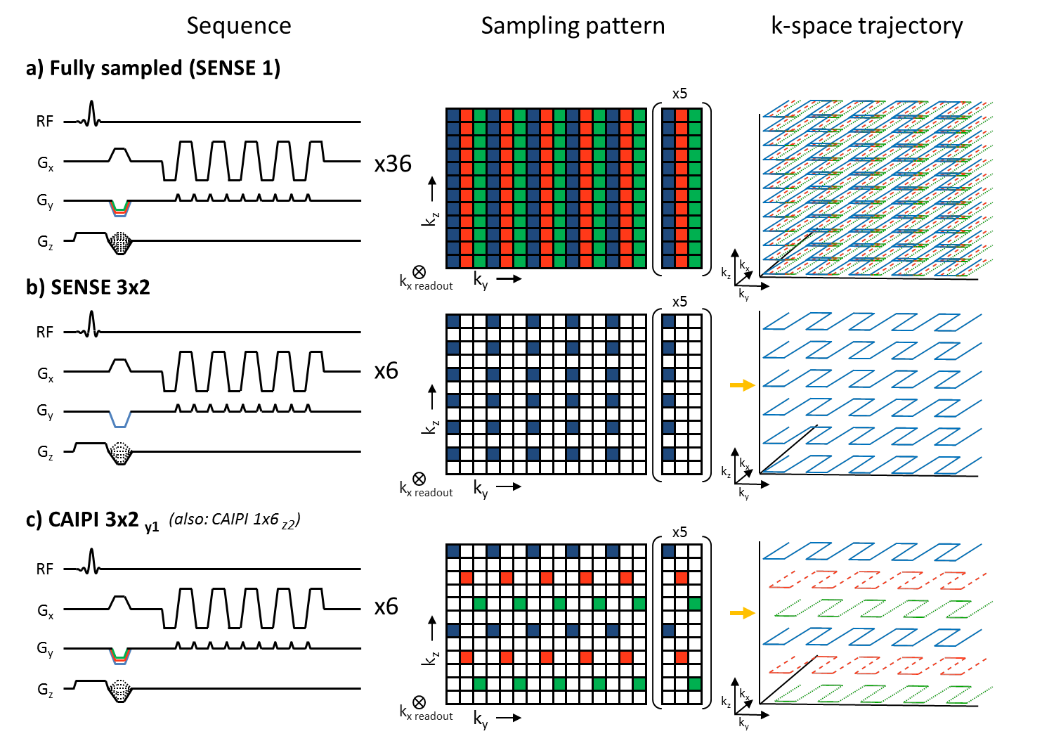
Recent advances in parallel imaging approaches, such as CAIPIRINHA (CAIPI) and compressed sensing, may be used to reduce scan time even further [5-7]. Here we show that the scan time of T2* weighted 3D EPI scans can be reduced considerably, while maintaining high image quality, by using a shot selective 2D CAIPIRINHA acquisition pattern. A shot selective 2D CAIPIRINHA sequence was implemented for multi-shot 3D EPI scans, which, instead of adding extra gradients, leaves them out (Figure 1). The sequence was applied to sub-millimeter T2* weighted 3D EPI anatomical imaging at 7T.
Methods
Two healthy participants were scanned in a 7 Tesla Achieva system (Philips, Best, the Netherlands). Whole brain T2* weighted multi-shot 3D EPI anatomical scans were acquired with a 32 channel headcoil [8] (Nova Medical, USA). The following scan parameters were used:
- 1 mm T2* weighted imaging. TE/TR= 27/72 ms, 16° flip angle, 1x1x1mm3 voxels, 240x196x150mm3 FOV, 150 slices, matrix size 240x195, EPI factor 13, CAIPIRINHA factor 5 shift 2, total acquisition time 0:33 min for CAIPI and 2:43 min for the fully sampled SENSE 1 dataset.
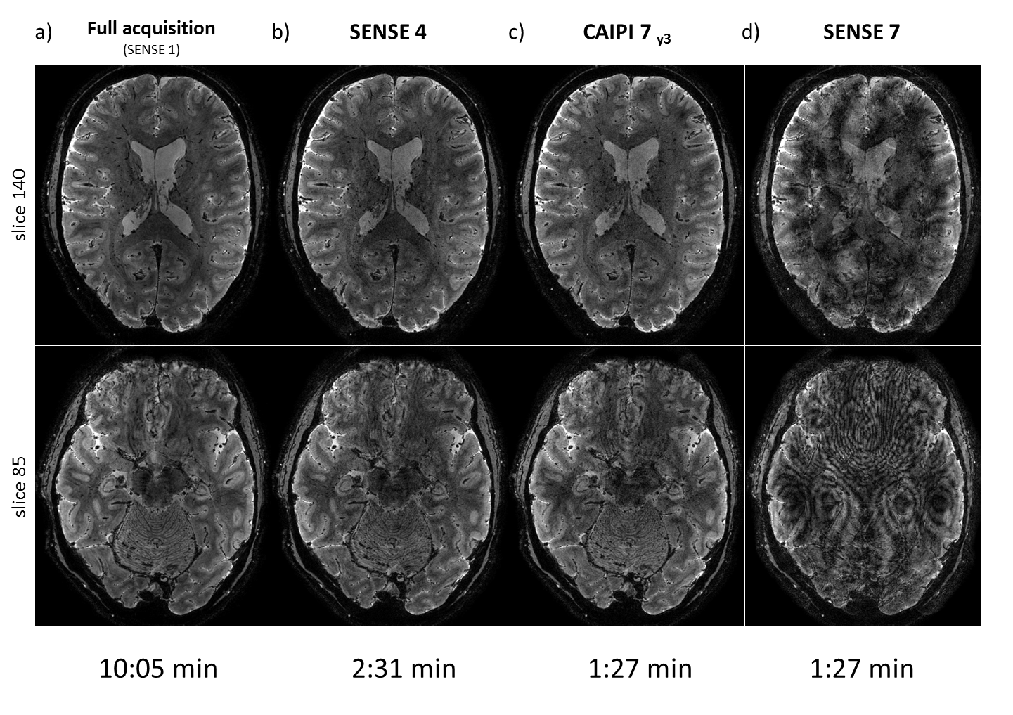
- 0.5 mm T2* weighted imaging. TE/TR= 27/72 ms, 19° flip angle, 0.5x0.5x0.5mm3 voxels, 240x186x150mm3 FOV, 300 slices, matrix size 480x364, EPI factor 13, CAIPIRINHA factor 7 shift 3, total acquisition time 1:27 min. For reference, an identical scan was made using SENSE 1 (10:05 min), SENSE 4 (2:31 min) and SENSE 7 (1:27 min).
The scans were reconstructed offline in a modified Philips Recon2.0 environment.
Results
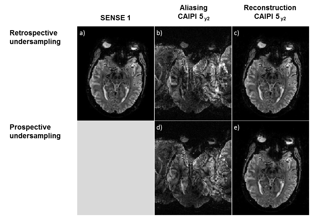
First the sequence implementation was evaluated by comparing prospectively and retrospectively undersampled datasets, as displayed in Figure 2. Note that both the aliasing patterns and reconstructions are close to identical, which demonstrates good performance of the implemented sequence.
The sub-millimeter T2* weighted anatomical scans are displayed in Figure 3, both fully sampled (Figure 3a) and undersampled with: SENSE 4 (Figure 3b), CAIPIRINHA 7 (Figure 3c) and SENSE 7 (Figure 3d). These scans have an isotropic resolution of 0.5 mm. Note that the CAIPIRINHA implementation can be used to shorten the total scan time substantially, while preserving the anatomy and structures of the brain.
Discussion and conclusion
The results show that the implementation of shot selective 2D CAIPIRINHA patterns for 3D EPI sequences can be used to significantly shorten scan time of sub-millimeter anatomy scans. For whole-brain anatomical imaging with 0.5 mm isotropic resolution the achieved scan time is about 1.5 minute, a reduction by a factor of 4, as compared to the existing implementation of 6 min [4].
The current results are acquired at a field strength of 7T, however, the applied method can be easily translated to more clinically available 3T scanners. This holds promising perspectives for future application in routine examinations among patients.
Acknowledgements
This work was supported by the Netherlands Organization for Scientific Research (NWO), specifically research grants: 040.11.581, ALW-834.14.004 and Vidi Grant 13339 (Petridou).References
[1] Haase et al. (1986) FLASH imaging: Rapid NMR imaging using low flip-angle pulses. Journal of Magnetic Resonance 67, pp. 258–266.
[2] J.H. Duyn et al. (2007) High-field MRI of brain cortical substructure based on signal phase. Proc Natl Acad Sci USA. Jul 10;104(28):11796-801.
[3] M.J. Versluis (2010) et al. Origin and reduction of motion and f0 artifacts in high resolution T2*-weighted magnetic resonance imaging: application in Alzheimer's disease patients. Neuroimage 51, 1082–1088.
[4] J.J.M. Zwanenburg et al. (2011) Fast high resolution whole brain T2* weighted imaging using echo planar imaging at 7T. NeuroImage 56 1902–1907
[5] F.A. Breuer et al. (2006) Controlled Aliasing in Volumetric Parallel Imaging (2D CAIPIRINHA). Magn Reson Med 55:549–556
[6] B.A. Poser et al. (2014) Accelerated 3D EPI using 2D blipped-CAIPI for high temporal and/or spatial resolution. Proceedings of the 22th Annual Meeting of the ISMRM Milan. pg.#1506.
[7] M. Narsude et al. (2016) Three-Dimensional Echo Planar Imaging with Controlled Aliasing: A Sequence for High Temporal Resolution Functional MRI. Magn Reson Med 75:2350–2361
[8] P.J. Ledden et al. (2007) 32 Channel Receive-Only SENSE Array for Brain Imaging at 7T. Proceedings of the 15th ISMRM Berlin pg.#0242
Figures