0245
Inversion Recovery Zero Echo Time (IR-ZTE) Imaging for Direct Myelin Detection in Human Brain1Department of Radiology, University of California San Diego, San Diego, CA, United States, 2GE Healthcare, San Diego, CA, United States, 3Radiology Service, VA San Diego Healthcare System, San Diego, CA, United States
Synopsis
In MRI, direct myelin imaging is challenging due to the short T2* decay (less than 0.5ms) and very low proton density. In the literature, it has been reported that ultrashort echo time (UTE) imaging can directly capture the fast decaying myelin signal. To further enhance the dynamic range, adiabatic inversion recovery (IR) preparation can be utilized so that the white matter signal can be suppressed. Moreover, dual echo UTE imaging scheme can suppress the remaining gray matter signal. In this study, we explore the feasibility of IR prepared zero echo time (IR-ZTE) imaging for direct myelin imaging in the human brain.
Introduction
In MRI, direct myelin imaging is challenging due to the short T2* decay (<0.5ms) and very low proton density. In our previous work, we have shown that ultrashort echo time (UTE) imaging can directly capture the fast decaying signal from the myelin lipid proton1–4. To enhance the dynamic range, adiabatic inversion recovery (IR) preparation is utilized so that the white matter signal can be suppressed. In this study, we explore the feasibility of IR-prepared zero echo time (IR-ZTE) imaging for direct myelin imaging in the human brain.Methods
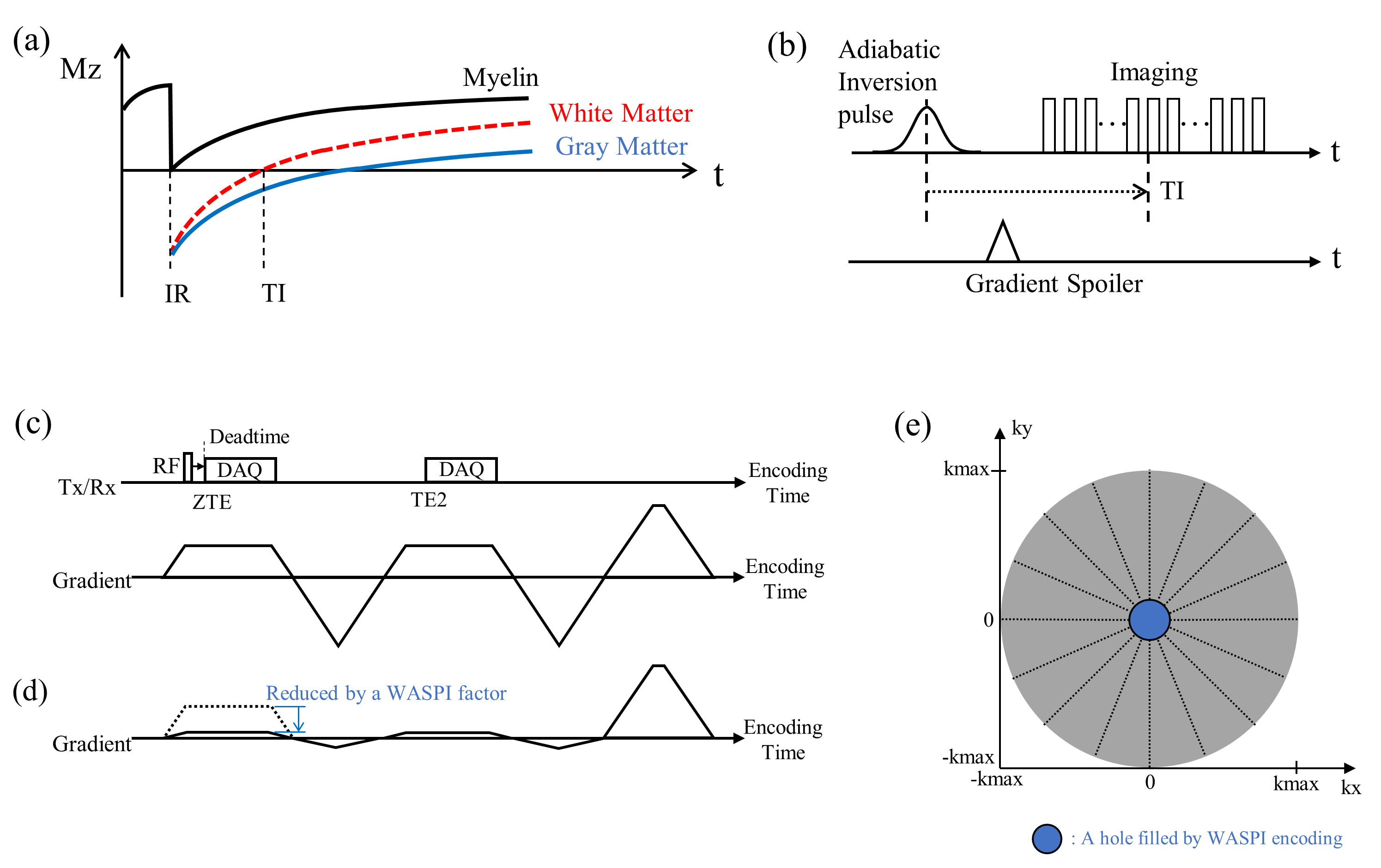
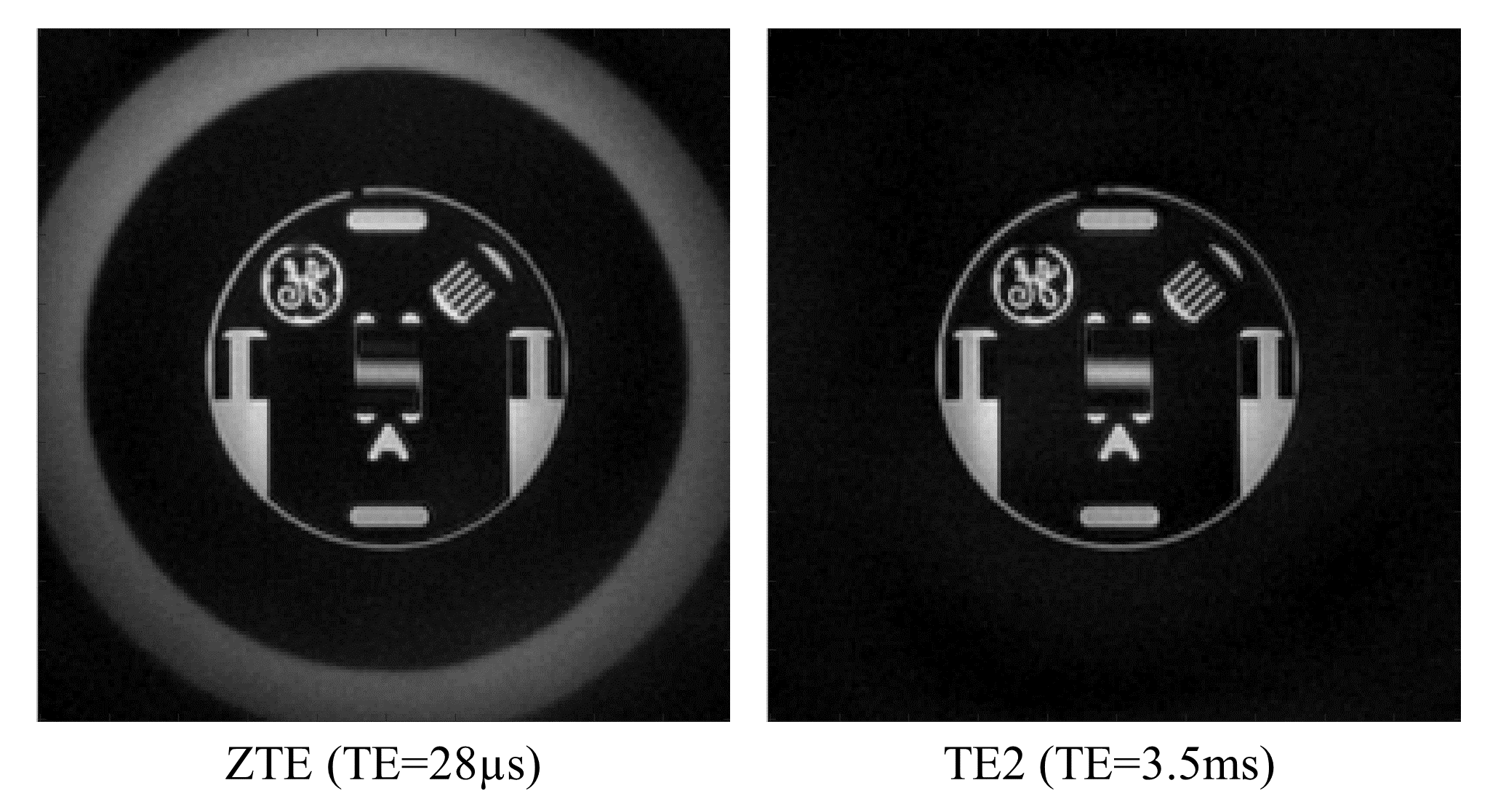
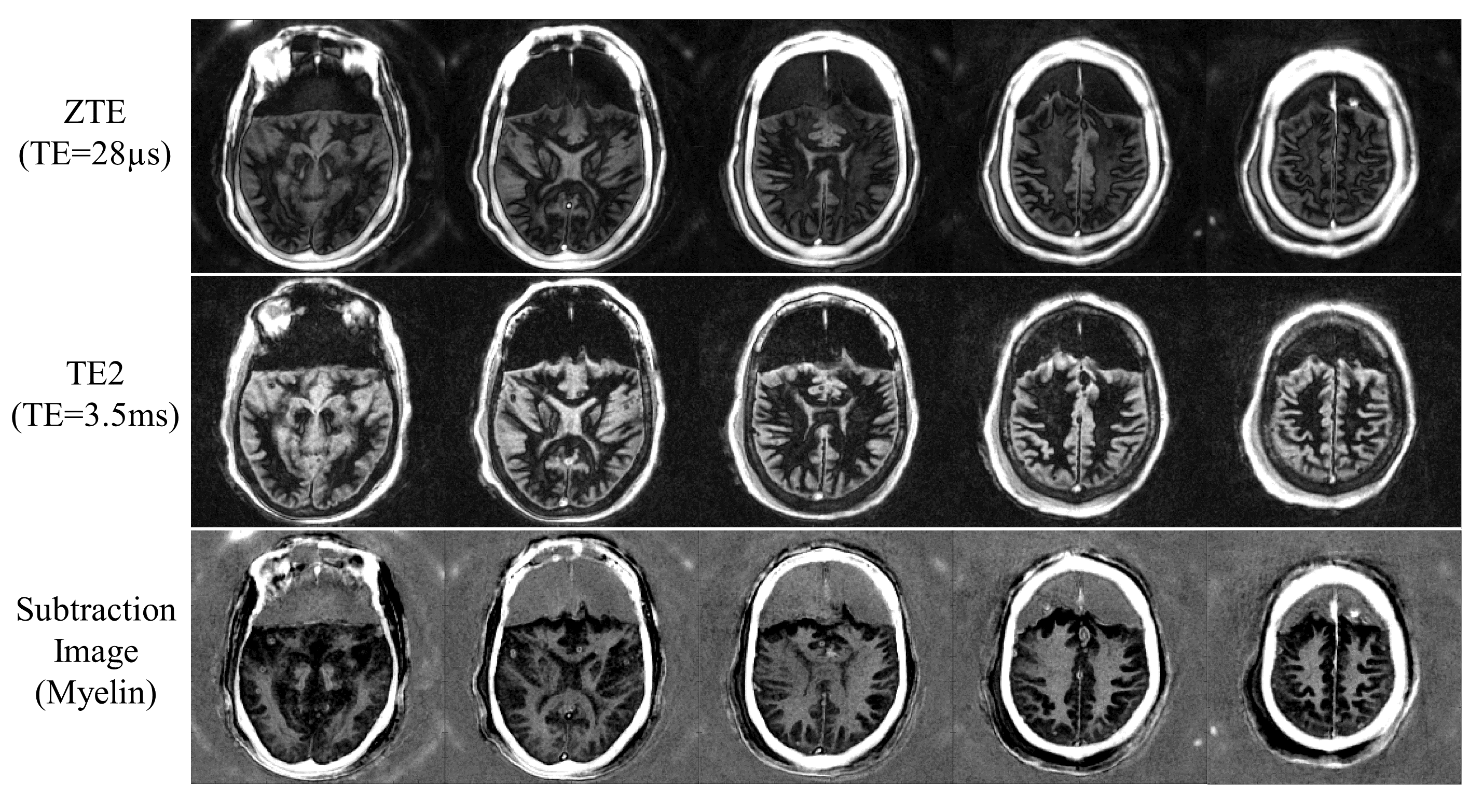
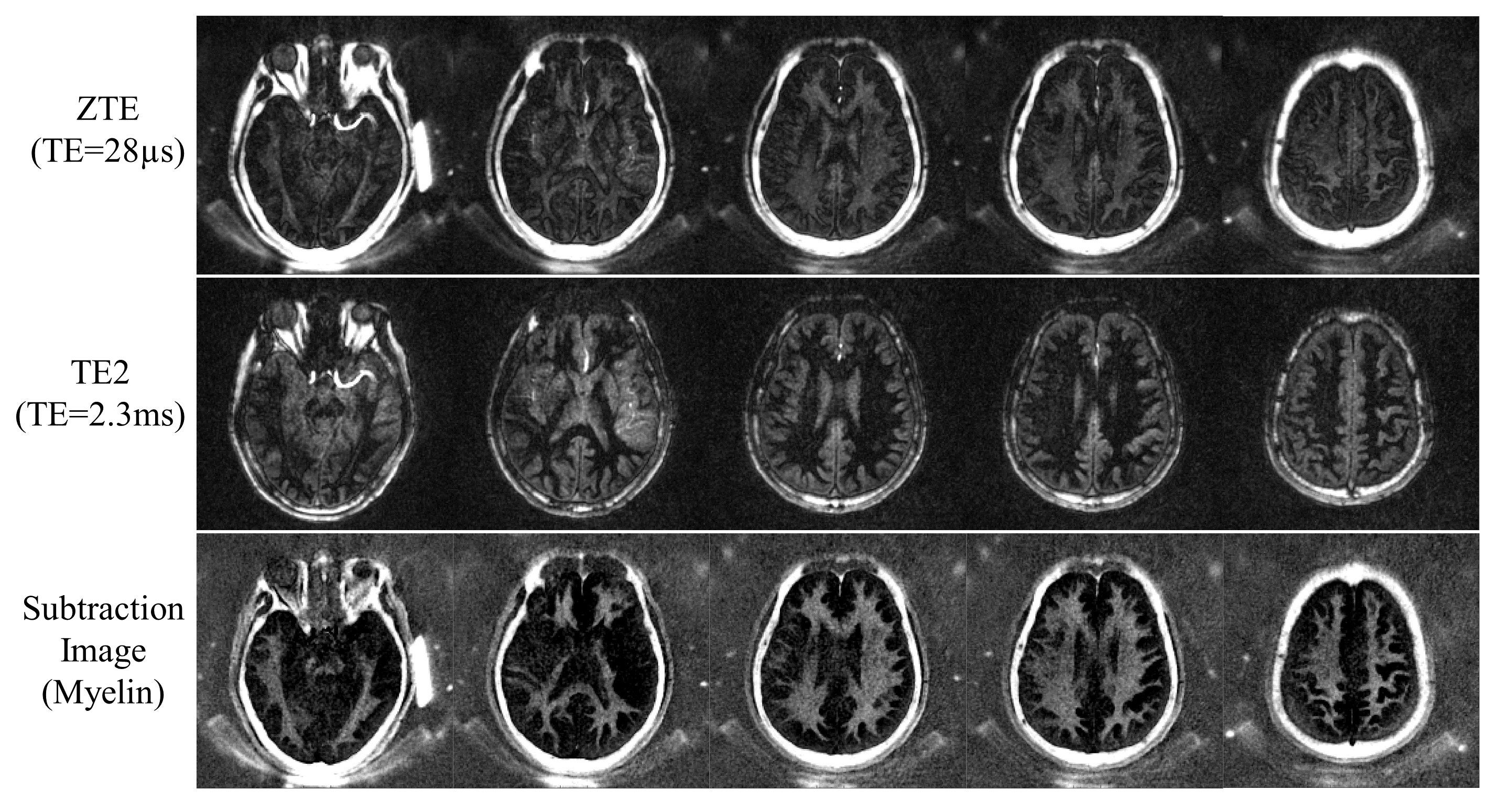
Figure 1-a shows typical inversion recovery curves of the components in the human brain at 3T. After IR-preparation the signal from long T2 white matter can be suppressed by starting ZTE data acquisition at its nulling point. Note that multiple spokes are acquired after each IR-preparation to efficiently acquire the k-space data (as shown in Figure 1-b), which is essential to reduce the scan time for 3D ZTE imaging. Each rectangular block in Figure 1-b represents an imaging sequence to acquire one spoke, based on ZTE imaging as described in Figure 1-c. ZTE imaging is performed using the rotating readout gradients to cover a 3D k-space sphere. Note that the RF pulse is applied during the plateau of the readout gradient, which leaves a hole (missing data) in the central region of the k-space due to the RF coil deadtime (blind time during transmitter/receiver switching). To fill the missing data, encoding with a derated readout gradient amplitude is performed as in WASPI5, as shown in Figure 1-d. The k-space is acquired, as shown in Figure 1-e. In this study, a WASPI factor of 8x was used. In our IR-ZTE framework, the clinical ZTE sequence was modified to implement a dual-echo ZTE sequence to acquire an image at the later TE (TE2) so that the remaining gray matter signal can be suppressed by subtracting the second echo image from the ZTE image. To evaluate the feasibility of IR-ZTE for direct myelin imaging, we performed a phantom experiment with a GE-resolution phantom, ex vivo imaging with a cadaveric brain (56-year-old female donor), and in vivo imaging with three healthy volunteers (36-, 30-, and 36-year old males). The experiments were performed on a 3T GE-MR750 scanner using a 12-ch receive-only HNU coil. The imaging parameters for the phantom experiment are as follows: a hard pulse with flip angle (FA)=4° (pulse width=20µs), readout BW=±31.25kHz, matrix size=200x200x40, FOV=220x220x160mm3, inter-spoke TR=8ms, TE=28µs/2.4ms, # of WASPI encoding=384, # of radial frequency encoding=22560, and scan time=4min 5sec. The ex vivo experiment was performed with the parameters matched above except for the following parameters: adiabatic inversion pulse applied (Silber Hoult pulse, pulse width=8.64ms), TR=1000ms, TI=310ms, TE=28µs/3.5ms, # of WASPI encoding=544, # of radial frequency encoding=33856, # of spokes per IR preparation=16, inter-spoke TR=10ms, and scan time=35min 54sec. In vivo experiment was performed using the same parameters as in the ex vivo experiment except for matrix size=190x190x36, FOV=220x220x144mm3, TI=330ms, TE=28µs/2.3ms, # of WASPI encoding=528, # of radial frequency encoding=15648, # of spokes per IR=30, inter-spoke TR=8ms, and scan time=11min 18sec. All images were reconstructed using online reconstruction based on GE Orchestra-SDK (v1.7.1).Results
Figure 2 shows the dual echo IR-ZTE images obtained with a GE-resolution phantom, where no manifest imaging artifacts are exhibited in the images without IR preparation. Figure 3 shows the IR-ZTE image obtained with the ex vivo cadaveric human brain. In the ZTE image (top), myelin signal is visible but with poor contrast due to high signals from the surrounding gray matter. In the second echo image (middle), the myelin signal decays to near-zero, which assures that there is no remaining white matter water signal in the ZTE image. What we observe, then, is nearly pure myelin lipid. In the subtraction image (bottom), the myelin is detected with high contrast, with other surrounding long T2 tissues suppressed. Figure 4 shows in vivo results of a healthy volunteer (36-year-old male), including a high contrast myelin image with the proposed IR-ZTE method.Discussion and Conclusion
In this study, we demonstrated the clinical feasibility of IR-prepared dual-echo ZTE for direct myelin imaging in the human brain. Compared to other UTE techniques utilizing ramp-sampling, ZTE is capable of capturing a shortly decay signal due to the use of ramped, constant readout gradient, which can be beneficial in the direct imaging of myelin. Moreover, the feature of the constant gradient sampling makes ZTE robust to eddy current-induced gradient distortion. In future work, we will evaluate the proposed IR-ZTE based myelin imaging technique in clinical applications, such as multiple sclerosis and traumatic brain injury.Acknowledgements
The authors acknowledge research support from GE Healthcare, NIH (R01NS092650), and VA Clinical Science and Rehabilitation R&D Awards (I01CX001388 and I01RX002604).References
1. Du J, Ma G, Li S, et al. Ultrashort echo time (UTE) magnetic resonance imaging of the short T2 components in white matter of the brain using a clinical 3T scanner. Neuroimage 2014;87:32–41 doi: 10.1016/j.neuroimage.2013.10.053.
2. Sheth V, Shao H, Chen J, et al. Magnetic resonance imaging of myelin using ultrashort Echo time (UTE) pulse sequences: Phantom, specimen, volunteer and multiple sclerosis patient studies. Neuroimage 2016;136:37–44 doi: 10.1016/j.neuroimage.2016.05.012.
3. Sheth VR, Fan S, He Q, et al. Inversion recovery ultrashort echo time magnetic resonance imaging: A method for simultaneous direct detection of myelin and high signal demonstration of iron deposition in the brain – A feasibility study. Magn. Reson. Imaging 2016;38:87–94 doi: 10.1016/j.mri.2016.12.025.
4. Waldman a, Rees JH, Brock CS, Robson MD, Gatehouse PD, Bydder GM. MRI of the brain with ultra-short echo-time pulse sequences. Neuroradiology 2003;45:887–92 doi: 10.1007/s00234-003-1076-z.
5. Wu Y, Dai G, Ackerman JL, et al. Water- and fat-suppressed proton projection MRI (WASPI) of rat femur bone. Magn. Reson. Med. 2007;57:554–567 doi: 10.1002/mrm.21174.
Figures