0243
Single-shot Diffusion-weighted Spiral Imaging in the Brain on a Clinical Scanner1Tomographic Imaging Systems, Philips Research, Hamburg, Germany, 2Radiology, LUMC, Leiden, Netherlands, 3MRI, Philips Healthcare, Best, Netherlands
Synopsis
Single-shot diffusion-weighted imaging is predominantly performed with echo planar imaging today. Spiral imaging allows shorter echo times and thus promises higher signal-to-noise ratio, but is sensitive to various system imperfections. While previous work resorted to using a field camera for this reason, this work demonstrates the feasibility of single-shot diffusion-weighted spiral imaging in the brain on a clinical scanner without extra hardware for field monitoring. Good image quality was generally achieved in volunteers for different diffusion gradient directions up to high b-values using the demand trajectory for gridding, parallel imaging for acceleration, and static main field inhomogeneity mapping for deblurring.
Purpose
Diffusion has emerged as one of the most important contrasts in MRI1. By applying strong, pulsed magnetic field gradients, the micro-environment of the imaged water protons can be probed, serving important applications such as tumor diagnostics, stroke detection, and fiber tracking. However, these gradients render the acquisition sensitive to physiological motion, including respiration and heartbeat. Therefore, single-shot imaging is generally preferred1. Moreover, these gradients inevitably reduce the available signal, among others by prolonging the echo time (TE), which increases the transverse relaxation time constant (T2) decay. Spiral imaging allows shorter TEs and thus promises higher signal-to-noise ratio (SNR). However, it is demanding in terms of gradient system performance and fidelity. Therefore, the use of dedicated hardware for field monitoring was promoted in previous work2. The aim of the present work is to study the feasibility of single-shot diffusion-weighted spiral imaging in the brain on a clinical scanner without such extra hardware.Methods
Single-shot spin-echo diffusion-weighted spiral imaging with fat suppression was implemented on a 3 T Ingenia Elition scanner (Philips Healthcare, Best, Netherlands), equipped with multi-time scale active gradient demand-to-field control, and was evaluated in phantoms and 8 healthy volunteers. Typical sequence parameters included a field of view of 230 mm, an in-plane resolution of 1.3 mm, a slice thickness of 4 mm, an acquisition window (AQ) of 48 ms, and a parallel imaging reduction factor (R) of 3. For a b-value up to 1000 s/mm2, a TE of 60 ms was chosen, whereas a TE of 93 ms was selected for higher b-values up to 5000 s/mm2. The rather long AQ is challenging, in particular for the deblurring. But the AQ still remains shorter than the T2 reported for white matter3 , it therefore does not compromise resolution by significant T2* blurring and it is advantageous in terms of SNR. The spiral gradient waveform was numerically optimized4 and provided uniform undersampling of k-space in radial direction. Acquired data were processed with a regularized, iterative spiral sensitivity encoding reconstruction5, followed by a field map-based deblurring6 with integrated Maxwell correction7. The main field inhomogeneity (ΔB0) was estimated from a separate preparation scan performed in advance. To analyze gradient system fidelity, k-space trajectory and zeroth order eddy current mapping were performed using the thin slice approach8. Measured and demand k-space trajectories were compared, as were images reconstructed with them. In addition, the required accuracy of the resonance frequency determination and the ΔB0 mapping were assessed.Results and Discussion
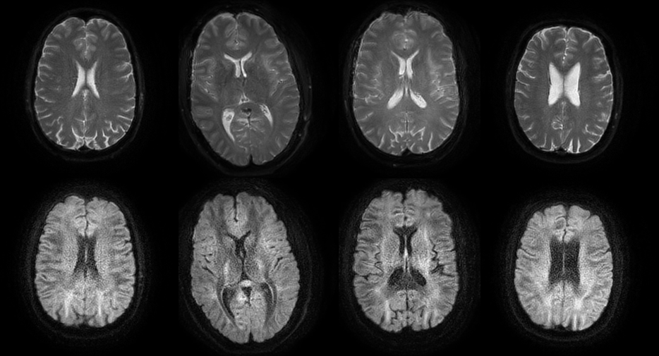
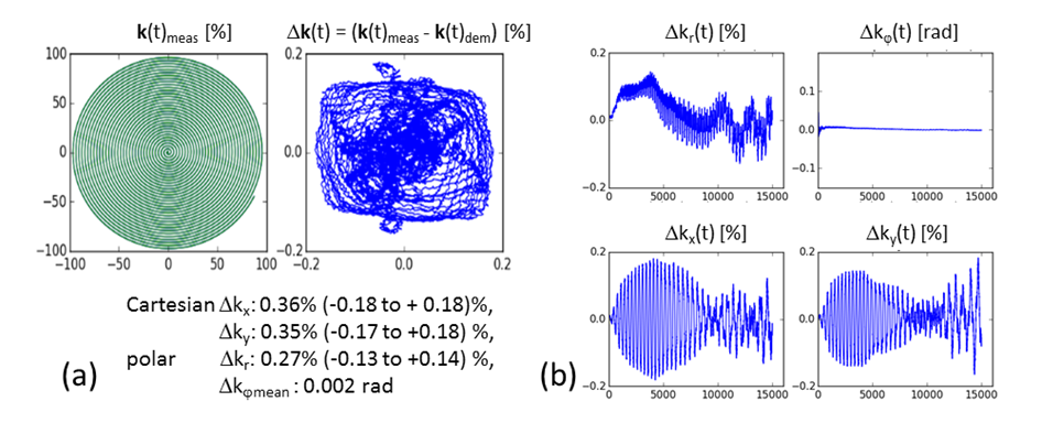
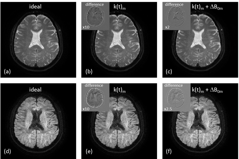
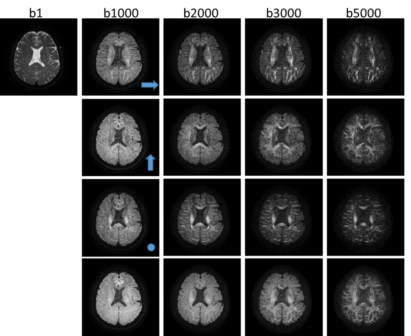
Phantom and volunteer experiments demonstrated the basic feasibility of single-shot diffusion-weighted spiral imaging on a clinical 3 T system. Good image quality was achieved using the demand k-space trajectory in gridding for both non-diffusion-weighted and diffusion-weighted scans. Figure 1 shows selected, representative results obtained in four of the volunteers. These indicate a sufficiently high fidelity of the gradient system. According to Fig. 2, the maximum deviation between realized and demand trajectory was smaller than 20% of one k-space increment (2 kmax / matrix size). Differences in resulting images, compiled in Fig. 3, are minor and seemed negligible for the moment. Furthermore, the accuracy and reproducibility of the resonance frequency determination and of the ΔB0 mapping were found to be sufficient to prevent blurring in this rather sensitive spiral acquisition with long AQ. However, patient motion-induced field fluctuations represent the lower limit for the accuracy of a separate, static mapping. Finally, image quality was not severely compromised by the application of strong diffusion sensitizing gradients up to b-values of 5000 s/mm² in three orthogonal diffusion gradient directions, as illustrated in Fig .4.Conclusion
Single-shot diffusion-weighted spiral imaging in the brain is feasible on a clinical 3 T scanner. The performance and fidelity of the employed gradient system was found to be sufficient to dispense with extra hardware for field monitoring. Further work will have to benchmark spiral imaging with echo planar imaging in this application.Acknowledgements
Thanks to Jim Pipe, Klaas Pruessmann and Bertram Wilm for helpful discussions and suggestions.References
1 Le Bihan D, Mangin JF, Poupon C, Clark CA, Pappata S, Molko N, Chabriat H. Diffusion tensor imaging: concepts and applications. J Magn Reson Imaging 2001; 13:534-546.
2 Wilm BJ, Barmet C, Gross S, Kasper L, Vannesjo SJ, Haeberlin M, Dietrich BE, Brunner DO, Schmid T, Pruessmann KP. Single-shot spiral imaging enabled by an expanded encoding model: Demonstration in diffusion MRI. Magn Reson Med 2017; 77:83-91.
3 Lee CE, Baker EH, Thomasson DM. Normal regional T1 and T2 relaxation times of the brain at 3T. Proc ISMRM 2006; 959.
4 Pipe JG, Zwart NR. Spiral trajectory design: A flexible numerical algorithm and base analytical equations. Magn Reson Med 2014; 71:278–285.
5 Pruessmann KP, Weiger M, Börnert P, Boesiger P. Advances in sensitivity encoding with arbitrary k-space trajectories. Magn Reson Med 2001; 46:638-51.
6 Wang D, Zwart NR, Pipe JG. Joint water–fat separation and deblurring for spiral imaging. Magn Reson Med 2018; 79:3218–3228.
7 Börnert P, Schomberg H, Aldefeld B, Groen J. Improvements in spiral MR imaging. MAGMA 1999; 9:29-41.
8 Brodsky EK, Klaers JL, Samsonov AA, Kijowski R, Block WF. Rapid measurement and correction of phase errors from B0 eddy currents: Impact on image quality for non-Cartesian imaging. Magn Reson Med 2013; 69:509–515.
Figures