0232
Improved gSlider Reconstruction for Isotropic High-Resolution DTI Using A Model-Based Method and Virtual Coil Concept1Center for Biomedical Imaging Research, Department of Biomedical Engineering, School of Medicine, Tsinghua University, Beijing, China, 2Department of Radiology, Stanford University, Stanford, CA, United States, 3A. A. Martinos Center for Biomedical Imaging, Department of Radiology, Massachusetts General Hospital, Charlestown, MA, United States, 4Department of Electrical Engineering and Computer Science, MIT, Cambridge, MA, United States
Synopsis
Recently, generalized SLIce Dithered Enhanced Resolution (gSlider) has been proposed as a new acquisition strategy for isotropic high-resolution DTI. In this study, to further boost the SNR performance and reconstruction accuracy, the model-based DTI reconstruction method was merged into the gSlider reconstruction procedure. Moreover, virtual coil (VC) concept was also integrated into the proposed method to further improve the SNR of gSlider reconstruction. Compared with conventional GRAPPA, the superiority of the model-based method has been demonstrated by in vivo results, especially when in combination with the virtual coil concept.
Introduction
The generalized SLIce Dithered Enhanced Resolution (gSlider) is a newly proposed technique for isotropic high-resolution dMRI1. It’s more SNR efficient than 2D ssEPI acquisition, but still suffers the intrinsic low SNR of dMRI. The problem is more obvious when using in-plane parallel imaging (PI) to reduce distortion. Therefore, a reconstruction method with high SNR maintaining capacity is desired. For under-sampled DTI data, the model-based method has shown improved accuracy and SNR than conventional compressed sensing (CS) or GRAPPA2-4. The virtual coil (VC) concept has also been demonstrated to further improve PI reconstruction5-7. In this work, we combined the model-based method and virtual coil concept to improve the SNR and accuracy of the gSlider-DTI reconstruction.Methods
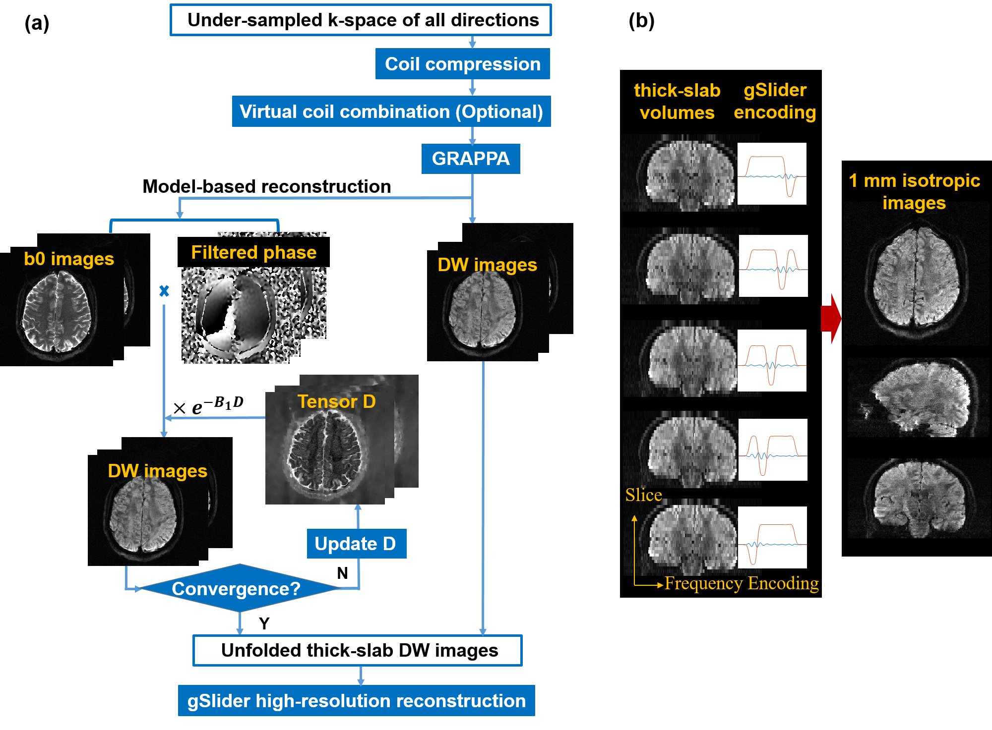
Data Acquisition: This study was approved by the Institutional Review Board and written informed consent was obtained from the participant. Whole-brain DTI data was acquired on a Philips 3.0T Achieva TX MR scanner with a 32-channel head coil. The gSlider-EPI-DTI sequence was performed with following parameters: gSlider factor=5, 1 mm isotropic resolution, GRAPPA=2, partial Fourier=0.6, FOV=216×216×120 mm3, 2 b0 images, b=800 s/mm2, 6 diffusion directions, TE/TR=76/10000 ms, NSA=1, acquisition time=7 min. A 6-sec low resolution reference data was acquired for GRAPPA calibration. We used the linear phase gSlider RF pulses designed by the SLR algorithm8 (https://github.com/wgrissom/gSliderRF). To mitigate inter-slab striping artifact, we 1) used 15% thicker refocusing pulse and ensured the same slab selection gradient amplitude of excitation and refocusing to minimize the influence of off-resonance effect; 2) used a long TR to minimize signal saturation of adjacent slabs.
Reconstruction: Figure 1 shows the reconstruction diagram. The in-plane under-sampled thick-slab volumes were firstly unfolded by GRAPPA or the model-based procedure. In model-based reconstruction, the k-space signal of the m-th diffusion direction Km is modeled by2
$$\mathbf {K}_m(\mathbf {D})=F(\mathbf {f}\cdot \mathbf {p}_m\cdot e^{-\mathbf {B}_m\mathbf {D}}),m=1,2,...,N_{direction}[1]$$
where D is the six-element diffusion tensor, f is the magnitude of unfolded b0 image, pm represents the motion-induced phase of diffusion images, Bm is the B-matrix of m-th diffusion direction, F is the Fourier transformation operator, and Ndirection is the total number of diffusion directions. Note that the channel dimension is omitted in the equation, where f, p and K include multi-channel information. With the acquired under-sampled k-space signal K’, f and pm can be obtained by GRAPPA, then the only unknown D can be solved by2
$$arg\min_{\mathbf {D}}\frac{1}{2}\parallel \mathbf {K'}-R\mathbf {K}(\mathbf {D})\parallel ^2_2+\lambda \cdot TV(\mathbf {D})[2]$$
where K(D) is the k-space data calculated by Equation [1] using the estimated D, R is the operator to select data of K at the same location of K’. Finally, DWI images can be obtained with the model in [1]. As model-based reconstruction is computation intensive, we carried out coil compression9 to compress data from 32 into 8 channels with maximum coil information preserved, and applied parallel computation for further computational acceleration.
The five unfolded thick-slab volumes were then combined to obtain the final isotropic images by solving a regularized linear equation, with encoding information from Bloch Simulation1. Real-valued thick-slab data were used to avoid magnitude bias1.
We compared the proposed VC+model-based method, model-based method and conventional GRAPPA method on DW images, FA and color-coded FA maps.
Results
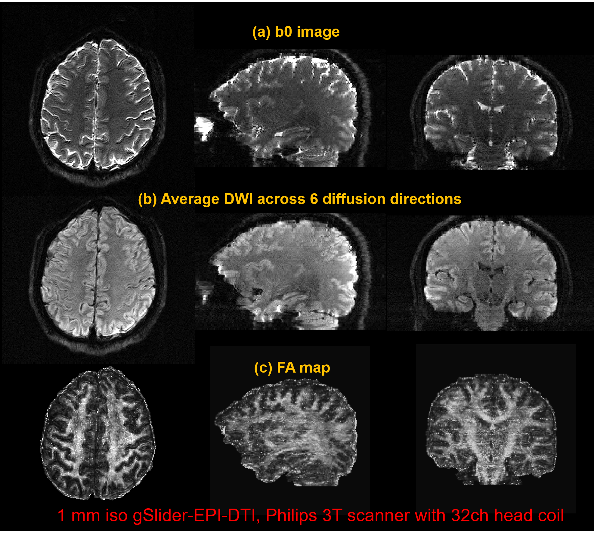
Figure 2 shows the reconstructed 1 mm isotropic b0 images, average DW images and FA maps by VC+model-based reconstruction.
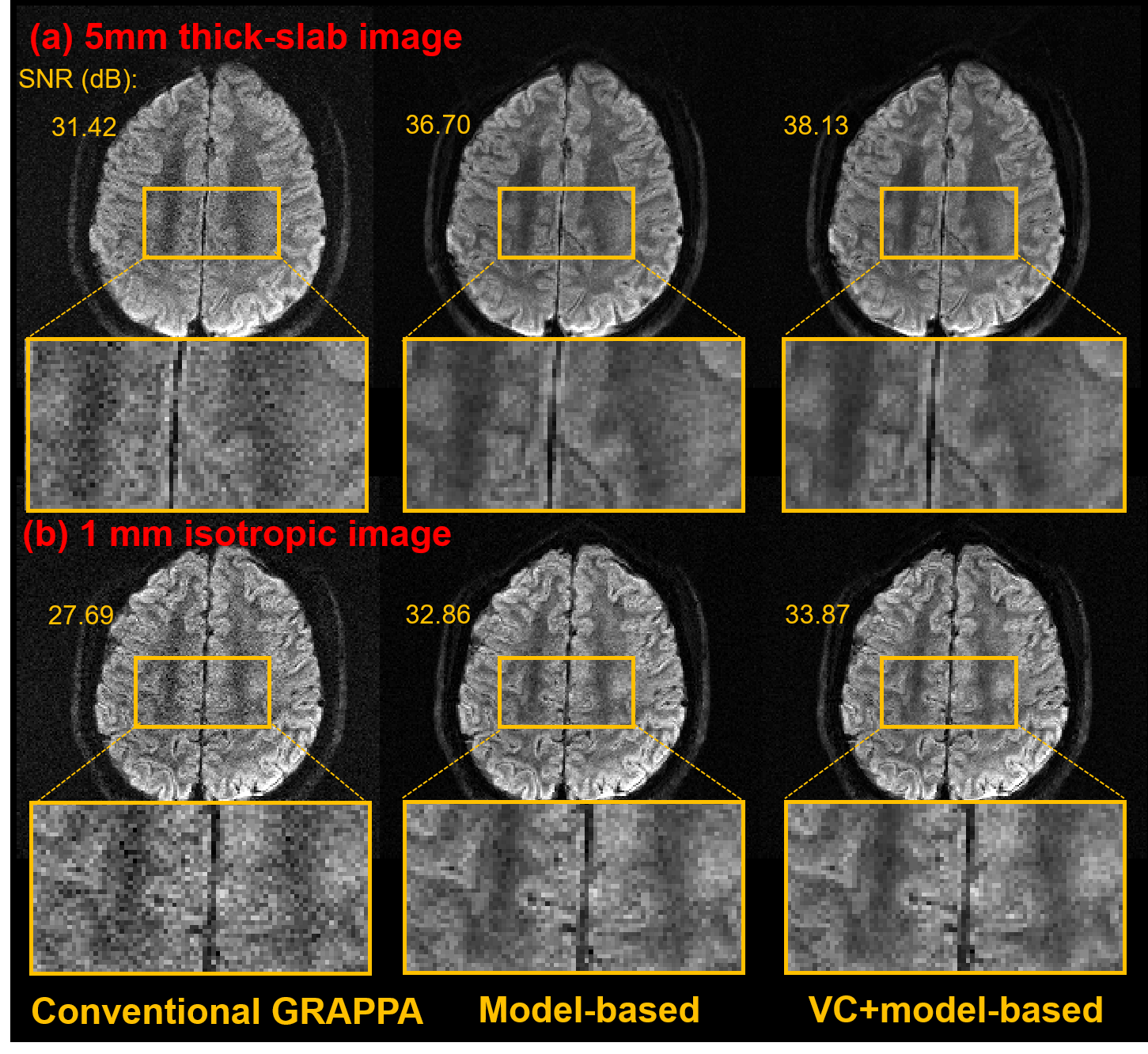
Figure 3(a) compares the SNR performance of conventional GRAPPA, model-based and VC+model-based reconstruction. Model-based procedure significantly improved the image quality compared to GRAPPA, and can achieve additional SNR gain when in combination with VC. Higher SNR of slab image can benefit the subsequent high-resolution through-slice reconstruction, as illustrated in Figure 3(b).
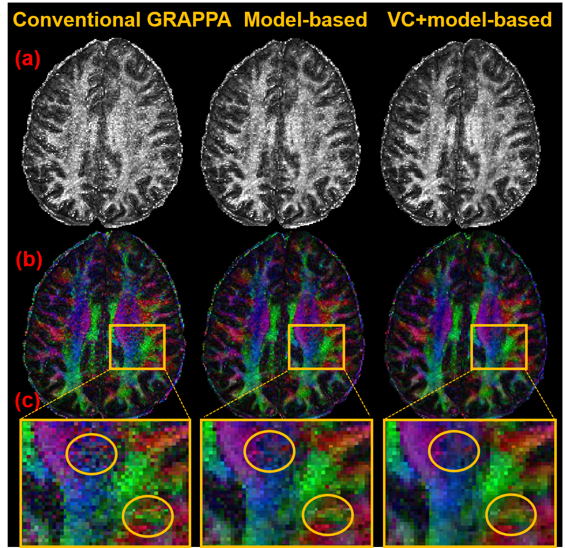
Figure 4 shows the FA and color-coded FA map calculated from the isotropic images. The result also demonstrated the improved SNR of model-based and VC+model-based reconstruction. The performance of VC+model-based reconstruction was even better.
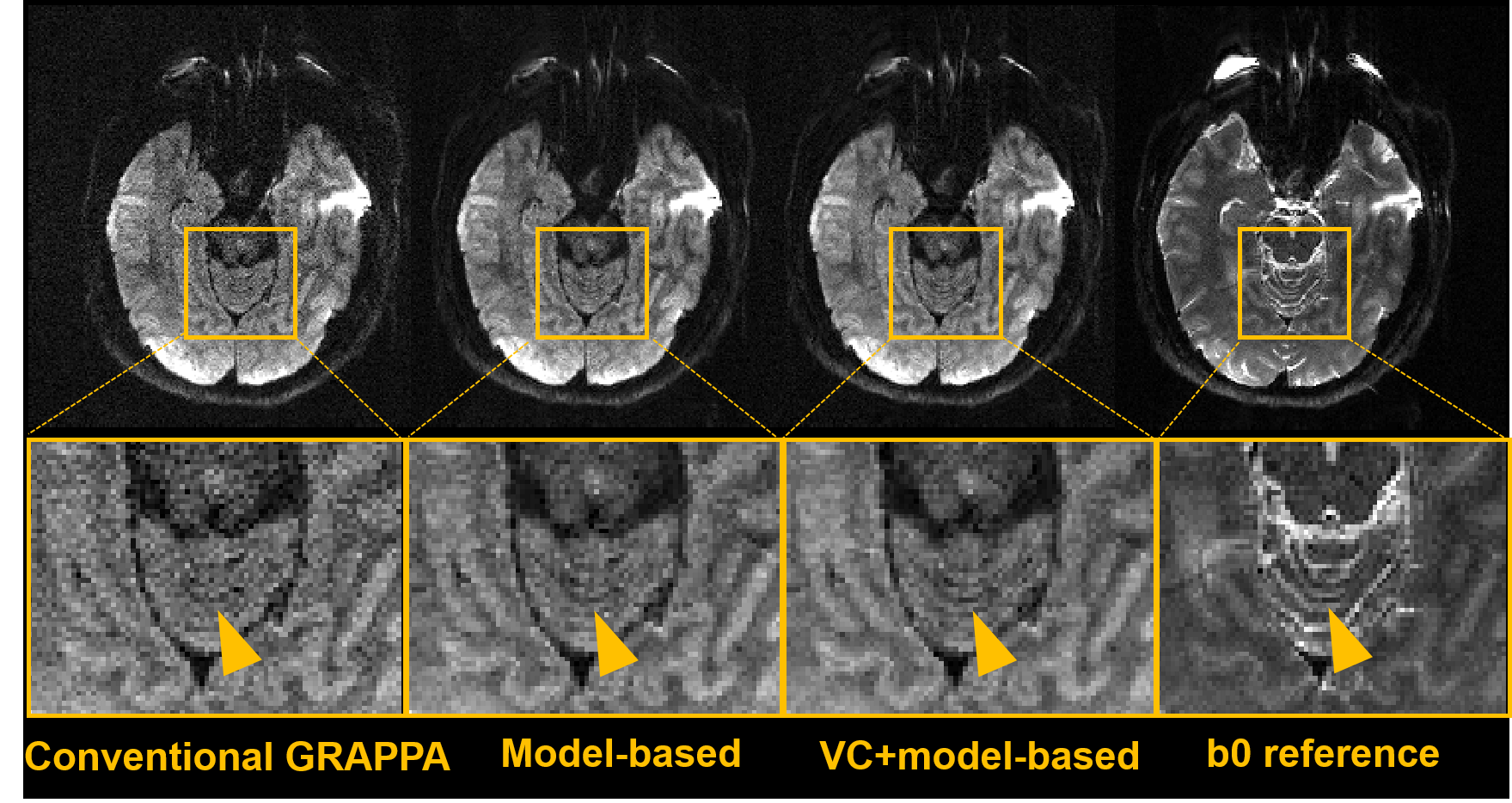
Figure 5 shows the improved reconstruction accuracy of model-based procedure than conventional GRAPPA. Model-based reconstruction may help depict detailed information more clearly.
Discussion and Conclusion
Model-based formulation uses the correlation across different diffusion directions, while conventional GRAPPA reconstructed images of each direction individually. Thus model-based reconstruction can improve accuracy and SNR. With virtual coil, additional image and phase information can be provided, thus are beneficial to the reconstruction of under-sampled data.
In this work, we proposed a model-based reconstruction method combined with virtual coil concept for the parallel imaging accelerated isotropic gSlider-EPI-DWI. It shows improved SNR and accuracy compared to conventional GRAPPA. Moreover, VC+model-based method can achieve additional SNR gain than none-VC model-based method.
Acknowledgements
The authors would like to thank Drs. William A Grissom and Jun Ma in Vanderbilt University for providing gSlider RF pulses, thank Drs. Kawin Setsompop and Congyu Liao in MGH for suggestions on dealing with gSlider striping artifact.References
[1] Setsompop K, Fan Q, Stockmann J, et al. High-resolution in vivo diffusion imaging of the human brain with generalized slice dithered enhanced resolution: Simultaneous multislice (gSlider-SMS). Magn Reson Med 2017.
[2] Dong Z, Dai E, Wang F, et al. Model‐Based Reconstruction for Simultaneous Multislice and Parallel Imaging Accelerated Multishot Diffusion Tensor Imaging. Medical physics 2018.
[3] Welsh CL, DiBella EVR, Adluru G, Hsu EW. Model-based reconstruction of undersampled diffusion tensor k-space data. Magnetic Resonance in Medicine 2013;70(2):429-440.
[4] Zhu Y, Peng X, Wu Y, et al. Direct diffusion tensor estimation using a model-based method with spatial and parametric constraints. Medical Physics 2017;44(2):570-580.
[5] Blaimer M, Gutberlet M, Kellman P, Breuer FA, Köstler H, Griswold MA. Virtual coil concept for improved parallel MRI employing conjugate symmetric signals. Magnetic Resonance in Medicine 2009;61(1):93-102.
[6] Blaimer M, Jakob PM, Breuer FA. Regularization method for phase-constrained parallel MRI. Magnetic Resonance in Medicine 2014;72(1):166-171.
[7] Kettinger AO, Kannengiesser SAR, Breuer FA, Vidnyanszky Z, Blaimer M. Controlling the object phase for g-factor reduction in phase-Constrained parallel MRI using spatially selective RF pulses. Magnetic Resonance in Medicine 2018;79(4):2113-2125.
[8] Jun Ma, Thomas Witzel, William A Grissom, and Kawin Setsompop. Minimum peak power root-flipped gSlider-SMS RF pulses for high-resolution in vivo diffusion imaging. Proc ISMRM 2017.
[9] Zhang T, Pauly JM, Vasanawala SS, Lustig M. Coil compression for accelerated imaging with Cartesian sampling. Magnetic Resonance in Medicine 2013;69(2):571-582.
Figures