0227
Diffusion acquisition methods for unfixed ex vivo neonatal brain scan at 7T: sequence optimisation and preliminary experiments1Wellcome Centre for Integrative Neuroimaging, University of Oxford, Oxford, United Kingdom, 2Department of Experimental Psychology, University of Oxford, Oxford, United Kingdom, 3Sir Peter Mansfield Imaging Centre, School of Medicine, University of Nottingham, Nottingham, United Kingdom, 4Centre for the Developing Brain, King's College London, London, United Kingdom, 5Biomedical Engineering Department, King's College London, London, United Kingdom
Synopsis
In this work we present preliminary investigations into the use of diffusion-weighted steady state free procession (DW-SSFP) sequences and conventional diffusion-weighted spin-echo (DW-SE) sequences for unfixed neonate brains. We conduct an exploratory experiment to demonstrate the feasibility of existing dMRI methods on an unfixed porcine brain. We then propose a framework to match DW-SE and DW-SSFP contrast in a way that predicts optimal performance of each, in order to compare between methods.
Introduction
Diffusion MRI (dMRI) of post-mortem brains can provide a link between in vivo imaging and anatomical dissection or microscopy, as well as the opportunity to acquire very high-resolution data unimpeded by subject compliance over long scan times. However, alterations of post-mortem tissue mean that straightforward adaptation of in-vivo imaging is severely compromised.
Post-mortem imaging can be done in fixed or unfixed tissue. Most studies use fixed tissue, in which T1, T2 and diffusivity are severely reduced. Conventional diffusion-weighted spin-echo (DW-SE) imaging has extremely low signal-to-noise ratio (SNR) in fixed tissue, while diffusion-weighted steady state free procession (DW-SSFP) provides significant improvements in SNR1-2. Unfixed brain tissue has been less well studied, but provides benefits including flexibility in tissue handling and the ability to scan in situ. Although unfixed tissue is more similar to in vivo tissue, MR properties are still altered by temperature, cessation of physiological processes and tissue atrophy.
Here, we present preliminary investigations into the use of DW-SSFP and DW-SE sequences for unfixed neonate brains. We conduct an exploratory experiment to demonstrate the feasibility of existing dMRI methods on an unfixed porcine brain. We then propose a framework to match DW-SE and DW-SSFP contrast in a way that predicts optimal performance of each, in order to compare between methods.
Methods
Post-mortem brain scan
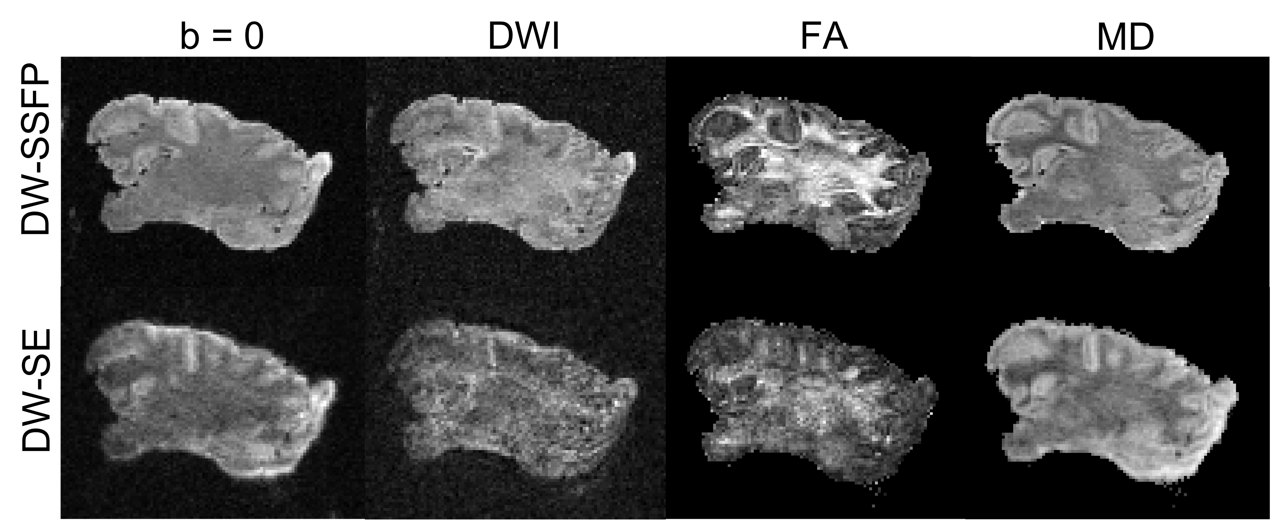
We conducted a feasibility study to establish whether in-vivo or fixed-tissue dMRI protocols would be more appropriate for unfixed tissue. We extracted a porcine brain from an animal that was sacrificed for an independent experiment conducted under a British Home Office license. The brain was packed in a cylinder filled with susceptibility-matched fluid and scanned on a Siemens 7T scanner 28 hours after death. Scanning used a vendor-provided DW-SE sequence optimised for neonatal tissue properties, and a custom DW-SSFP sequence that was previously optimised for fixed tissue. DW-SE used a readout-segmented EPI sequence that reduces image distortion and blurring3-4. Scan parameters: TR/TE=19s/82ms, 11 segments, GRAPPA=2, b=4000s/mm2. DW-SSFP used5-6:TR/TE=28/19ms, flip angle=35°, diffusion gradient amplitude 52mT/m and duration 10.4ms (q=230cm-1). Both sequences acquired 47 directions, FOV=196x148x96mm3, 0.8mm isotropic resolution. Quantitative T1, T2 and B1 maps were also acquired.
Sequence optimisation
One challenge of matching DW-SE and DW-SSFP protocols for predicting optimal performance is that contrast differs based on signal mechanisms. To establish target contrast, we extracted a diffusion tensor of a voxel in the corpus callosum from a dHCP neonatal brain7 for simulation. The tensor was scaled to the predicted ADC value of unfixed post-mortem neonatal brain. Tissue parameters (T1=2126ms, T2=135ms, ADC=2.72×10⁻³ mm2/s) were predicted for unfixed post-mortem neonates at 7T8-9, including temperature and field strength corrections10-14.
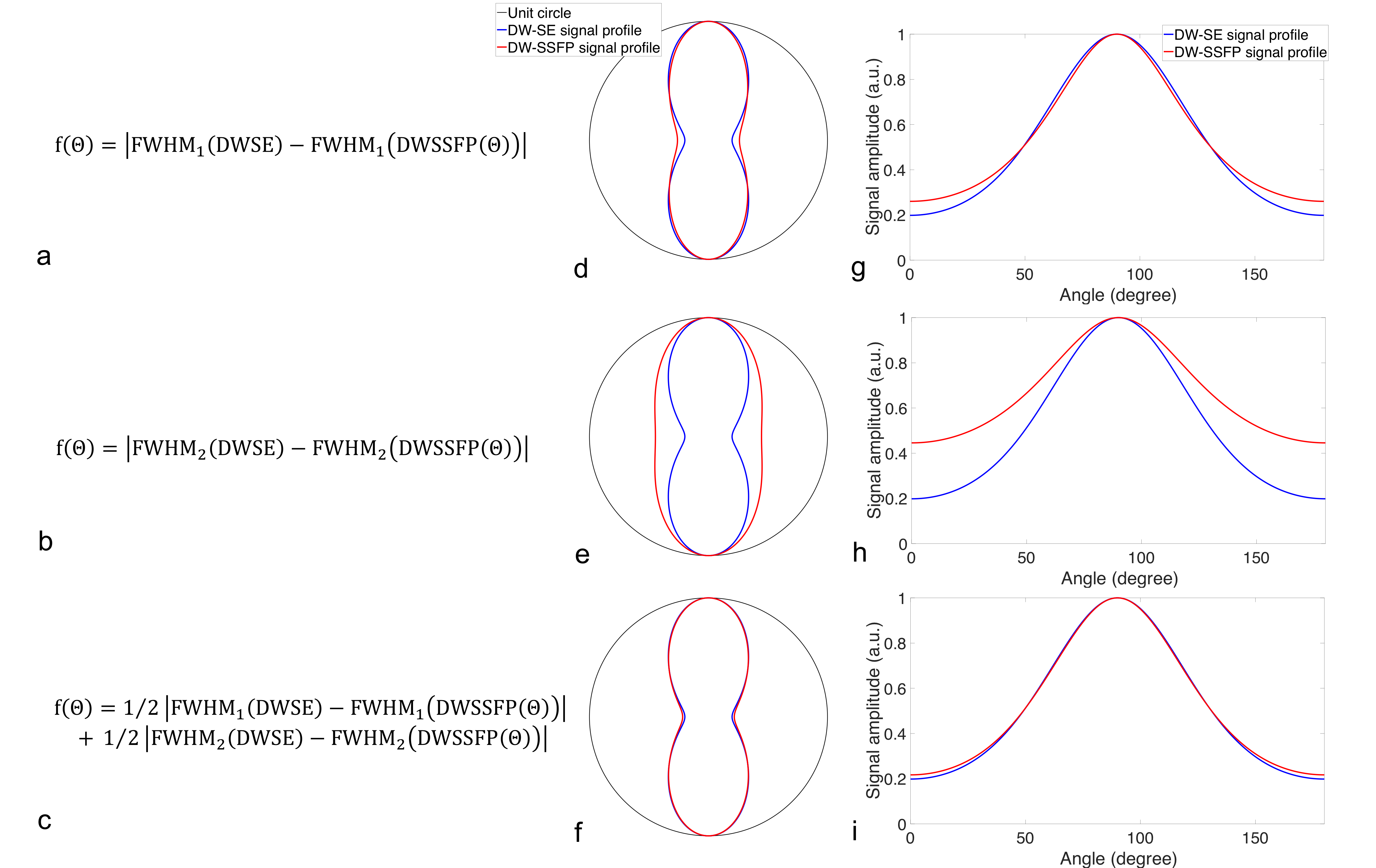
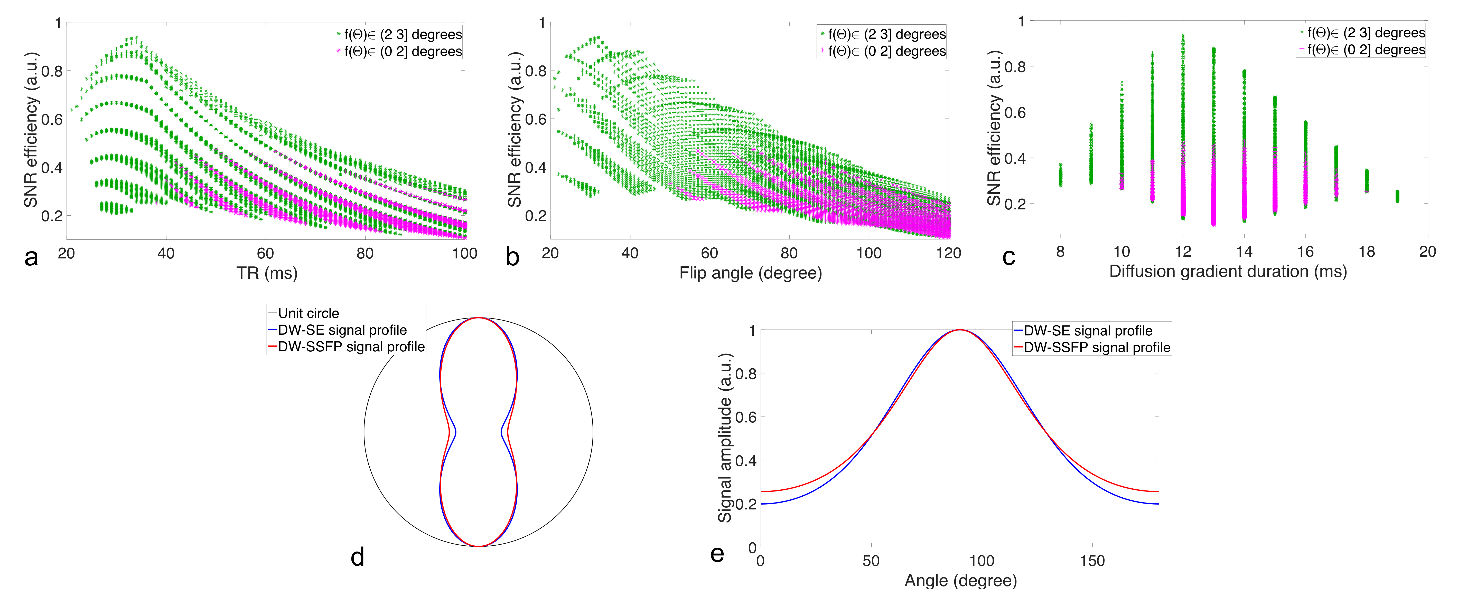
The DW-SE signal profile was simulated for this tensor with b=4000s/mm2 and used as a target for DW-SSFP signal. Signal profiles were simulated for combinations of diffusion gradient duration=0-99ms, TR=0-99ms, flip angle 0-120°, diffusion gradient amplitude 52mT/m. Readout time was limited to 20ms to limit blurring and distortion. We then optimised the DW-SSFP sequence parameters to match the DW-SE signal profile within some tolerance. The cost function was based on a generalisation of the concept of full-width at half-maximum (FWHM), using two terms (Fig. 2c). The best parameter set was identified that was within the set tolerance and provided the highest SNR efficiency (Fig. 3d,e).
Results
In the porcine brain experiment, DW-SSFP acquisition out-performed DW-SE, which suffers from distortion, blurring and low SNR. These results suggest that there is value in utilising custom sequences in unfixed tissue, similar to the approaches previously developed for fixed tissue.
The contrast optimisation identified a broad range of DW-SSFP parameters that produce nearly identical signal profile to the DW-SE sequence, with the inclusion of a tolerance on exact profile matching further enabling the identification of protocols that provide high SNR efficiency (Fig. 3). These results suggest that, even with optimal conditions for DW-SE requiring acquisition schemes not generally available, DW-SSFP should out-perform DW-SE by a significant margin (with SNR efficiencies of 0.08 for DW-SE compared to 0.94 for DW-SSFP).
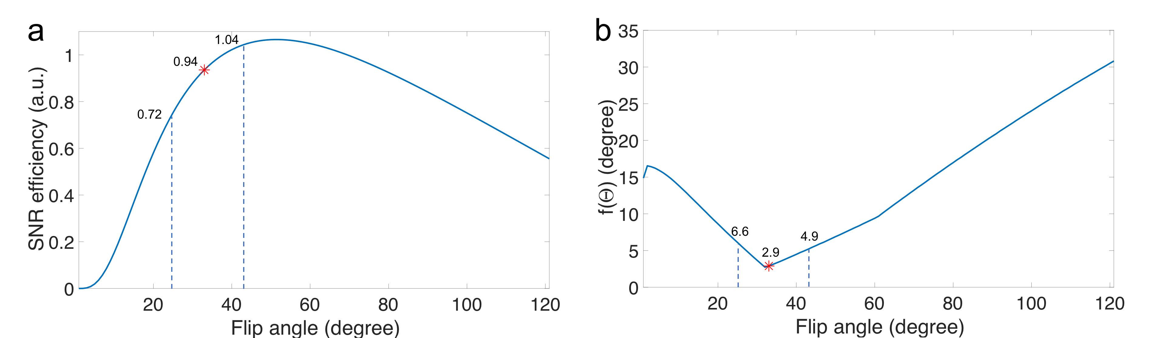
Fig.4 shows SNR efficiency and FWHM errors (Fig.2c) for different flip angles, which may vary substantially at 7T. With 30% flip angle variation (bounded by dashed lines), the SNR efficiency varies between 70% and 110% of the value corresponding to the optimal sequence parameters, with little change in the FWHM metric.
Discussion and Conclusions
We present preliminary results of high-resolution dMRI scan of unfixed brains at 7T using DW-SSFP and DW-SE sequences. Compared with conventional (vendor-supplied) DW-SE sequences, DW-SSFP provides superior SNR efficiency and image fidelity. Future work will investigate potential optimisations of DW-SE beyond conventional acquisitions, validity of the predicted tissue properties of unfixed, neonatal brain tissue at 7T, and the overall feasibility of high-resolution dMRI in neonatal brain.
Acknowledgements
Funding for this work was provided by Wellcome Trust fellowship (202788/Z/16/Z, KM). The Wellcome Centre for Integrative Neuroimaging is supported by core funding from the Wellcome Trust (203139/Z/16/Z).References
1. McNab, J.A., Jbabdi, S., Deoni, S.C., Douaud, G., Behrens, T.E. and Miller, K.L., 2009. High resolution diffusion-weighted imaging in fixed human brain using diffusion-weighted steady state free precession. Neuroimage, 46(3), pp.775-785.
2. Miller, K.L., Stagg, C.J., Douaud, G., Jbabdi, S., Smith, S.M., Behrens, T.E., Jenkinson, M., Chance, S.A., Esiri, M.M., Voets, N.L. and Jenkinson, N., 2011. Diffusion imaging of whole, post-mortem human brains on a clinical MRI scanner. Neuroimage, 57(1), pp.167-181.
3. Holdsworth, S.J., Skare, S., Newbould, R.D., Guzmann, R., Blevins, N.H. and Bammer, R., 2008. Readout-segmented EPI for rapid high resolution diffusion imaging at 3T. European journal of radiology, 65(1), pp.36-46.
4. Porter, D.A. and Heidemann, R.M., 2009. High resolution diffusion‐weighted imaging using readout‐segmented echo‐planar imaging, parallel imaging and a two‐dimensional navigator‐based reacquisition. Magnetic Resonance in Medicine, 62(2), pp.468-475.
5. Foxley, S., Jbabdi, S., Clare, S., Lam, W., Ansorge, O., Douaud, G. and Miller, K., 2014. Improving diffusion-weighted imaging of post-mortem human brains: SSFP at 7 T. Neuroimage, 102, pp.579-589.
6. Pallebage-Gamarallage, M., Foxley, S., Menke, R.A., Huszar, I.N., Jenkinson, M., Tendler, B.C., Wang, C., Jbabdi, S., Turner, M.R., Miller, K.L. and Ansorge, O., 2018. Dissecting the pathobiology of altered MRI signal in amyotrophic lateral sclerosis: A post mortem whole brain sampling strategy for the integration of ultra-high-field MRI and quantitative neuropathology. BMC neuroscience, 19(1), p.11.
7. Bastiani, M., Andersson, J., Cordero-Grande, L., Murgasova, M., Hutter, J., Price, A.N., Makropoulos, A., Fitzgibbon, S.P., Hughes, E., Rueckert, D. and Victor, S., 2018. Automated processing pipeline for neonatal diffusion MRI in the developing Human Connectome Project. NeuroImage.
8. Thayyil, S., De Vita, E., Sebire, N.J., Bainbridge, A., Thomas, D., Gunny, R., Chong, K., Lythgoe, M.F., Golay, X., Robertson, N.J. and Cady, E.B., 2012. Post-mortem cerebral magnetic resonance imaging T1 and T2 in fetuses, newborns and infants. European journal of radiology, 81(3), pp.e232-e238.
9. McDowell, A.R., Shelmerdine, S.C., Carmichael, D.W. and Arthurs, O.J., 2018. High resolution isotropic diffusion imaging in post-mortem neonates: a feasibility study. The British journal of radiology, 91, p.20180319.
10. Cox, E.F. and Gowland, P.A., 2008. Measuring T2 and T2’ in the brain at 1.5 T, 3T and 7T using a hybrid gradient echo-spin echo sequence and EPI. In Proceedings of the 16th annual meeting of ISMRM, Toronto, Canada (p. 1411).
11. Wright, P.J., Mougin, O.E., Totman, J.J., Peters, A.M., Brookes, M.J., Coxon, R., Morris, P.E., Clemence, M., Francis, S.T., Bowtell, R.W. and Gowland, P.A., 2008. Water proton T 1 measurements in brain tissue at 7, 3, and 1.5 T using IR-EPI, IR-TSE, and MPRAGE: results and optimization. Magnetic Resonance Materials in Physics, Biology and Medicine, 21(1-2), p.121.
12. Birkl, C., Langkammer, C., Haybaeck, J., Ernst, C., Stollberger, R., Fazekas, F. and Ropele, S., 2014. Temperature‐induced changes of magnetic resonance relaxation times in the human brain: A postmortem study. Magnetic resonance in medicine, 71(4), pp.1575-1580.
13. Arthurs, O.J., Price, G.C., Carmichael, D.W., Jones, R., Norman, W., Taylor, A.M. and Sebire, N.J., 2015. Diffusion-weighted perinatal postmortem magnetic resonance imaging as a marker of postmortem interval. European radiology, 25(5), pp.1399-1406.
14. Papadopoulou, I., Langan, D., Sebire, N.J., Jacques, T.S. and Arthurs, O.J., 2016. Diffusion-weighted post-mortem magnetic resonance imaging of the human fetal brain in situ. European journal of radiology, 85(6), pp.1167-1173.
Figures